Diffusion-Weighted Imaging (DWI) and Diffusion Tensor Imaging (DTI)
Why diffusion imaging matters
Brain function depends critically on connectivity, not just isolated cortical regions. White-matter tracts enable communication between distributed brain areas, and disruption of these pathways underlies many neurological and neuropsychological conditions.
Diffusion MRI tracks how water molecules move inside the brain. In white matter, water tends to move along nerve fibers, not across them. By measuring this movement, scientists can infer the direction, integrity, and organization of white-matter pathways, basically mapping the brain’s wiring while a person is alive.
Regular (structural) MRI, by contrast, mostly shows anatomy: the shape, size, and location of brain structures. It can tell you where white matter is, but not how well it’s connected or which regions are talking to each other.
What diffusion is
Diffusion refers to the random movement of molecules due to thermal energy. Molecules naturally move from areas of higher concentration to lower concentration.
In the brain:
Higher diffusivity means water molecules move more freely
Lower diffusivity means movement is restricted by tissue structure
Diffusion MRI measures how freely water molecules move within brain tissue.
Isotropic vs anisotropic diffusion
Isotropic diffusion
Diffusion is equal in all directions.
This occurs in:
cerebrospinal fluid (CSF),
grey matter (to a lesser extent).
There is no preferred direction of movement.
Anisotropic diffusion
Diffusion does not occur equally in all directions.
In white matter, the movement of water is directionally constrained.
This happens because:
Axons are organized into parallel bundles
Myelin sheaths and internal cellular structures limit water movement across fibers
Water moves most freely along the length of axons and is hindered in directions perpendicular to them.
This directional dependence of diffusion is known as anisotropy, and it is the fundamental property that diffusion MRI measures and uses to characterize white-matter structure.
Diffusion properties in brain tissue
Different tissues show distinct diffusion characteristics:
CSF shows very high diffusivity and very low anisotropy
Grey matter shows moderate diffusivity and low anisotropy
White matter shows moderate diffusivity but high anisotropy
These differences allow diffusion MRI to distinguish tissue types and infer white-matter organisation.
Diffusion-Weighted Imaging (DWI)
DWI is the basic diffusion MRI acquisition. It measures how much the MRI signal is attenuated by diffusion in different directions.
Key concepts:
Diffusion sensitivity can be applied in any direction
Signal attenuation depends on how freely water moves in that direction
Acquiring data in multiple gradient directions allows inference about tissue structure
What DTI is
Diffusion Tensor Imaging (DTI) is a type of MRI that uses water movement to learn about white matter in the brain.
It does not see nerve fibres directly.
It infers their organisation from how water moves.
What DTI measures
DTI measures how water diffuses inside brain tissue, focusing on:
How much water moves (overall diffusion)
Whether water moves more in one direction than others (anisotropy)
The main direction of water movement inside each voxel
From this, we calculate common measures:
FA (fractional anisotropy) – how directional the diffusion is
MD (mean diffusivity) – how freely water moves overall
Principal direction – the dominant orientation of diffusion
How DTI works (step by step)
The MRI applies diffusion sensitivity in many directions.
Water movement reduces the MRI signal differently depending on direction.
These differences are measured inside each voxel.
A tensor model (a mathematical shape) is fitted to summarise diffusion.
The tensor describes:
diffusion strength
diffusion direction
how directional it is
The main axis of the tensor is assumed to line up with fibre orientation.
What DTI is good at (strengths)
Works in living humans
Non-invasive and safe
Good at showing large, well-organised white-matter tracts
Sensitive to:
development
injury
stroke
tumours
FA and MD are easy to calculate and compare across people
Forms the basis for tractography
DTI is especially useful for group comparisons and clinical applications.
What DTI cannot do (limitations)
Voxel size is large (mm), axons are tiny (µm)
Measures are indirect and reflect many tissue features at once
Sensitive to damage, but not specific to the cause
Cannot show direction of information flow
Fails when fibres cross or fan within a voxel
Assumes only one main fibre direction per voxel
Tractography based on DTI can produce:
false connections
missing real connections
What MRI is (baseline)
MRI is a way of making images using:
a strong magnetic field
radio waves
signals from hydrogen atoms (mostly in water)
Standard MRI (like T1 or T2 scans) shows:
anatomy
shapes and boundaries of tissues
It does not look at water motion.
What DWI is, and why it’s still MRI
DWI (Diffusion-Weighted Imaging) is MRI with an extra trick added.
How:
the scanner applies special magnetic gradients
these gradients make the signal sensitive to water movement
What changes:
if water moves a lot → signal drops
if water is restricted → signal stays stronger
Why it’s MRI:
same scanner
same physics
just different gradient settings
So DWI is:
MRI that measures water motion instead of just structure
What DTI is, and how it relates to DWI
DTI (Diffusion Tensor Imaging) is not a new scan.
It is:
a way of analysing DWI data
How:
DWI images are acquired in many directions
a mathematical model (the tensor) is fitted to those measurements
What DTI gives:
FA (directionality)
MD (overall diffusion)
main diffusion direction
So:
DWI = how the data are acquired
DTI = how the data are modelled and summarised
Why DWI and DTI exist at all
Standard MRI shows:
what tissue looks like
DWI/DTI show:
how tissue is organised at a microscopic level
especially white matter structure
They are used because:
they detect stroke earlier than conventional MRI
they are sensitive to white matter damage
they allow tractography
One clean way to remember it
MRI = the general imaging method
DWI = MRI made sensitive to water movement
DTI = a model applied to DWI to describe direction and structure
The key idea
Normal (structural) MRI sees shape and structure.
DWI sees microscopic water movement.
Some problems change water behaviour first, long before they change visible structure.
Why a normal MRI can miss early stroke
What happens in acute stroke (minutes–hours)
Blood flow stops
Cells lose energy
Ion pumps fail
Cells swell (cytotoxic oedema)
Water gets trapped inside cells
At this point:
Brain tissue has not fallen apart
No bleeding
No big swelling you can see
So on normal MRI:
Shape looks normal
Tissue boundaries look normal
Nothing obvious to “see”
But at the microscopic level:
Water movement has changed dramatically
DWI is sensitive to that immediately.
Why normal MRI sees tumours but not early damage
Tumours change structure
Tumours usually cause:
extra tissue
mass effect
displacement of normal anatomy
blood–brain barrier leakage
These are macroscopic changes.
Structural MRI is very good at:
seeing abnormal shapes
detecting contrast enhancement
spotting mass effects
That’s why tumours are often obvious.
Why cellular swelling is invisible to normal MRI
Cellular swelling means:
cells get a bit bigger
extracellular space gets smaller
But:
this happens at the micrometre scale
MRI voxels are millimetres wide
So the voxel still looks “full of brain” either way.
Normal MRI averages everything together and sees:
“Still looks like brain.”
DWI sees:
“Water can’t move like it used to.”
Why DWI sees things before anatomy changes
Normal MRI relies on:
tissue breakdown
fluid shifts
bleeding
structural distortion
Those take hours to days.
DWI relies on:
water motion inside cells
That changes in minutes.
So:
Function changes before structure does.
Tumour characteristics and treatment response
Tumours are a special case because both methods help.
Structural MRI:
shows size and location
DWI:
shows how tightly packed the cells are
shows whether treatment is killing cells
A tumour can:
look the same size
but have very different diffusion properties
So DWI gives extra information, not redundant information.
One sentence to remember
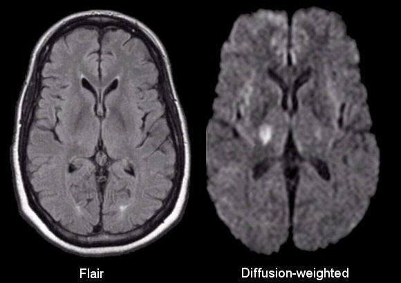
Normal MRI and DWI look at different kinds of changes in the brain. A normal MRI shows structure and shape, so it is good at seeing things like tumours, bleeding, or major swelling, but it only changes when the tissue itself looks different. In conditions like an early stroke, the brain tissue still looks normal, but the cells stop working properly and swell, which traps water inside them. Normal MRI cannot see this because the changes are too small. DWI, however, is sensitive to how water moves, so it can detect these microscopic changes within minutes, long before the brain’s structure changes. That’s why doctors often use both scans together: one shows what the brain looks like, and the other shows how healthy the tissue is at a very small scale.
b-value
The b-value tells us how strongly the MRI is made sensitive to water movement.
Low b-value → weak sensitivity to diffusion
High b-value → strong sensitivity to diffusion
When the b-value is higher:
differences in water movement are easier to see
but the image becomes noisier (less signal)
That’s the trade-off:
better contrast, but poorer signal quality.
How b-values are used in practice
A diffusion scan usually includes:
b = 0 images (no diffusion weighting, like a normal MRI)
one or more higher b-values (to measure diffusion)
The scanner compares these images to work out diffusion.
Scan times are relatively short (minutes), making DWI practical in clinical settings.
Clinical usefulness of DWI
DWI is highly sensitive to acute tissue changes.
Key applications:
Acute ischaemic stroke: restricted diffusion appears within minutes to hours, earlier than conventional MRI changes
Tumours and oedema: diffusivity is altered by changes in cellular density and extracellular space
Monitoring treatment response: changes in diffusivity can reflect tumour progression or regression
Because diffusion responds quickly to microstructural change, DWI is a powerful clinical tool.
Artefacts and preprocessing in DWI
DWI uses fast imaging techniques, which makes it vulnerable to artefacts.
Common issues include:
Susceptibility-induced distortion at tissue–air boundaries
Eddy currents from rapidly switching gradients
Patient motion, particularly relevant in paediatric imaging
Standard preprocessing tools (e.g. TOPUP and EDDY) are used to correct these distortions and recover anatomically meaningful data.
From DWI to DTI
DWI provides diffusion-weighted images.
DTI (Diffusion Tensor Imaging) adds a mathematical model to describe diffusion within each voxel.
The diffusion tensor models diffusion as an ellipsoid, characterised by:
the amount of diffusion,
the degree of anisotropy,
the principal direction of diffusion.
DTI therefore summarises diffusion information rather than just displaying raw signal attenuation.
DTI summary parameters
Fractional anisotropy (FA)
FA quantifies how directional diffusion is.
FA ≈ 0 → isotropic diffusion
FA closer to 1 → highly anisotropic diffusion
High FA is typically associated with organised white-matter tracts.
FA (Fractional Anisotropy) is important because it tells us how organised and directional white-matter tissue is.
In white matter, axons are bundled together and wrapped in myelin. This structure forces water to move mainly along the fibres rather than in all directions. FA measures how strongly water movement is constrained to one direction. When white matter is healthy and well organised, FA is high.
When something disrupts that organisation, FA changes. If axons are damaged or lost, if myelin breaks down, if there is swelling, or if fibres cross within the same voxel, water movement becomes less directional and FA decreases. Because of this, FA is very sensitive to changes in white-matter microstructure, even when the brain still looks normal on standard MRI.
Mean diffusivity (MD)
MD reflects the overall magnitude of diffusion, regardless of direction.
Increased MD can reflect oedema, tissue loss, or inflammation
Decreased MD can reflect acute cytotoxic oedema (e.g. stroke)
MD (Mean Diffusivity) is important because it tells you how freely water can move in brain tissue, which reflects tissue health at a microscopic level.
Water moves differently depending on what the tissue is like. When tissue is healthy and intact, cell membranes, axons, and myelin act as barriers, so water movement is partly restricted and MD stays in a normal range. When tissue is acutely injured, such as in an early stroke, cells swell and trap water, so water movement is reduced and MD decreases. When tissue is damaged or breaking down over time, barriers are lost, water moves more freely, and MD increases.
Principal diffusion direction (PDD)
The PDD represents the direction of greatest diffusion within a voxel.
By assuming this aligns with the dominant fibre orientation, we can estimate local white-matter direction.
PDD (Principal Diffusion Direction) is important because it tells us the main orientation of white-matter fibres in each small region of the brain.
In white matter, water moves most easily along the length of axons. When DTI measures diffusion in many directions, it finds the direction where water moves the most. That direction is the principal diffusion direction.
This matters because:
it tells us the local orientation of white-matter bundles
it is the building block of tractography (connecting directions across voxels)
PDD assumes one main fibre direction per voxel, so it struggles where fibres cross or fan.
PDD is important because it gives the main fibre direction in each voxel, enabling mapping and tracking of white-matter pathways.
FA colour maps
FA colour maps encode the orientation of white matter:
Red = left–right
Green = anterior–posterior
Blue = superior–inferior
Importantly, these maps show orientation only, not direction of information flow.
Tractography
Tractography reconstructs white-matter pathways by:
selecting seed points,
following the principal diffusion direction voxel-to-voxel,
stopping when diffusion becomes isotropic or curvature is excessive.
This allows visualisation and segmentation of major white-matter tracts.
Applications include:
white-matter mapping,
studying effects of lesions or tumours,
surgical planning and neuronavigation,
tract-based group analyses of FA and MD.
Noise, uncertainty, and ROIs
Tractography accumulates local errors as it progresses, and trajectories can jump incorrectly between fibre bundles.
To manage this:
probabilistic tractography estimates uncertainty,
regions of interest (ROIs) are used to constrain tracking,
waypoint, exclusion, and termination masks improve anatomical specificity.
ROI definitions are not easily transferable between individuals, but standardised protocols (“recipes”) can be applied.
Limitations of DWI and DTI
Diffusion MRI has important limitations:
Spatial resolution is relatively low (2–3 mm voxels)
Diffusion measures are sensitive but not specific
Changes may reflect many processes (degeneration, inflammation, oedema)
Direction of communication cannot be inferred
Tractography works best for large, coherent tracts
False positives and false negatives are common
The crossing-fibre problem
DTI assumes a single dominant fibre orientation per voxel.
In reality, many voxels contain crossing or kissing fibres.
When fibres cross:
there is no true single principal direction,
FA can be misleading,
tractography may fail.
More advanced models (e.g. HARDI, multi-fibre models) attempt to address this, but introduce new trade-offs.
Integrated take-home messages
Diffusion MRI exploits the directional movement of water to probe white-matter structure.
DWI measures diffusion sensitivity; DTI adds a tensor model.
FA, MD, and PDD summarise different aspects of diffusion.
Diffusion MRI is clinically powerful but biologically non-specific.
Tractography visualises connectivity but must be interpreted cautiously.
Developmental stage critically shapes diffusion measures.
Final synthesis sentence
Diffusion MRI provides a uniquely sensitive window into white-matter organisation and development, but meaningful interpretation requires understanding the physics, modelling assumptions, developmental context, and limitations of DWI, DTI, and tractography.
Tractography: what it is
Tractography is a method for reconstructing white-matter pathways using diffusion MRI.
It works by:
following the main diffusion direction from voxel to voxel
linking these directions together to form lines called streamlines
When many streamlines run together, they form fibre bundles, which often correspond to known white-matter tracts.
This allows researchers and clinicians to:
visualise white matter
isolate specific tracts
measure values like FA or MD within those tracts
Important point: tractography is a model-based reconstruction, not a direct image of axons.
Noise and uncertainty in tractography
Diffusion measurements are noisy, and each voxel has some uncertainty.
As tractography follows directions step by step:
small local errors add up
streamlines may slowly drift away from the true pathway
paths can jump from one tract to a nearby one
Probabilistic tractography addresses this by:
running tracking many times
showing a range of possible paths
giving an idea of uncertainty rather than one “correct” line
Regions of interest (ROIs)
ROIs are used to constrain tractography and make results more anatomically meaningful.
They can:
define where streamlines start (seed)
force streamlines to pass through certain areas (waypoints)
stop or exclude streamlines from unwanted regions
Using ROIs:
reduces false connections
is standard practice in applications like surgical planning
Complex ROI strategies
Often, one ROI is not enough.
Multiple ROI types may be combined:
seed regions
waypoint regions
exclusion masks
termination regions
Challenges:
ROIs are hard to transfer directly between people
brains differ in size, shape, and development
However:
standardised ROI “recipes” can be reused across studies
Development and ageing: why diffusion measures change
White matter does not stay the same across life — it develops, matures, and later declines. Diffusion MRI is sensitive to these changes.
During childhood and adolescence
The brain is still building and refining its white matter.
What happens biologically:
Myelination increases (axons get more insulation)
Fibres become more tightly packed and better aligned
Connections become more efficient
What diffusion MRI sees:
FA increases
Water is pushed more strongly along fibres as organisation improves.MD decreases
Extra barriers from myelin and cells restrict water movement.
Many white-matter tracts mature at the same time, so their diffusion measures often change together.
During ageing
With ageing, some of these processes reverse.
What happens biologically:
Myelin breaks down
Axons may be lost or become disorganised
Tissue becomes less dense
What diffusion MRI sees:
FA decreases
Water movement becomes less directional.MD increases
Water moves more freely as barriers are lost.
Limitations of diffusion MRI and tractography
Key limitations include:
relatively low spatial resolution (millimetres)
diffusion measures reflect many tissue properties at once
sensitive to damage, but not specific to the cause
degeneration, inflammation, oedema can look similar
cannot show direction of information flow
tractography works best in large, well-organised tracts
false positives and false negatives are common
Limitations of DTI specifically
DTI uses a single tensor per voxel.
This works well when fibres:
are parallel
dominate the voxel
But it fails when:
two or more fibre bundles cross within one voxel
there is no single true principal direction
In these cases, DTI oversimplifies the anatomy.
The crossing-fibre problem
In real brains:
multiple fibre directions in one voxel are very common
Because the tensor model can only represent one direction:
crossing fibres are misrepresented
some tracts are missed or incorrectly reconstructed
This is a fundamental limitation of DTI, not a scanning mistake.
Improved models beyond DTI
More advanced diffusion models:
use more diffusion directions
often use higher b-values
allow multiple fibre orientations per voxel
Advantages:
better handling of crossings
more realistic fibre orientation estimates
Disadvantages:
longer scan times
lower signal
more false-positive connections
diminishing returns beyond a point
Big-picture summary
Water diffusion in the brain is shaped by tissue structure.
Diffusion MRI measures this indirectly.
Tractography follows preferred diffusion directions to estimate white-matter pathways.
Diffusion measures are useful for studying development, stroke, tumours, and surgical planning.
All results require careful interpretation, because models simplify complex biology.
Fractional Anisotropy (FA): what changes mean
Change in white matter | What happens to FA | Simple explanation |
|---|---|---|
Increased myelination | FA increases | Myelin strongly blocks water across fibres, so water mainly moves in one direction |
Axonal loss | FA decreases | Fewer fibres mean fewer barriers, so water moves more freely in all directions |
Oedema (swelling) | FA decreases | Extra water makes diffusion less directional |
Fibre crossing | FA decreases | Water moves in multiple directions within the same voxel |
One line to remember
FA is high when water movement is strongly guided in one direction, and low when movement becomes more mixed or unrestricted.
White matter and water movement (very simple)
White matter is made of long nerve fibres (axons) wrapped in myelin.
These fibres run together in bundles, like straws in a box.
Because of this structure:
water can move more easily along the fibres
water has difficulty moving across the fibres
So water movement is directionally constrained in white matter.
What “water diffusion step size” means
Water molecules are always moving randomly.
The water diffusion step size means:
the average distance a water molecule travels in a short time
In the brain, this distance is:
only a few micrometres (µm)
roughly the size of a cell or smaller
At this tiny scale, water molecules:
constantly bump into axons and myelin
are guided by how the fibres are arranged
What a structural MRI voxel is
MRI does not look at single cells.
Instead, it divides the brain into many tiny 3D boxes called voxels.
A structural MRI voxel is:
a small cube of brain tissue, usually about 1 millimetre (1000 µm) across
Each voxel contains:
thousands of axons
lots of water
different types of tissue all mixed together
MRI measures the average signal from everything inside one voxel.
Why size matters
This is the key idea.
Water moves over a few micrometres
MRI measures signals from millimetre-sized voxels
So:
MRI cannot see individual axons
MRI cannot see the tiny paths water takes
But the overall pattern of water movement inside a voxel is affected by those tiny structures.
What DWI actually measures
Diffusion-weighted imaging (DWI) measures:
how freely water moves inside each voxel
Even though the fibres are too small to see:
their presence changes how water moves
this change shows up in the diffusion signal
So:
diffusion properties reflect the microstructure statistically
That means:
not directly
but as an average effect across many molecules and fibres
One analogy to lock it in
Imagine shaking a sealed box:
inside are thousands of long sticks
you can’t see them
but the box resists motion more in some directions
From that resistance, you infer: “There must be aligned objects inside.”
DWI does the same thing using water motion.
One sentence to remember
Water moves over micrometre distances, MRI measures millimetre-sized boxes, and diffusion MRI uses the average water motion to infer structures that are too small to see directly.
Pros and Cons of MRI, DWI, DTI, and Tractography
Method | What it measures | Best used for | Main strengths | Main limitations |
|---|---|---|---|---|
Structural MRI | Tissue relaxation properties (T1, T2) | Brain anatomy and large-scale pathology | High spatial resolution, good tissue contrast, clear structural detail | Limited sensitivity to microstructure; white matter may look normal despite injury |
DWI | Degree of water diffusion | Detecting restricted diffusion | Very sensitive to acute pathology and microscopic tissue changes | Lower spatial resolution |
DTI | Direction and magnitude of diffusion (tensor model) | White matter organisation | Provides directional information; basis for FA and tractography | Assumes one main fibre direction per voxel; performs poorly where fibres cross |
Tractography | Modelled fibre pathways based on DTI directions | Visualising white matter tracts | Intuitive view of pathways; useful for surgical planning | Model-based, not direct anatomy; prone to false positives and false negatives |