The Cytoskeleton
Structural and Functional Classification of the Cytoskeleton

Overview of Cytoskeletal Architecture:
The eukaryotic cytoskeleton provides structural integrity, defined cell morphology, intracellular mechanical support, tracks for directional transport of organelles and vesicles, machinery for cell motility, and force generation during cell division.
The cytoskeleton is a dynamic, non-static network whose filaments undergo precisely regulated assembly and disassembly rather than protein degradation and de novo synthesis.
The Three Principal Filament Classes:
Microfilaments (MFs):
Subunit: Monomeric globular actin (G-actin), polymerizing into filamentous actin (F-actin).
Filament Diameter: .
Nucleotide Specificity: Binds and hydrolyzes ATP (-dependent).
Structural Geometry: Polarized, right-handed two-stranded double helix with a helical repeat of .
Dynamics & Mechanics: Highly dynamic; forms linear bundles, two-dimensional branched networks, and three-dimensional gels.
Motor Proteins: Serves as tracks exclusively for the myosin superfamily of molecular motors.
Subcellular Distribution: Distributed throughout the cytoplasm, highly enriched in the cell cortex directly beneath the plasma membrane, in specialized protrusions (microvilli, filopodia, lamellipodia), and inside the nucleus.
Microtubules (MTs):
Subunit: -tubulin heterodimer.
Filament Diameter: outer diameter (hollow cylinder enclosing an inner lumen).
Nucleotide Specificity: Binds and hydrolyzes GTP.
Structural Geometry: Polarized cylinder typically composed of 13 parallel protofilaments aligned in a ring.
Dynamics & Mechanics: Stiff, rigid hollow tubes that resist compressive forces; display dynamic instability.
Motor Proteins: Serves as tracks for plus-end-directed kinesins and minus-end-directed dyneins.
Subcellular Distribution: Radiates from a centralized microtubule organizing center (MTOC/centrosome) toward the cell periphery; completely excluded from the interior of the nucleus during interphase.
Intermediate Filaments (IFs):
Subunit: Heterogeneous family of tissue-specific fibrous proteins (e.g., nuclear lamins, keratins, vimentin, neurofilaments).
Filament Diameter: Approximately .
Nucleotide Specificity: Does not bind or hydrolyze nucleotides.
Structural Geometry: Non-polarized, flexible rope-like polymers assembled from antiparallel tetramers.
Function & Subcellular Distribution: Provides high tensile strength and mechanical resistance against shear stress; nuclear lamins form a dense meshwork directly lining the inner nuclear membrane, supporting the nuclear envelope until phosphorylated during mitosis.
Microfilament-Based Morphological Structures:
Microvilli: Highly ordered, non-motile parallel microfilament bundles projecting the apical membrane outward in polarized epithelial cells (e.g., intestinal enterocytes) to dramatically expand surface area for nutrient transporters and digestive enzymes.
Cell Cortex: A dense, two-dimensional microfilament meshwork underlying the inner leaflet of the plasma membrane, determining cell shape and surface mechanical resilience.
Adherens Belt: A circumferential belt of contractile microfilaments associated with cadherins, linking adjacent epithelial cells together mechanically.
Filopodia: Slender, finger-like transient linear bundles extending at the leading edge of migrating cells, functioning as sensory probes.
Lamellipodia: Broad, sheet-like projections filled with a highly branched, cross-linked microfilament meshwork driving the forward protrusion of the leading edge.
Stress Fibers: Contractile antiparallel bundles of actin filaments cross-linked by myosin II, anchored at focal adhesions, allowing cells to exert isometric tension and stretch across extracellular substrates.
Contractile Ring: A transient, dynamic actomyosin band assembled during late anaphase that constricts the plasma membrane inward during cytokinesis to separate daughter cells.
Microfilament Architecture, Actin Biochemistry, and Assembly Dynamics

Actin Abundance and Evolutionary Conservation:
Actin is the most abundant protein in eukaryotic cells, accounting for of total cellular protein by weight in non-muscle cells () and up to of total protein by weight in muscle cells.
Actin evolved from an ancient bacterial ancestral homolog, MreB, which maintains prokaryotic cell shape.
Highly conserved across eons: human actin shares amino acid sequence identity with the actin of single-celled amoebae, reflecting intense purifying selective pressure.
Rapid remodeling of the actin cytoskeleton occurs via shifting equilibrium between monomeric globular actin (G-actin) and filamentous actin (F-actin), without altering total protein levels through synthesis or degradation.
Molecular Structure of the G-Actin Monomer:
G-actin is a globular, nearly spherical protein divided into four structural subdomains (designated I, II, III, and IV).
A deep cleft is situated between subdomains II and IV on one side and subdomains I and III on the other.
This central cleft forms a high-affinity binding pocket that accommodates one molecule of ATP complexed with a divalent magnesium ion ().
Actin is an ATPase; it slowly hydrolyzes bound ATP into ADP and inorganic phosphate ().

F-Actin Filament Polarity:
Polymerization of G-actin occurs head-to-tail, generating an inherently polar filament with two structurally distinct ends: the plus (+) end and the minus (-) end.
Minus (-) End: Corresponds to the end of the monomer displaying the exposed, open ATP-binding cleft.
Plus (+) End: Corresponds to the opposite face of the monomer where the ATP-binding cleft is buried against the adjacent monomer.
Myosin S1 Decoration Assay:
Filament polarity can be verified experimentally by incubating actin microfilaments with purified, decapitated myosin motor heads (subfragment S1).
Myosin S1 fragments bind stoichiometrically along the filament at an angle, producing a characteristic "arrowhead" pattern visible via electron microscopy.
The arrowheads consistently point toward the minus (-) end; hence, the minus end is termed the pointed end.
The opposite plus (+) end is termed the barbed end.

Phases of In Vitro Polymerization:
In a cell-free system containing purified G-actin and physiological salt (, ), assembly proceeds through three distinct phases:
Nucleation (Lag Phase): The initial, rate-limiting step. Unstable G-actin dimers form transiently; addition of a third monomer yields a stable actin trimer ("nucleus"). Prior to nucleus formation, polymer mass remains near zero.
Elongation Phase: Rapid addition of G-actin monomers onto both ends of the pre-formed nuclei, causing a steep, linear rise in total filament mass.
Steady-State Phase: As free monomer concentration falls, the rate of monomer addition at the filament ends becomes exactly balanced by the rate of monomer dissociation. Net polymer mass reaches a constant plateau.
Spiking the reaction at time zero () with pre-formed actin nuclei or filament fragments entirely eliminates the nucleation lag phase, shifting kinetics immediately into rapid elongation.
Actin Dynamics: Critical Concentration, Treadmilling, and Regulatory Proteins
Critical Concentration ():
The critical concentration () is the threshold concentration of free G-actin monomers at which the rate of monomer addition equals the rate of monomer dissociation (, meaning ).
If the free G-actin concentration is below , no polymerization occurs, and existing filaments depolymerize.
If free G-actin is above , net filament assembly occurs until the remaining free monomer concentration drops exactly to .
Kinetic Bias and Differential Critical Concentrations:
Actin polymerization does not require ATP hydrolysis; ATP-G-actin readily polymerizes.
Hydrolysis of ATP occurs only after the monomer is incorporated into the filament lattice, followed by slow release of inorganic phosphate ().
Because monomers are preferentially added to the plus end, newly incorporated subunits at the plus end retain ATP, forming an ATP cap with a low critical concentration ().
Monomers at the older minus end have had time to hydrolyze ATP and release , leaving them in the ADP-bound state; this generates an ADP cap with a high critical concentration ().
Kinetic Constants:
Plus (+) end: on-rate constant ; off-rate constant .
Minus (-) end: on-rate constant ; off-rate constant .
When monomer concentration exceeds , both ends grow, but the plus end elongates approximately 12-fold faster than the minus end due to its higher association rate constant.
If non-hydrolyzable ATP analogs are utilized during assembly, the nucleotide gradient is abolished, and the kinetic bias between the two ends is significantly reduced.
Actin Treadmilling:
At intermediate free G-actin concentrations between the two critical concentrations (, i.e., ), a steady-state kinetic phenomenon known as treadmilling occurs.
Net assembly occurs exclusively at the plus end while net disassembly occurs simultaneously at the minus end.
Individual actin subunits move steadily down the length of the filament from the plus end toward the minus end without any net change in overall filament length.
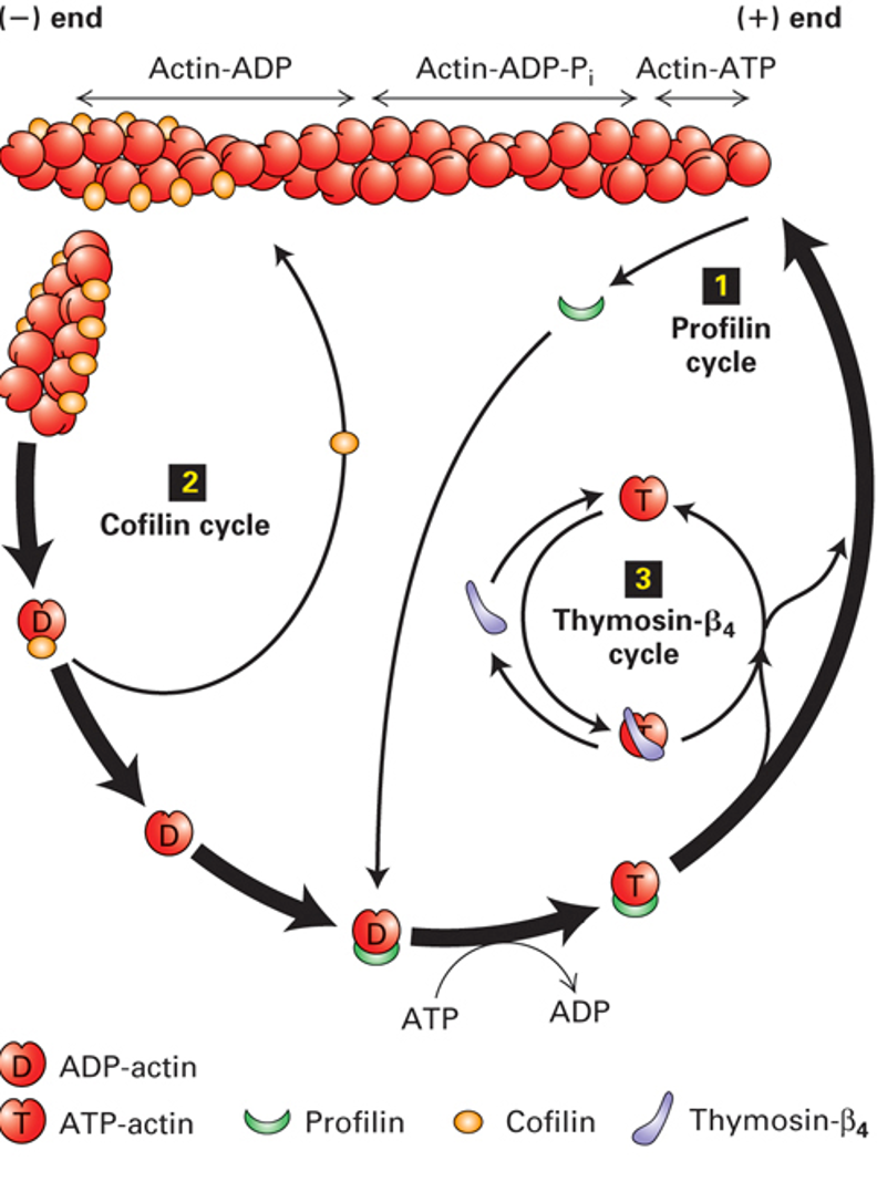
Regulation of Actin Filament Dynamics in Cells:
The total actin concentration in eukaryotic cells is high, ranging between and . Because this is orders of magnitude above both () and (), all cellular actin would theoretically polymerize into F-actin, leaving zero free G-actin.
In vivo, approximately of total actin remains unpolymerized as G-actin. Cells control the availability and assembly of actin through specific regulatory proteins:
End-Capping Proteins:
CapZ: Heterodimeric protein that binds with high affinity to the plus (+) end, blocking both assembly and disassembly. When CapZ caps the plus end, polymerization is restricted to the minus end, which requires a monomer concentration above .
Tropomodulin: Caps the minus (-) end of actin filaments, preventing subunit dissociation. In stable structures such as muscle sarcomeres or erythrocyte cortical skeletons, tropomodulin locks the minus end against depolymerization, ensuring permanent filament length.
Actin Monomer Sequestration:
Thymosin-: Binds specifically to ATP-G-actin in a 1:1 complex, sterically preventing monomer addition to either the plus or minus ends. Thymosin- buffers the cellular monomer pool, maintaining a large reservoir of unpolymerized G-actin without requiring alterations in total actin protein synthesis.
Nucleotide Exchange and Targeted Assembly:
Profilin: Acts as an actin nucleotide exchange factor. Profilin binds the ADP-G-actin released from disassembled filaments, accelerating the displacement of ADP by ATP. Profilin binds the face of actin opposite the ATP cleft, physically blocking incorporation onto the minus end while leaving the binding site for the plus end fully accessible. Profilin-ATP-actin can assemble exclusively onto free plus (+) ends, converting the basal 12-fold kinetic bias into an absolute plus-end assembly specificity.
Filament Severing and Depolymerization:
Cofilin (Actin Depolymerizing Factor): Binds specifically along the sides of older ADP-F-actin filaments, inducing mechanical twisting and conformational strain that severs the filament into small fragments. Severing dramatically multiplies the number of free minus (-) ends available for rapid depolymerization while simultaneously generating new filament fragments that serve as nucleation seeds.
Directed Cell Migration and Rho Family GTPase Signaling Networks

Extracellular Guidance and Receptor Distribution:
Directed cell migration occurs in response to localized extracellular chemical signals (chemoattractants), such as platelet-derived growth factor (PDGF), fibroblast growth factor (FGF), epidermal growth factor (EGF), inflammatory cytokines, or bacterial surface components (e.g., lipoprotein A).
Cell surface receptors (e.g., receptor tyrosine kinases and G-protein coupled receptors) are distributed uniformly across the entire 360-degree periphery of the plasma membrane.
Spatial localization of the intracellular migratory response occurs because only the subset of receptors directly facing the highest concentration of the extracellular chemoattractant gradient binds ligand and activates downstream effectors.
Rho Family Small Monomeric GTPases:
The intracellular signals are transduced by the Rho family of monomeric small G proteins: Cdc42, Rac, and Rho.
Like all G proteins, they alternate between an inactive GDP-bound cytosolic state and an active GTP-bound state anchored to the inner leaflet of the plasma membrane via prenyl/lipid anchors.
Distinct GTPases orchestrate specialized actin architectures across migrating cells:
Cdc42 and Filopodia Formation:
Localized activation of Cdc42 at the cell front induces the formation of filopodia (slender sensory projections) and establishes front-to-back polarity via Par6.
WASp Activation: Active Cdc42-GTP binds the Rho-binding domain (RBD) of autoinhibited Wiskott-Aldrich Syndrome protein (WASp).
Clinical Pathology of WASp: Wiskott-Aldrich syndrome is a rare X-linked primary immunodeficiency characterized by recurrent infections, immune dysregulation, and microthrombocytopenia (small platelets); defects stem from the inability of white blood cells and platelets to properly assemble branched actin and migrate toward infection sites.
WASp recruitment also requires interaction with the membrane phospholipid phosphatidylinositol 4,5-bisphosphate () via its basic (B) domain.
Unfolding of WASp exposes its acidic (A), connector (C), and WASp-homology (W/V) domains, which bind an ATP-actin monomer and recruit the Arp2/3 complex.
Arp2/3 Nucleation: Actin-related proteins 2 and 3 (Arp2 and Arp3) form a heterodimer mimicking an actin filament plus end. When bound to the side of an existing mother filament, Arp2/3 nucleates the rapid growth of a daughter microfilament at a fixed, invariable branching angle.
Rac and Lamellipodia Formation:
Rac-GTP activation across the leading edge recruits and activates WAVE (WASP-family verprolin-homologous protein).
WAVE similarly activates the Arp2/3 complex, nucleating extensive branched dendritic actin meshworks that form broad lamellipodial sheets. These branched meshworks push against the plasma membrane across a wide front, driving efficient forward protrusion.
Rho and Stress Fiber Formation / Rear Retraction:
Active Rho-GTP coordinates linear filament assembly and actomyosin contraction.
Formin Pathway:
In its inactive cytosolic state, formin is folded into an autoinhibited conformation through an intramolecular interaction between its N-terminal and C-terminal domains.
Rho-GTP binds the Rho-binding domain (RBD) of formin, unfolding the protein.
The active formin FH2 domains form a dimeric, doughnut-shaped collar encircling the plus (+) end of an actin filament.
The adjacent flexible, proline-rich FH1 domains act as recruitment landing sites that bind Profilin-ATP-actin complexes from the cytosol and feed them directly to the growing plus end.
As monomers are added, the FH2 ring rocks and steps processively toward the growing plus end without dissociating, shielding the plus end from CapZ while driving the assembly of long, unbranched linear actin bundles.
Rho Kinase (ROCK) and Actomyosin Contraction:
Rho-GTP simultaneously activates Rho-associated kinase (Rho kinase / ROCK).
Rho kinase phosphorylates and activates the regulatory light chain of myosin II while phosphorylating and inhibiting myosin light chain phosphatase.
The resulting accumulation of phosphorylated myosin light chains () activates the motor activity of non-muscle myosin II bipolar filaments.
Myosin II filaments cross-link adjacent antiparallel linear actin filaments, exerting contractile tension within stress fibers and within the rear cell cortex.
Spatial Coordination of Cell Translocation (The Four-Step Crawling Cycle):
Protrusion: Rac and Cdc42 drive localized Arp2/3-dependent branched actin assembly at the leading edge, pushing the lamellipodium and filopodia forward.
Adhesion: The advancing lamellipodium attaches to the extracellular matrix (ECM, composed of collagen and fibronectin) via transmembrane integrin receptors clustered into focal adhesions.
Translocation (Cell Body Rolling): The forward-pushing branched network and cytoplasmic flow draw the cell mass forward. Contractile stress fibers stretch between focal contacts, pulling the nucleus and cellular contents forward. Microfilaments pulling on the nucleus can create a mechanical "slingshot" or piston effect that propels the nucleus and cytoplasm into the newly protruded domain.
De-adhesion and Tail Retraction: At the trailing edge (rear), Rho activation dominates, suppressing Rac activity through mutual antagonism. Rho kinase stimulates myosin II contraction, pulling the cell rear forward. Concurrently, integrin-ECM attachments at the rear are dismantled, and cofilin actively severs and disassembles rear actin filaments, recycling G-actin monomers to the leading edge.
Experimental Dissection of Cell Migration: Dominant Mutations and Wound Closure Assays

Mechanisms of Dominant Mutations in Small GTPases:
Dominant Negative Mutants:
Possess point mutations that permanently lock the G protein into an inactive, GDP-bound conformation or prevent guanine nucleotide exchange factor (GEF) binding.
Act dominantly by irreversibly binding and sequestering endogenous GEFs, depleting the cellular pool of GEFs required to activate endogenous wild-type G proteins. This phenocopies haploinsufficiency (where a single functional allele is insufficient to maintain normal cellular function).
Dominant Active Mutants:
Possess point mutations that abolish intrinsic GTPase activity (analogous to oncogenic Ras mutations), preventing GTP hydrolysis.
Lock the G protein permanently in the GTP-bound, active state, driving continuous signaling through downstream effectors regardless of extracellular cues.
In Vitro Wound Healing (Scratch) Assays:
Normal, non-transformed epithelial or fibroblast cells grown in culture proliferate until forming a continuous, single-cell-thick monolayer, at which point contact inhibition arrests further cell division and migration.
In a wound assay, a mechanical stylus or rubber spatula scrapes a cell-free gap ("scratch") through the monolayer.
Cells bordering the scrape rapidly detect growth factors (FGF, PDGF) released locally and migrate into the open space until confluent contact is re-established.
Effects of Dominant Negative GTPases:
Control cells achieve wound closure within a standard 3-hour assay.
Cells expressing Dominant Negative Rac show severely restricted lamellipodial protrusion, resulting in less than wound closure.
Cells expressing Dominant Negative Cdc42 fail to orient toward the chemoattractant gradient, resulting in approximately closure.
Cells expressing Dominant Negative Rho completely fail to retract their trailing edge and cannot assemble stress fibers, resulting in virtually wound closure.
Phenotypic Morphology of Dominant Active Mutants:
Dominant Active Rho: Cells fill with an excessive density of thick, rigid, parallel linear stress fibers spanning across the cytoplasm, preventing dynamic morphological remodeling and leading to cellular tearing.
Dominant Active Rac: Induces massive, continuous, 360-degree peripheral lamellipodia formation around the entire cell boundary.
Dominant Active Cdc42: Causes spontaneous, multi-directional projection of filopodia in every direction simultaneously, completely abolishing directional cell polarity.
In Vivo Cell Migration and Quantitative Model Systems:
During embryogenesis (e.g., zebrafish gastrulation), coordinated cell migrations drive morphological patterning, as demonstrated by live imaging of GFP-tagged histone markers tracking somatic and neural crest cell migrations from the animal pole toward the vegetal plate to establish the body axis.
The 2012 World Cell Race: A standardized international competition evaluating the intrinsic migration speeds of varied normal and cancer cell lines migrating along standardized fibronectin-coated micropatterned tracks ( or wide) over a linear measurement distance of , highlighting that metastatic cancer cells exhibit elevated autonomous migration velocities compared to non-malignant controls.
Microtubule Structure, Centrosome Organization, and -TuRC Nucleation

The Tubulin Heterodimer:
The structural building block of microtubules is the -tubulin heterodimer, composed of one -tubulin subunit and one -tubulin subunit.
-Tubulin: Contains a non-exchangeable GTP-binding site at the intradimer interface. The GTP bound to -tubulin is permanently buried and never hydrolyzed.
-Tubulin: Contains an exchangeable GTP-binding site exposed at the surface of the dimer. The GTP bound to -tubulin is hydrolyzed to GDP following polymerization.
Structural Polarity: Dimers assemble in a head-to-tail orientation along longitudinal protofilaments. Because of this uniform orientation, the plus (+) end terminates with exposed -tubulin subunits, whereas the minus (-) end terminates with exposed -tubulin subunits.
Protofilament Arrangements and Microtubule Classes:
Singlet Microtubules: Composed of 13 parallel protofilaments arranged in a hollow cylinder ( diameter); standard structural organization found in the cytoplasm of non-neuronal cells.
Doublet Microtubules: Composed of one complete 13-protofilament A tubule fused to an incomplete 10-protofilament B tubule; found specifically in the axonemes of cilia and flagella.
Triplet Microtubules: Composed of one complete 13-protofilament A tubule fused to two incomplete 10-protofilament tubules (B and C tubules); found specifically in centrioles and basal bodies.
Centrosome Architecture and Microtubule Nucleation:
In interphase non-neuronal cells, virtually all microtubules originate from a single, centralized Microtubule Organizing Center (MTOC), the centrosome.
The centrosome consists of a pair of mutually perpendicular, barrel-shaped centrioles (designated the older mother centriole and the newly formed daughter centriole) surrounded by an amorphous protein matrix termed the pericentriolar material (PCM).
Each centriole consists of nine triplet microtubules arranged radially in a pinwheel configuration.
Centrioles duplicate semi-conservatively during S phase: the mother and daughter centrioles separate, and each templates the orthogonal assembly of a new daughter centriole through a protein-templated assembly mechanism lacking nucleic acids.
-Tubulin Ring Complex (-TuRC):
In animal cells, the pericentriolar material contains specialized -tubulin ring complexes (-TuRC).
The -TuRC forms an open ring of 13 -tubulin subunits that matches the diameter and architecture of a 13-protofilament microtubule.
-TuRC binds the exposed -tubulin subunits of incoming -tubulin heterodimers, nucleating protofilament assembly and permanently capping the minus (-) end.
Consequently, in non-neuronal cells, all microtubule minus (-) ends are anchored within the centrosome, and dynamic growth and shortening occur exclusively at the plus (+) ends extending outward toward the cell periphery.
An interphase cell contains a defined population of approximately 700 microtubules radiating from the centrosome.
Dynamic Instability of Microtubules: The GTP Cap, Catastrophe, and Rescue

Microtubule Assembly Kinetics and Critical Concentration:
In cell-free systems, microtubule assembly requires exceeding the critical concentration (). At the plus end, .
The physiological concentration of tubulin heterodimers in eukaryotic cells is maintained at , which is far higher than the plus-end critical concentration.
Despite this thermodynamic surplus, cellular microtubules do not fully polymerize; rather, individual microtubules coexist in a continuous, non-equilibrium dynamic steady state characterized by alternating phases of rapid elongation and catastrophic depolymerization, termed dynamic instability.
Molecular Mechanism of the GTP Cap and Protofilament Conformation:
Free -tubulin heterodimers enter the polymerizing plus (+) end in the GTP-bound state ().
Microtubule protofilaments containing adopt a straight, linear conformation that packs tightly with adjacent protofilaments through lateral subunit interactions, stabilizing the cylindrical microtubule wall.
As new dimers add to the growing tip, the -tubulin of the newly incoming dimer acts as a GTPase-activating protein (GAP) for the -tubulin of the preceding dimer, stimulating rapid GTP hydrolysis to GDP.
Protofilaments composed of are thermodynamically unstable and strained within the microtubule cylinder; in isolation, GDP-protofilaments naturally curve outward.
In an actively growing microtubule, the rate of dimer addition exceeds the rate of GTP hydrolysis, maintaining a protective ring of subunits at the plus end, termed the GTP cap.
The GTP cap constrains the interior GDP-tubulin core in a forced linear geometry under mechanical compression.
The Catastrophe Mechanism:
If the rate of subunit addition pauses or falls below the rate of GTP hydrolysis, the terminal -tubulins at the plus end hydrolyze their bound GTP to GDP, resulting in the loss of the GTP cap.
Without the straight GTP cap to constrain them, the mechanical strain within the interior GDP-protofilaments is released.
The protofilaments curve rapidly outward, splaying apart like peeling a banana or splitting frayed hair ends.
Lateral protofilament interactions are broken, and the microtubule undergoes rapid depolymerization into small protofilament curls and free tubulin dimers, an event termed catastrophe.
The rate of depolymerization during catastrophe is significantly faster than the rate of polymerization during growth.
The Rescue Mechanism:
During rapid shrinkage, catastrophic disassembly can be halted if a shrinking plus end re-binds multiple dimers, re-establishing a stable, linear GTP cap.
This transition from depolymerization back to elongation is termed rescue.
Dynamic instability allows the cellular microtubule network to explore three-dimensional cytoplasmic space continuously via search-and-capture mechanisms.
Pharmacological Modulation (Taxol):
Taxol (Paclitaxel): A pharmacological compound that binds specifically along the interior wall of the microtubule and prevents -tubulin from hydrolyzing its bound GTP.
By locking tubulin subunits in a stable, straight conformation mimicking the GTP-bound state, Taxol freezes microtubules in a permanent polymer state, completely abolishing dynamic instability.
Taxol-stabilized microtubules are used in in vitro motility assays to study motor proteins without spontaneous filament depolymerization.
Microtubule-Associated Proteins: End-Binding Proteins and Axonal Spacers
Plus-End Tracking Proteins (+TIPs):
A specialized family of microtubule-associated proteins (MAPs) specifically tracks and binds the growing plus (+) ends of microtubules.
EB1 (End-Binding Protein 1):
Binds with high affinity to the structural transition zone at the plus end between newly incorporated GTP-tubulin and hydrolyzed GDP-tubulin.
EB1 increases the catastrophe frequency by promoting protofilament curvature and splaying.
Functions as a molecular hitchhiking platform: diverse non-secretory cytosolic cargo proteins and signaling complexes bind to EB1, riding the advancing plus end of polymerizing microtubules out toward the cell cortex.
EB3 (End-Binding Protein 3):
Acts antagonistically to EB1; binds the plus end and stabilizes lateral protofilament interactions, functioning as a molecular brace that inhibits catastrophe and promotes rescue.

Structural Microtubule-Associated Proteins (MAPs):
Cytosolic proteins that bind longitudinally along the outer wall of microtubules, bridging the seam between adjacent protofilaments to stabilize the cylinder against dynamic instability.
MAP2 (Microtubule-Associated Protein 2):
Possesses a basic microtubule-binding domain and an extended, rigid projection arm that extends perpendicularly outward by approximately .
MAP2 is localized primarily to the neuronal cell body (soma), dendrites, and the wide proximal axon initial segment.
The long projection arm acts as a physical spacer, maintaining broad parallel separation () between adjacent microtubules to provide ample cytoplasmic space for multi-directional trafficking of bulky organelles and vesicles.
Tau Protein:
Possesses homologous microtubule-binding domains but a significantly shorter, more compact projection arm.
Enriched in the distal axon, where the axonal diameter narrows.
Tau cross-links microtubules into tightly packed, parallel bundles with narrow inter-filament spacing ().
This close packing provides high mechanical stability along long distances while maintaining sufficient clearance for motor proteins to walk along individual outer protofilaments.
Molecular Pathogenesis of Alzheimer's Disease: Tauopathies, Isoforms, and Neurodegeneration

Tau Hyperphosphorylation and Tangle Formation:
Alzheimer's disease (AD) is characterized pathologically by two distinct proteinopathies: extracellular amyloid-beta () plaques derived from amyloid precursor protein (APP) cleavage, and intracellular neurofibrillary tangles (NFTs) composed of aggregated, hyperphosphorylated Tau.
The Tau P301S Mutation:
A missense mutation in the MAPT gene converting proline at position 301 to serine (P301S).
This mutation creates an aberrant phosphorylation consensus sequence that allows cellular kinases to hyperphosphorylate Tau.
Hyperphosphorylated Tau detaches from the microtubule surface, losing its stabilizing and spacing capacity.
Free cytosolic hyperphosphorylated Tau misfolds into insoluble paired helical filaments that aggregate into neurofibrillary tangles.
Consequences of Microtubule Collapse:
Loss of Tau destabilizes axonal microtubules, causing their progressive depolymerization and breakdown.
Axonal transport of neurosecretory vesicles and mitochondria halts, accumulating stalled organelles and proteinaceous debris within the axon.
The cellular degradative machinery is overwhelmed: the ubiquitin-proteasome system cannot clear insoluble Tau aggregates, and lysosomes fail to process the accumulated debris, leading to neuroinflammation, synaptic loss, and neurodegeneration.
Apolipoprotein E (ApoE) Biochemistry and Neuronal Lipid Delivery:
Neurons consume large amounts of cholesterol to maintain axonal membrane integrity, sustain synaptic vesicle turnover, and build myelin sheaths, but mature central nervous system neurons cannot synthesize adequate cholesterol de novo.
Supporting astrocytes synthesize and secrete high-density lipoprotein (HDL)-like particles containing Apolipoprotein E (ApoE).
Astrocyte-derived ApoE binds to neuronal-specific cell surface receptors (ApoE/HDL receptors), delivering cholesterol and phospholipids via endocytosis.
The Three Human ApoE Isoforms:
The human APOE gene exists as three major polymorphic alleles (, , ), encoding proteins of 299 amino acids that differ by single amino acid substitutions at positions 112 and 158:
ApoE2: Contains Cys112 and Cys158. Present in of the global population. Confers significant protection against Alzheimer's disease, delaying the age of onset, although homozygous inheritance predisposes to familial type III hyperlipoproteinemia.
ApoE3: Contains Cys112 and Arg158. The most common allele, carried by of the global population, conferring average baseline risk.
ApoE4: Contains Arg112 and Arg158. Present in roughly of the general population (with carrying two copies), yet found in nearly of all Alzheimer's patients. ApoE4 is the single strongest genetic risk factor for late-onset sporadic Alzheimer's disease.
ApoE4 Exacerbates Tauopathy via Toxic Gain-of-Function:
Transgenic mouse models crossing human ApoE knock-in (KI) lines with Tau P301S transgenic mice demonstrate that ApoE4 directly accelerates and exacerbates Tau-mediated neurodegeneration independently of amyloid-beta () pathology.
By 3 months of age, P301S/E4 mice develop significantly elevated total brain Tau levels and widespread somatodendritic redistribution of Tau compared to P301S/E2, P301S/E3, or P301S/ApoE-knockout (EKO) mice.
By 9 months of age, P301S/E4 mice exhibit marked brain atrophy and neuroinflammation, whereas P301S/EKO mice are protected against neurodegeneration, demonstrating that ApoE4 acts through a toxic gain-of-function rather than a loss-of-function.
Microglia expressing ApoE4 secrete higher levels of pro-inflammatory tumor necrosis factor-alpha () upon immune challenge.
Co-culturing P301S neurons with ApoE4-expressing mixed glia markedly decreases neuronal viability, while glia lacking ApoE (EKO) support maximal neuronal survival with minimal release.
Epidemiological Risk Context:
Retrospective epidemiological analyses of deceased cohorts (such as the 2020 study of 5,000 deceased individuals) demonstrate a steep leftward shift in Kaplan-Meier dementia-free survival curves for homozygotes compared to and carriers.
In prospective studies that follow living, cognitively normal individuals over time, at least of homozygous individuals remain completely dementia-free at 85 years of age, indicating that ApoE4 represents a susceptibility factor requiring additional genetic, metabolic, or environmental triggers rather than a fully penetrant deterministic gene.
Axonal Transport: Extreme Geometries, Energetics, and Experimental Discovery

Morphological Extremes of Human Neurons:
In human central nervous system (CNS) neurons, total axon arbor lengths range from to over (e.g., human serotonergic neurons extend cumulative axonal arborizations estimated at up to ).
Neurons harbor between several hundred and over presynaptic boutons.
Each presynaptic site contains an active zone containing 40 to 850 synaptic vesicles that require continuous turnover and replenishment.
Because virtually all protein synthesis machinery, Golgi stacks, and endoplasmic reticulum networks are confined to the soma, all synaptic vesicle precursors (SVPs) must be transported down the axon over long distances.
Energetic Budget of Axonal Transport:
The cumulative energy expenditure of a single human cortical pyramidal neuron is approximately for ion channel flux and active synaptic transmission, plus dedicated solely to the ATPase to maintain the resting membrane potential.
Across human and rodent species, the total daily energy budget of a single cortical neuron is approximately (approximately 0.5 quadrillion ATP molecules per day), met by glycolysis and mitochondrial oxidative phosphorylation.
In basal forebrain cholinergic neurons, the total cumulative distance traveled daily by SVPs throughout the arbor is .
Because conventional kinesin-1 hydrolyzes exactly one ATP molecule per 16 nm step, the daily mechanical work of moving SVPs consumes to (approximately 0.4 to 0.7 trillion ATP/day).
Despite this high absolute value, transporting SVPs accounts for only to of the neuron's daily ATP budget.
Even when factoring in the movement of degradative endosomes, dense-core vesicles, and active zone proteins, total microtubule-based motor transport consumes less than of the total cellular energy expenditure.
Mitochondrial Transport as Distributed Energy Depots:
Random diffusion of ATP from the cell body along a 1-meter axon would require decades, resulting in an energy gradient and ATP depletion at distal synapses.
To supply local ATP, molecular motors transport whole mitochondria along axonal microtubules, positioning them at high-demand regions near presynaptic active zones.
The Squid Giant Axon as a Model System:
Molecular motor proteins were discovered experimentally using the giant axon of the Atlantic squid (Loligo pealeii), which measures () in length and up to in diameter.
Extrusion with a glass roller allows axoplasm (cytoplasm devoid of plasma membrane) to be extracted as an intact cylinder of jelly-like material.
Video-enhanced differential interference contrast (DIC) microscopy of extruded axoplasm demonstrated rapid, bidirectional motion of vesicular particles and elongated mitochondria along linear filaments.
Reconstitution experiments demonstrated that combining purified brain microtubules (stabilized with Taxol) with isolated synaptic vesicles yielded zero motion, even upon adding ATP.
Supplementing the reaction with clear soluble axoplasm restored vigorous vesicle translocation along microtubules.
Fractionation of this axoplasm identified the underlying motor proteins:
Kinesins: Plus-end-directed motor proteins responsible for anterograde transport (outward from soma toward the axon terminal).
Dyneins: Minus-end-directed motor proteins responsible for retrograde transport (inward from the axon terminal back toward the cell body).
Addition of non-hydrolyzable ATP analogs (e.g., AMP-PNP) locked the motor proteins tightly to both the vesicle and the microtubule, demonstrating that ATP binding allows microtubule attachment while ATP hydrolysis is required for translocation.
Molecular Motors: Structural Mechanisms of Kinesin and Dynein

Kinesin Superfamily Architecture:
Kinesin-1 (Conventional Kinesin):
A heterotetramer composed of two heavy chains and two light chains.
Motor Heads: Two globular N-terminal catalytic domains containing both a catalytic ATP-binding cleft and a microtubule-binding site.
Neck Linker: A flexible peptide segment connecting each head to the central coiled-coil dimerization stalk.
Tail Domain: Formed by the C-termini of the heavy chains complexed with two light chains, which bind to specific Rab GTPases and adaptor proteins on organelle membranes.
Specialized Kinesins:
Kinesin-2: A heterotrimeric plus-end-directed motor utilized for moving proteinaceous cargo within intraflagellar arrays and neural extensions.
Kinesin-5: A bipolar, homotetrameric motor containing four motor heads pointing in opposite directions; cross-links antiparallel overlapping polar microtubules during mitosis, sliding them apart to push spindle poles away from one another during anaphase B.
Kinesin-13: An immotile, non-transporting motor; uses ATP hydrolysis to bind microtubule plus ends and peel protofilaments apart, acting as an active catastrophe factor that drives microtubule depolymerization during mitosis.

The Kinesin Hand-over-Hand "Swivel-Step" Mechanism:
Kinesin-1 translocates processively toward the plus (+) end along a single protofilament without dissociating, taking hundreds of steps before falling off.
Step Size and Geometry: Each step advances the trailing head by , stepping from one -tubulin subunit over an intermediate -tubulin directly onto the next -tubulin. This advances the attached cargo stalk forward by .
The operational velocity of Kinesin-1 is approximately .
Step-by-Step Nucleotide Cycle:
Two-Head Bound State: The leading head is nucleotide-free and tightly bound to a -tubulin subunit; the trailing head has ADP bound in its pocket and interacts weakly with the microtubule.
ATP Binding: A molecule of ATP enters the catalytic pocket of the leading head. ATP binding induces a conformational change that causes its flexible neck linker to zip forward and dock against the catalytic core.
The Power Stroke (Swivel): Forward docking of the neck linker throws the trailing ADP-bound head forward by , swiveling it past the leading head to position it over the next available -tubulin subunit along the same protofilament.
Subunit Attachment and ADP Release: The new leading head lands onto the forward -tubulin, releasing its bound ADP to establish a tight, nucleotide-free attachment.
Hydrolysis and Undocking: The rear trailing head hydrolyzes its ATP to ADP and inorganic phosphate (). Release of undocks its neck linker, weakening its affinity for tubulin so it is ready to be pulled forward in the next step.
Each 16-nm step consumes exactly one molecule of ATP.
Cytoplasmic Dynein Architecture:
A multi-protein complex composed of two large heavy chains, intermediate chains, light-intermediate chains, and light chains.
Each heavy chain forms a ring of six AAA+ ATPase domains from which an extended coiled-coil stalk projects, ending in a globular microtubule-binding domain.
Dynein is recruited to membrane cargoes via the multi-protein Dynactin adaptor complex, which binds specific membrane Rab GTPases to direct transport exclusively toward the minus (-) end.
Organelle Trafficking Dynamics: Bidirectional Tug-of-War and Cortical Hand-Off
Rab GTPases Direct Motor Specificity:
Specific membrane-bound Rab family small GTPases mark individual organelle compartments and determine directional movement by recruiting the correct motor:
Post-Golgi secretory vesicles carry Rabs that selectively recruit Kinesin-1 or Kinesin-2 for plus-end-directed transport toward the plasma membrane.
Endocytic vesicles, late endosomes, and lysosomes carry specific Rabs that recruit dynactin/dynein complexes, driving minus-end-directed trafficking toward the cell center.
Mechanical Tug-of-War Dynamics:
Many intracellular organelles simultaneously display both kinesin and dynein motors.
Directional transport is determined by mechanical competition:
A single kinesin motor exerts a stall force of approximately , whereas an individual cytoplasmic dynein motor exerts approximately .
Because one kinesin out-pulls an opposing dynein, driving retrograde movement requires an organelle to recruit a team of multiple dyneins to overcome a single kinesin.
Physiological Switching: Pigment Granule Translocation in Melanophores:
Dermal melanophores in fish and amphibians alter skin pigmentation to match background lighting by redistributing melanin-filled pigment granules (melanosomes):
Dispersion (Dark Skin): Melanosomes are transported outward from the cell center to the cell margins along radiating microtubules via kinesin motors, darkening the cell's appearance.
Aggregation (Pale Skin): Intracellular signaling triggers the activation of dynein motors, pulling all pigment granules inward toward the centrosome into a tight, dense central cluster, making the cell appear pale.
Discontinuous Tracks and Microtubule Dynamics:
In vivo, microtubules do not form uninterrupted tracks spanning from the soma to distal terminals. Instead, the axonal cytoskeleton consists of overlapping arrays of short, non-contiguous microtubule segments.
As kinesin motors reach the end of an individual microtubule or encounter a filament undergoing catastrophic depolymerization, the motor-cargo complex stalls momentarily, detaches, and hops across to an adjacent microtubule to resume processive movement.
The Cortical Hand-Off (Microtubule-to-Actin Transition):
Microtubule plus (+) ends do not touch the plasma membrane; they terminate several hundred nanometers short, separated from the bilayer by the dense actin cortex.
As a secretory vesicle reaches the plus end of an axonal microtubule, it is too distant for vesicular Rab GTPases to engage their target t-SNARE/Rab-GAP effectors on the plasma membrane.
The Hand-Off:
At the microtubule terminal zone, kinesin releases the vesicle.
The vesicle engages an unconventional, plus-end-directed myosin motor (such as myosin V).
Myosin walks the vesicle through the dense cortical actin meshwork, delivering it to the active zone of the presynaptic plasma membrane for docking and SNARE-mediated exocytosis.
Questions & Discussion
Question: Does an end-binding protein like EB1 experience a delay before it starts moving along the microtubule after it binds?
Response: EB1 binds directly to the specific plus-end structure of a polymerizing microtubule. It does not actively translocate down the length of the filament using ATP hydrolysis; rather, it remains associated with the newly added tubulin subunits at the growing tip. As additional tubulin dimers add to the growing tip, EB1 dissociates and rebinds forward. If a microtubule is growing rapidly, tubulin addition can outpace the recruitment of EB1, but there is no prolonged latency; the protein simply associates dynamically with the plus end as long as elongation continues.
Question: When the animation describes adaptor proteins coupling cargo to motors, are those adaptors the Rab GTPases?
Response: Yes. The Rab family of small GTPases serves this coupling function. Membrane-anchored Rabs bind specific motor-receptor complexes (or dynactin adaptors) on their cytosolic face, recruiting the appropriate kinesin or dynein motor to the vesicle.