2. Intraocular Tumour - Retinal
Retinal Pigmentation
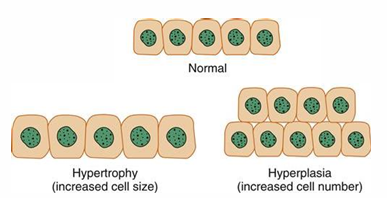
Hypertrophy vs Hyperplasia
Hypertrophy: Increase in size of individual cells without an increase in number.
Hyperplasia: Abnormal increase in the number of normal cells, characterized by reactive proliferation and migration of cells.
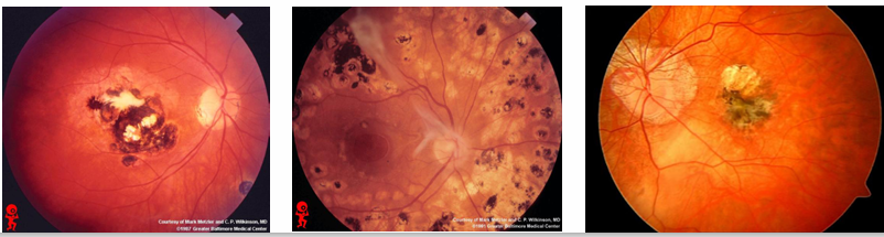
RPE (Retinal Pigment Epithelium) Hyperplasia:
Results in dense black pigmentation.
Response of retinal tissue due to prolonged insult/irritation.
Caused by various conditions, representing an attempt to repair alterations in retinal structure (e.g., chorioretinal scars, longstanding retinal detachment (RD)).
Often results in atrophy, disorganization of retinal architecture, and irregular margins.
Congenital Hypertrophy of the Retinal Pigment Epithelium (CHRPE)
A common finding in Caucasian fundi.
Present in approximately 1.2% of the population visiting optometry practices.
Appearance: Flat, black/brown/dark gray, well-circumscribed avascular lesions.
Location: Usually found in peripheral/midperipheral retina.
Nature: Benign hypertrophy of the RPE, usually asymptomatic.
Forms: Can appear as a solitary lesion or as multiple grouped lesions.
Some atypical forms of CHRPEs may be associated with systemic gastrointestinal malignancy
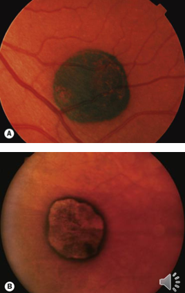
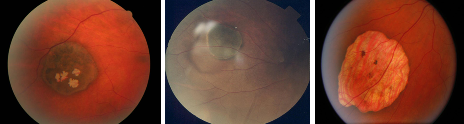
Solitary (Unifocal) CHRPE
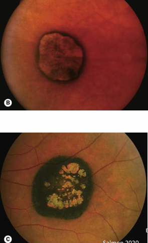
Shape: Typically round or oval, flat, darkly pigmented lesions occurring in the midperipheral or peripheral fundus.
Edges: Well-defined, smooth edges.
Additional Features:
May present with a depigmented halo just inside the margin.
Possible presence of depigmented lacunae (termed as “sunburst effect”) within the lesion.
Progression: Slight enlargement of the involved area over time is common.
Malignant potential: Very rarely can develop into an adenocarcinomatous nodule requiring treatment.
Association: No association systemic gastrointestinal malignancy
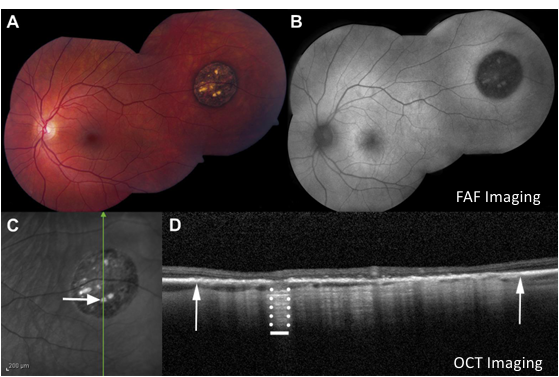
OCT (Optical Coherence Tomography) Appearance:
Imaging indicates thickened irregular RPE.
Often noted thinning or absence of outer retinal layers.
Fluorescence Imaging:
Lesions tend to be hypo-autofluorescent on FAF (Fundus Autofluorescence) imaging
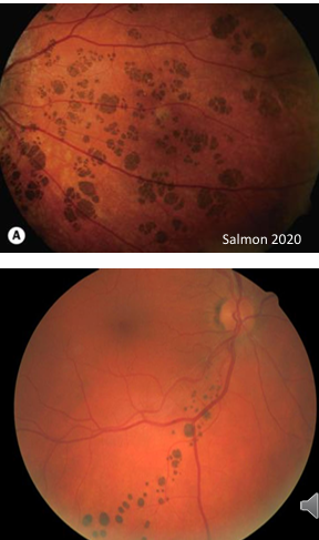
Grouped (Multifocal) CHRPE
Appearance: Multiple smaller lesions, presenting without holes or lacunae.
Pattern: Small patches grouped that resemble animal paw prints across the fundus, sometimes referred to as “bear tracks”.
They may appear depigmented, termed “Polar Bear Tracks”.
Prevalence: Usually unilateral.
Nature: Benign and not associated with gastrointestinal tract malignancy
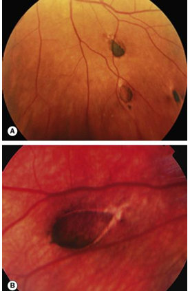
Atypical CHRPE
Appearance: Multiple oval, spindle, comma- or fishtail-shaped lesions of variable sizes, associated with irregularly hypopigmented margins and perilesional areas.
Involvement: Both eyes are typically involved.
Genetic Implications: Atypical CHRPE bilaterally may suggest Familial Adenomatous Polyposis (FAP) or Gardner’s syndrome.
Familial Adenomatous Polyposis (FAP):
Description: Presence of adenomatous polyps throughout the colon and rectum developing in adolescence.
Genetics: Often inherited in an autosomal dominant pattern.
Cancer Risk: Virtually all patients with FAP develop colorectal carcinoma by age 50.
Associated Findings: Over 80% of patients with FAP have atypical CHRPE at birth
Gardner’s Syndrome
Conditions:
Involves FAP along with osteomas of the skull and jaw.
Increased risk of adenocarcinoma of the colon.
Bilateral CHRPE: It serves as a marker for FAP among family members.
Atypical CHRPE: Generally bilateral, with lesions that are widely separated, oval or spindle-shaped.
Clinical Considerations:
Potential systemic association of CHRPE with FAP, although rare (occurring at a rate of approximately 1 per 100,000 in the population).
Should be considered in patients with suspicious lesions or a suggestive case history, and referral to a general practitioner is advised for further investigation.