Neuron
Homeostasis: The process by which biological systems maintain a stable internal environment to sustain life, despite changes in external conditions.
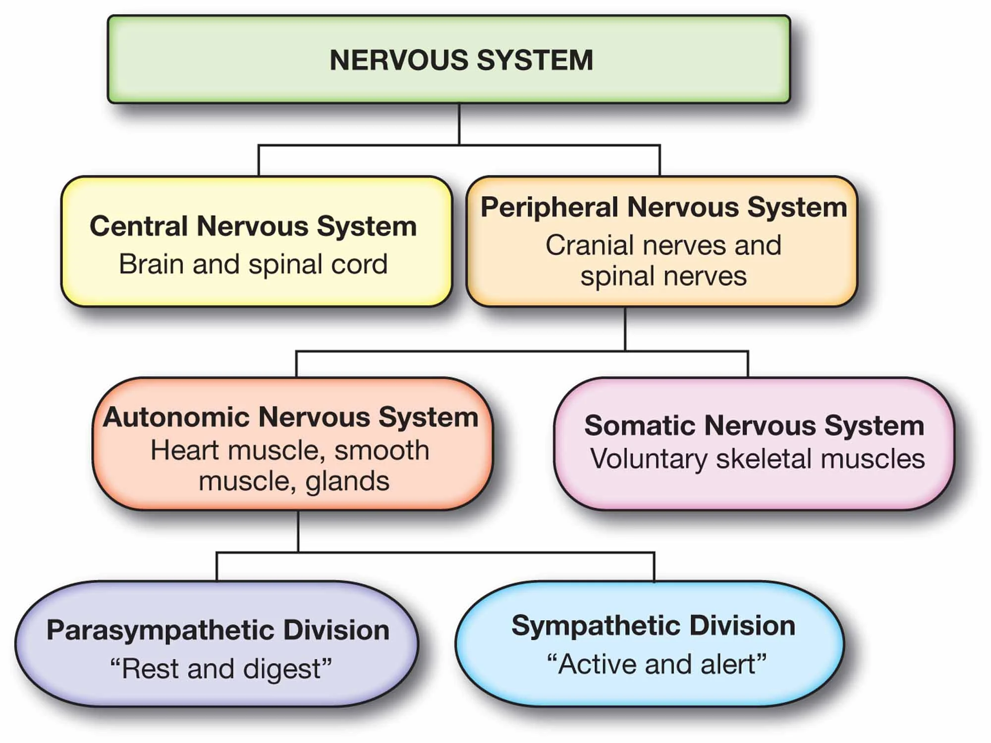
Nervous System: A complex network of neurons and cells that transmit signals between different parts of the body, enabling coordination and response to stimuli.
Central Nervous System (CNS): Part of the nervous system consisting of the brain and spinal cord, responsible for processing and interpreting sensory information and issuing instructions.
Peripheral Nervous System (PNS): The part of the nervous system outside the CNS, including all the nerves that branch out from the brain and spinal cord to the rest of the body.
Neurons: Specialized cells that transmit electrical and chemical signals in the nervous system; they are the functional units of the nervous system.
Glial Cells: Non-neuronal cells that provide support and protection for neurons in the central and peripheral nervous systems.
Nerves: Bundles of axons in the peripheral nervous system that transmit signals between the CNS and different parts of the body.
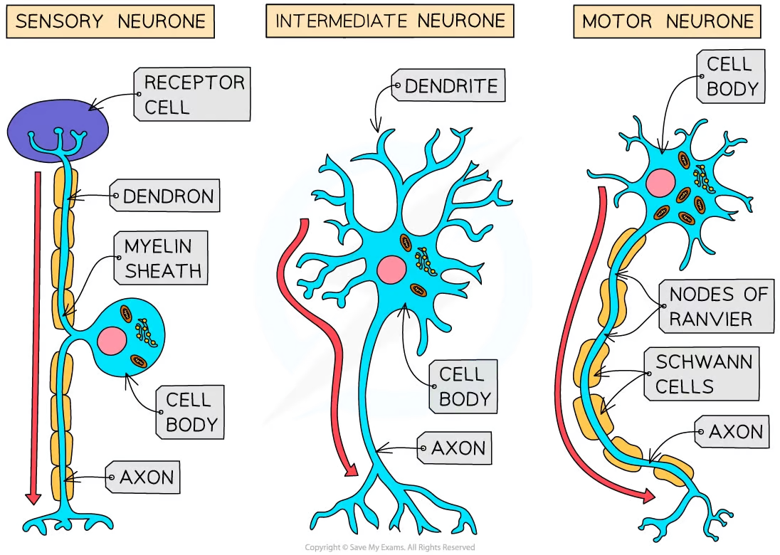
Reflex Arcs: The neural pathway that mediates a reflex action, involving sensory neurons, interneurons, and motor neurons.
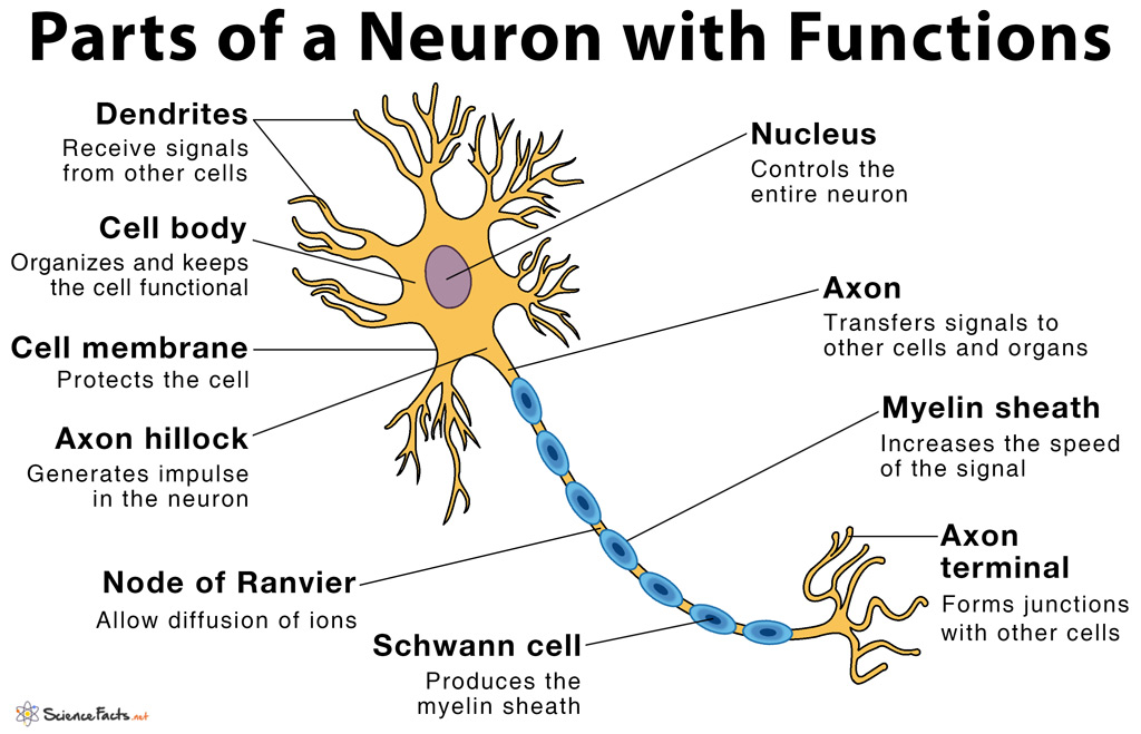
Dendrites: Branch-like extensions of a neuron that receive electrical messages from other cells.
Cell Body (Soma): The part of a neuron containing the nucleus and organelles; it processes incoming signals and generates outgoing signals to the axon.
Axon: A long, slender projection of a neuron that conducts electrical impulses away from the cell body to other neurons or effectors (muscles or glands).
Myelin Sheath: A fatty layer that covers the axons of some neurons, increasing the speed and efficiency of signal transmission.
Schwann Cells: Glial cells in the peripheral nervous system that produce the myelin sheath around neuronal axons.
Membrane Potential: The voltage difference across a cell's plasma membrane, due to the distribution of ions.
Resting Membrane Potential: The membrane potential of a neuron when it is not transmitting a signal, typically around -70 mV.
Polarization: The condition of a cell membrane where the inside of the cell is negatively charged relative to the outside.
Sodium-Potassium Exchange Pump: A membrane protein that uses ATP to transport sodium ions out of the cell and potassium ions into the cell, maintaining the resting potential.
Depolarization: The process during which the sodium channels open and sodium ions enter the cell, causing the membrane potential to become more positive.
Nodes of scher: Gaps in the myelin sheath along the axon, which allow for rapid conduction of nerve impulses through saltatory conduction.
Action Potential: A rapid rise and fall in membrane potential that results in the transmission of a nerve impulse along an axon.
Threshold Potential: The minimum membrane potential that must be reached to initiate an action potential.
Repolarization: The process of restoring the resting membrane potential following depolarization, primarily by the outflow of potassium ions.
Refractory Period: The time immediately following an action potential during which a neuron is unable to fire another action potential or requires a greater stimulus.
Synapse: The junction between two neurons, where neurotransmitters are released to transmit signals between the neurons.
Neuromuscular Junction: A synapse between a motor
White Matter: found deep in the brain and outer spinal cord
Grey Matter: the surface of the brain and inner spinal cord
Saltatory Conduction: a way that nerves send signals quickly by making the electrical impulses jump between gaps (Nodes of Ranvier) in the myelin covering of the axon instead of moving slowly along the entire length of the axon.
Synaptic Cleft: the small gap between two neurons where neurotransmitters are released to transmit signals from one neuron to another.
Neurotransmitters: Chemicals that transmit signals across a synapse from one neuron to another. They play a key role in communication within the nervous system and can either stimulate or inhibit the activity of the receiving neuron.
Axon Hillock: The region of a neuron where the axon begins, responsible for integrating incoming signals and generating action potentials when the threshold potential is reached.
Cell Membrane: A biological membrane that separates and protects the interior of a cell from its external environment. It is composed of a lipid bilayer with embedded proteins that regulate the movement of substances in and out of the cell.
Myelin Sheath: A fatty layer that covers the axons of some neurons, increasing the speed and efficiency of signal transmission.
Axon Terminal: The endpoint of an axon, where neurotransmitters are stored and released to transmit signals to other neurons or muscle cells across the synapse.
Steps of a Synapse
Here's how synapses work:
Structure of a Synapse: A synapse consists of three main parts:
Presynaptic Neuron: The neuron that sends the signal. It ends in a synaptic terminal or bouton.
Synaptic Cleft: A small gap between the presynaptic neuron and the postsynaptic cell.
Postsynaptic Cell: The cell that receives the signal, which could be another neuron, a muscle cell, or a gland cell.
Transmission of the Signal:
Action Potential Arrival: When an action potential (an electrical signal) reaches the synaptic terminal of the presynaptic neuron, it triggers the opening of voltage-gated calcium channels.
Calcium Influx: Calcium ions (Ca²⁺) enter the presynaptic terminal, which is crucial for the next step.
Neurotransmitter Release: The influx of calcium causes synaptic vesicles, which are small membrane-bound sacs containing neurotransmitters, to fuse with the presynaptic membrane. This process releases neurotransmitters into the synaptic cleft through exocytosis.
Signal Reception:
Binding to Receptors: The neurotransmitters diffuse across the synaptic cleft and bind to specific receptors on the postsynaptic membrane.
Postsynaptic Potential: This binding causes ion channels in the postsynaptic membrane to open or close, leading to changes in the membrane potential of the postsynaptic cell. This can result in either an excitatory or inhibitory postsynaptic potential, depending on the type of neurotransmitter and receptors involved.
Signal Termination:
Reuptake: Neurotransmitters may be taken back into the presynaptic neuron for reuse.
Enzymatic Degradation: Enzymes in the synaptic cleft can break down neurotransmitters, rendering them inactive.
Diffusion: Some neurotransmitters simply diffuse away from the synaptic cleft.
Types of Synapses:
Chemical Synapses: Use neurotransmitters to transmit signals, as described above.
Electrical Synapses: Allow direct passage of ions and electrical signals through gap junctions, enabling faster communication.
Synapses are essential for the complex processing and integration of information in the nervous system, allowing for everything from simple reflexes to complex behaviors and thoughts.