Scrotum and Testes Part 2
Learning Objectives
Describe the Sonographic Technique
Understand the methods used for examining the scrotum and testes through ultrasound.
Outline Needed Equipment
Familiarize with the tools required for conducting the examination.
Discuss Pre and Post Procedure Aspects
Understand the patient preparation and follow-up measures after the ultrasound.
Equipment Set-up
Warm Gel:
Essential to use warm gel to prevent the cremasteric reflex which can elevate the testes.
Additional Towels/Sheets:
Have extra towels or exam sheets available for patient comfort and cleanup.
High-Resolution Linear Transducer (7-18 MHz):
Necessary for detailed imaging of scrotum and testes.
Be prepared to switch to a curved probe as needed.
Doppler Capabilities:
Utilize low PRF (Pulse Repetition Frequency) and high gain for assessing small vessels inside the testes.
Disinfectant Wipes:
Required for cleaning the transducer post-examination.
Patient Preparation
Comfort and Consent:
Ensure the patient is comfortable; provide thorough explanation and obtain informed verbal consent.
Positioning:
Instruct patient to change in the room and lie supine.
Can use the patient's legs or towels to support the scrotum.
The penis should be positioned on the pelvis and held in place.
Transducer Handling:
Keep the transducer's hand positioned against the patient for support.
Post Procedure
Maintain Privacy:
Cover the patient's pelvis with a gown or towel post-examination.
Instructions:
Ensure the patient knows how to exit the room and understands procedures for receiving results.
Cleaning the Probe:
Disinfect using appropriate wipes or sterilize as per protocol.
Scanning Technique
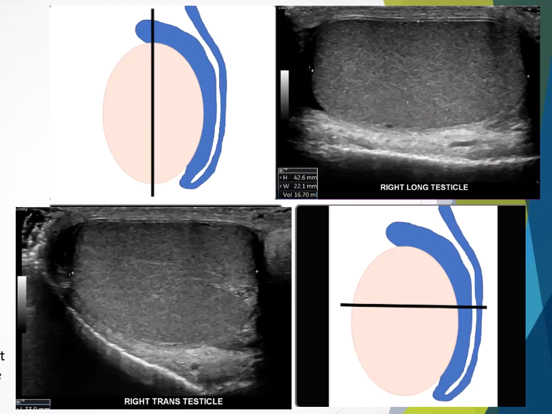
Imaging Planes:
Examine both testes in:
Transverse (upper/mid/lower)
Longitudinal (med/mid/lat)
Measurements:
Collect dimensions in both planes to calculate volume (using formula: ).
Assessment of Structures:
Evaluate both epididymis heads, bodies, and tails.
Image the spermatic cord.
Doppler Assessment:
Use color and PW (pulsed wave) Doppler to assess blood flow in testes for torsion or vascularity discrepancies.
Pathology Location:
Palpate and localize any lumps the patient feels for accurate imaging.
Valsalva Maneuver:
Recommended when evaluating for varicoceles or hernias.
Assess Other Areas
Groin Assessment:
Check for lymphadenopathy if necessary.
Kidney Evaluation:
Consider kidney assessment if a varicocele is detected to rule out renal tumor compression of the renal vein.
Normal Testes Ultrasound Findings
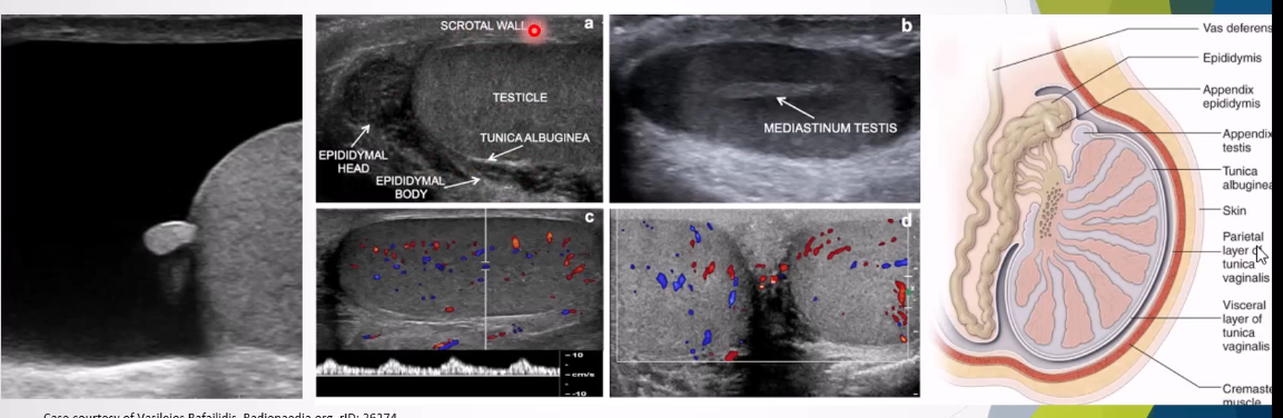
Appearance:
Testes appear oval, smooth, and homogeneous.
Lie:
Normally have a vertical lie unless a bell clapper deformity is present.
Structure:
Surrounded by a thin echogenic fibrous band (visceral tunica vaginalis).
Normal Volume:
Between 12-19 cc post-puberty, decreasing with age.
Dimensions:
Average length: , width: .
Normal Color Doppler Indications
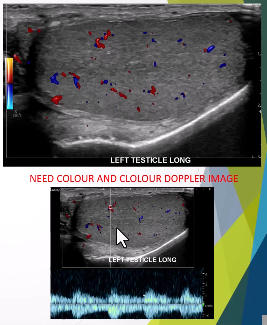
Assess for Torsion:
Ensure low PRF and high color gain set first on the asymptomatic side
Also need a spectral doppler trace of arterial and venous flow
Flow must be intratesticular to rule out torsion
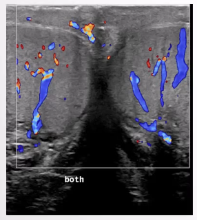
Normal Epididymis Imaging
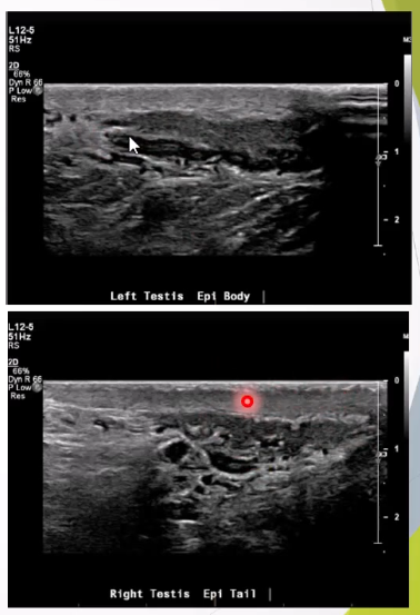
Transducer Movement:
Move posterior and laterally to assess the body and tail of the epididymis.
Variants in Anatomy
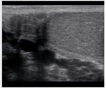
Rete Testis:
Tubular ectasia, characterized by dilated testicular mediastinal bubbles without specific shape or mass effect.
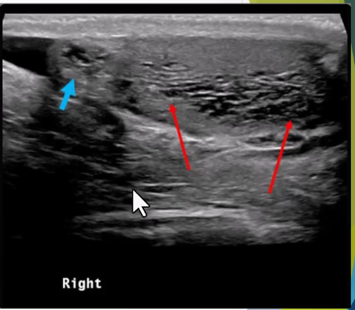
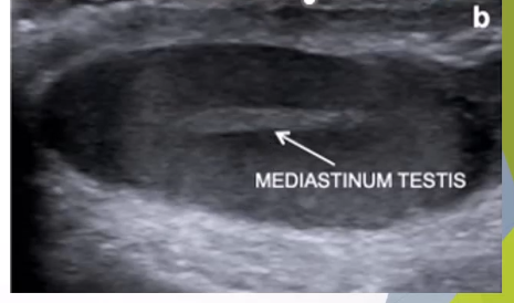
Mediastinum Testis:
Linear echogenic band at posterior part of the testis; supports the rete testis and blood vessels entering/exiting the testis.