Homework 7 Bailey McBride
Tibia
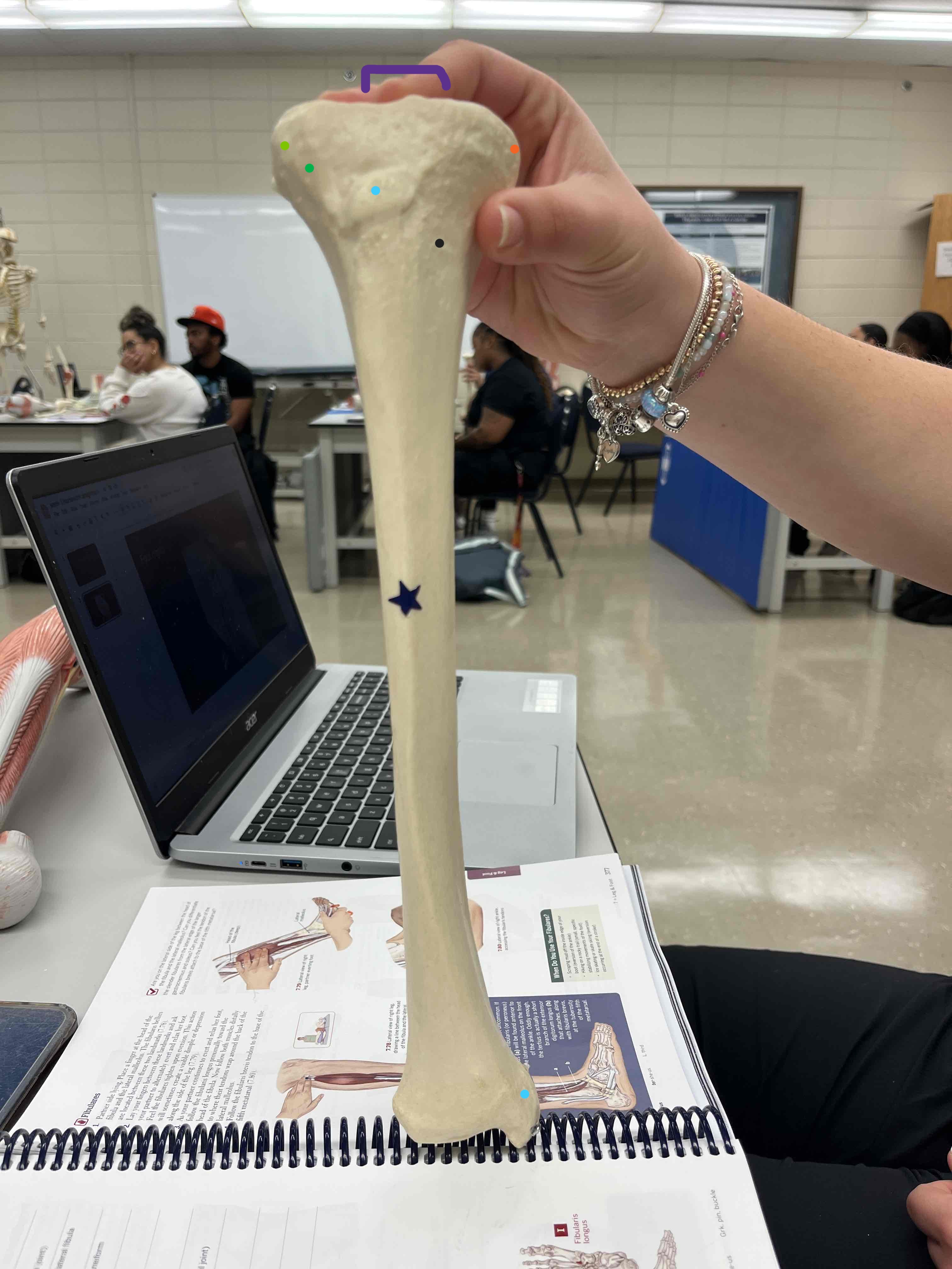
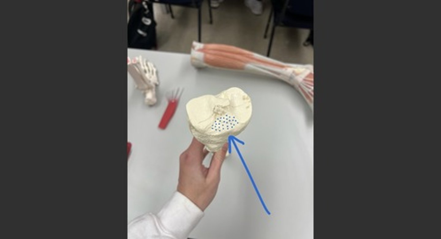
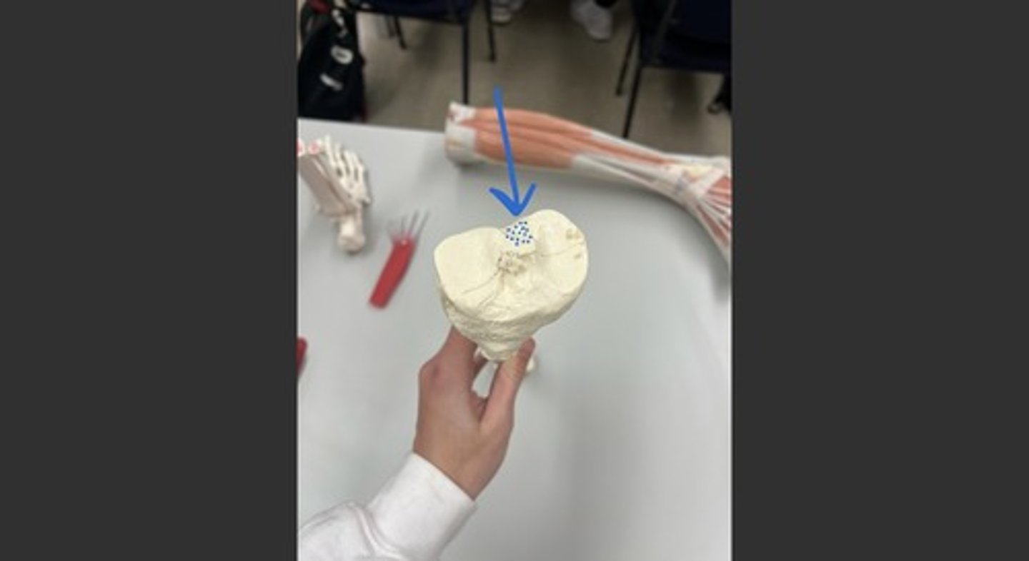
Anterior/Posterior Intercondylar Areas
Attachments:
ACL (Anterior Intercondylar Area)
PCL (Posterior Intercondylar Area)
Function: Stabilizes knee joint
Exercise: Squats
ADL: Walking downstairs
Medial & Lateral Condyles (orange/green)
Attachments:
Lateral Meniscus, Tibialis Anterior, Extensor Digitorum Longus (Lateral Condyle)
Medial Meniscus, Semimembranosus (Medial Condyle)
Function: Articulates with femur for knee movement
Exercise: Leg press
ADL: Standing up from a chair
Medial & Lateral Epicondyles (right below orange and green)
Attachments:
Tibialis Anterior, Extensor Digitorum Longus (Lateral Epicondyle)
Medial (Tibial) Collateral Ligament, Semimembranosus (Medial Epicondyle)
Function: Stabilizes knee joint
Exercise: Lunges
ADL: Stepping up onto a curb
PES Area (Pes Anserinus)-black
Attachments: Sartorius, Semitendinosus, Gracilis
Function: Knee flexion and stabilization
Exercise: Hamstring curls
ADL: Climbing stairs
Tibial Tuberosity(light blue)
Attachments: Patellar Ligament, Quadriceps Tendon
Function: Knee extension
Exercise: Leg extensions
ADL: Kicking a ball
Fibula
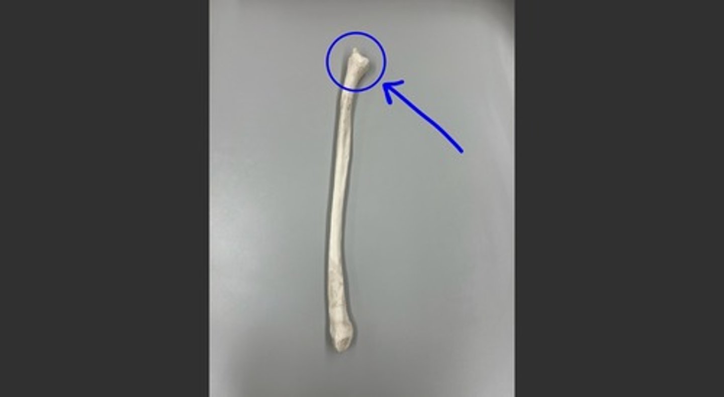
Head
Attachments: Lateral (Fibular) Collateral Ligament, Biceps Femoris (Long & Short Head)
Function: Supports knee stability
Exercise: Side lunges
ADL: Lateral stepping
Femur
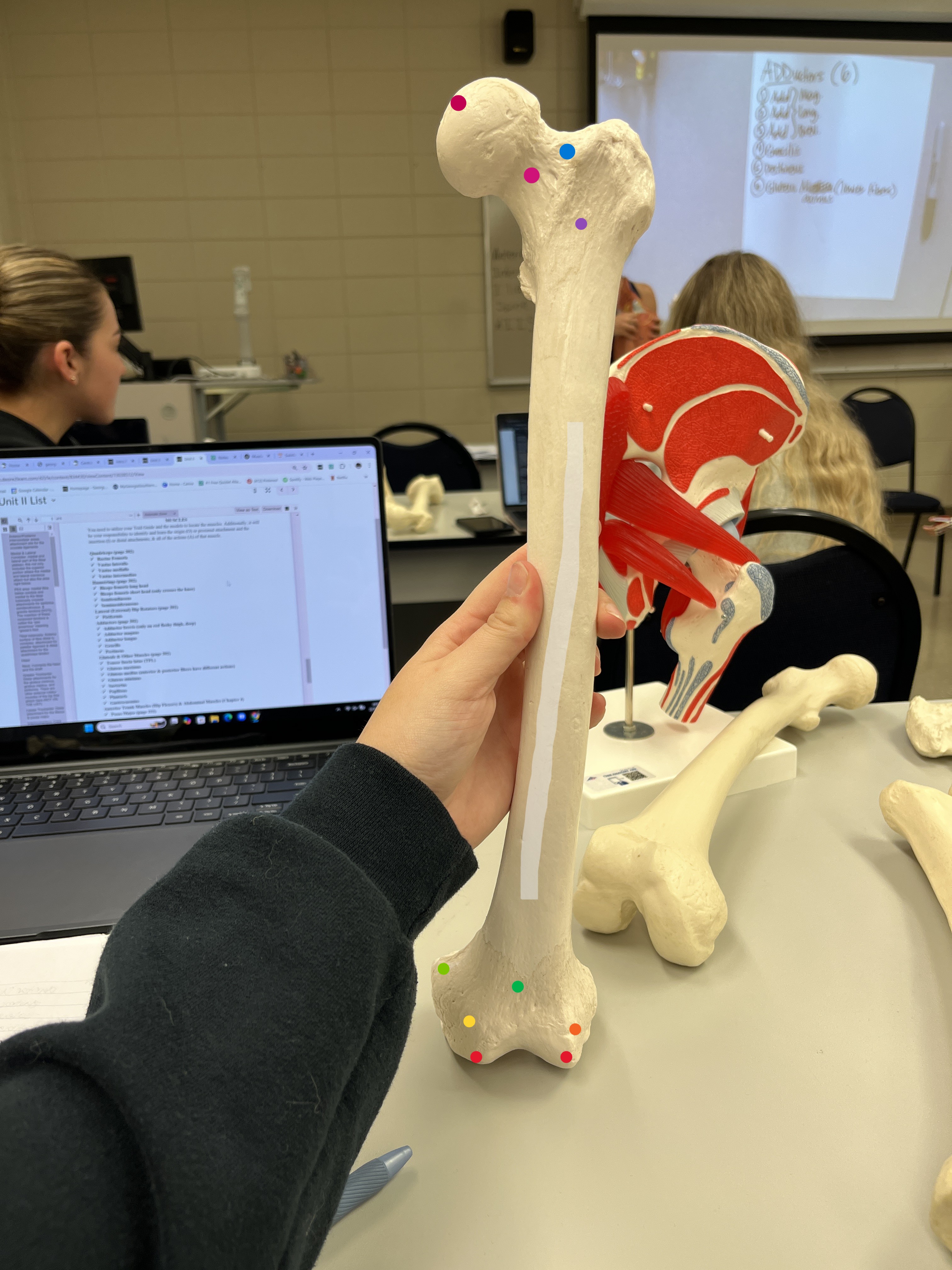
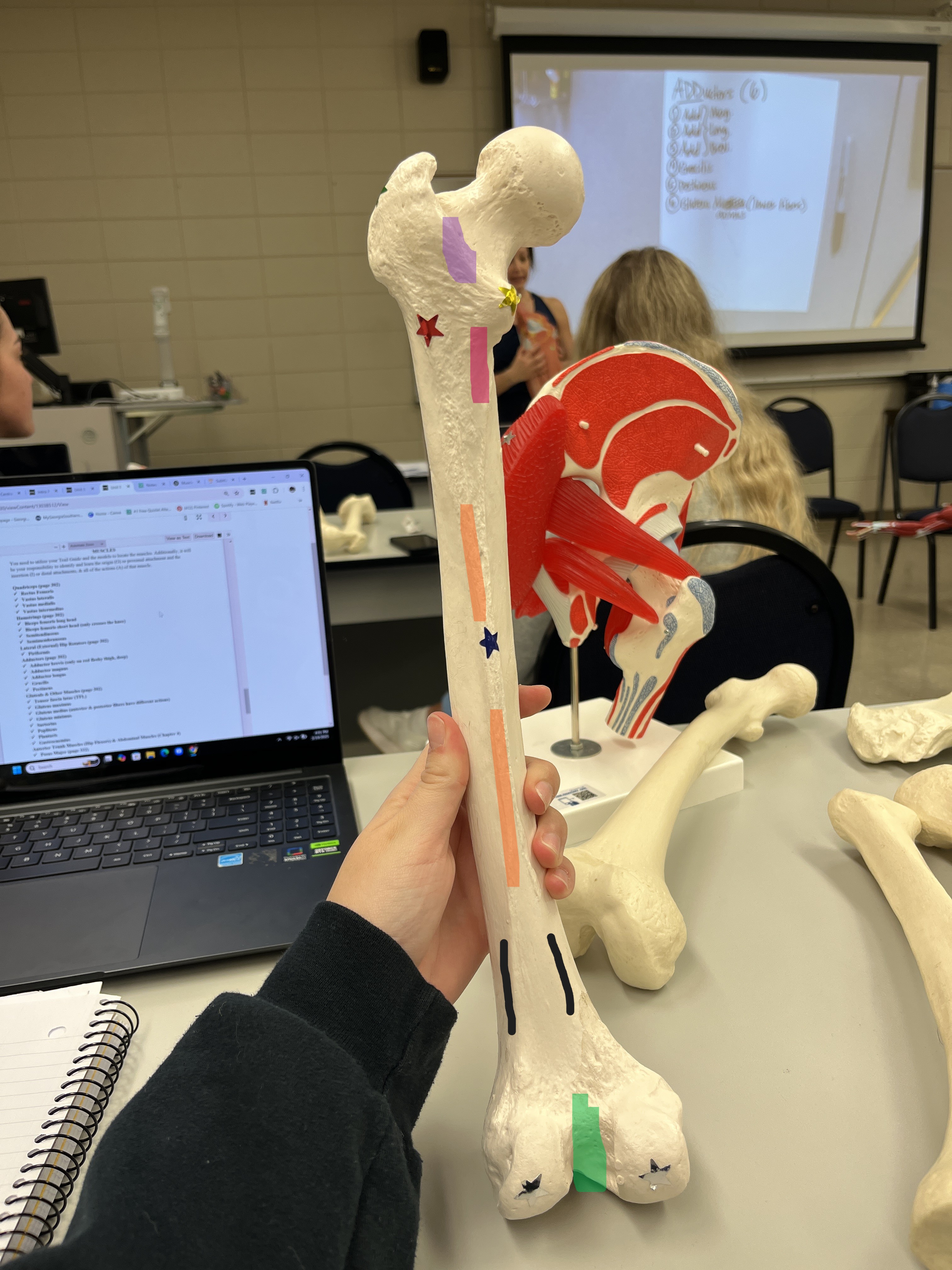
Head ( dark pink): The rounded proximal end of the femur that articulates with the acetabulum of the pelvis to form the hip joint.
Neck(pink): Connects the femoral head to the shaft, providing structural support and allowing for movement at the hip joint.
Greater Trochanter (green star): Large lateral projection where multiple muscles attach, including the gluteus minimus, gluteus medius, and piriformis. It serves as a key site for hip stabilization and rotation.
Lesser Trochanter(yellow Star): A smaller, posterior-medial projection that serves as the distal attachment for the iliacus and psoas major, playing a critical role in hip flexion.
Intertrochanteric Crest (purple highlight): A ridge on the posterior side between the greater and lesser trochanters, serving as an attachment site for muscles and stabilizing structures of the hip.
Intertrochanteric Line(purple): A ridge on the anterior side between the greater and lesser trochanters; provides attachment for the iliofemoral and pubofemoral ligaments, contributing to hip stability.
Intercondylar Fossa (Notch)Green Highlight: A deep groove between the medial and lateral condyles on the distal posterior femur; serves as an attachment site for the anterior and posterior cruciate ligaments, which help stabilize the knee.
Gluteal Tuberosity and/or Line (red star): Runs from the lip of the linea aspera to the base of the greater trochanter laterally. It may appear as a roughened depression or a prominent tuberosity. The gluteus maximus muscle distally attaches along this structure, contributing to hip extension and external rotation.
Linea Aspera(orange highlight): A prominent ridge along the posterior shaft of the femur that serves as an attachment site for multiple muscles, including:
Vastus lateralis
Vastus medialis
Adductor magnus
Adductor longus
Adductor brevis
Medial/Lateral Supracondylar Lines(black): Y-shaped ridges above the condyles that originate from the linea aspera and extend distally. The plantaris muscle originates from the lateral supracondylar line.
Lateral Epicondyle(dark orange) : A bony prominence on the lateral distal femur that serves as an attachment site for the lateral collateral ligament and the lateral head of the gastrocnemius muscle.
Medial Epicondyle(yellow): A bony prominence on the medial distal femur that serves as an attachment site for the medial collateral ligament and the medial head of the gastrocnemius muscle.
Adductor Tubercle(light green): Located on the distal end of the medial epicondyle; serves as the attachment site for the adductor magnus muscle.
Medial/Lateral Condyles(red): Large, rounded structures on the distal femur that articulate with the tibial condyles to form the knee joint. The gastrocnemius muscle attaches proximally here, while the popliteus originates on the lateral condyle.
Patellar Surface (lime green): A smooth anterior surface between the condyles where the patella glides during knee movement.
Pectineal Line(Pink highlight): A ridge extending from the linea aspera to the lesser trochanter; serves as the attachment site for the pectineus muscle.
Shaft of the Femur ( white highlight): The long, cylindrical portion of the femur. The anterior portion serves as the origin site for the vastus intermedius muscle, which assists in knee extension.
Trochanteric Fossa (blue): A deep depression on the medial surface of the greater trochanter, serving as an attachment site for several external rotator muscles of the hip.
Pelvis
Ilium (Superior Portion of the Os Coxae)
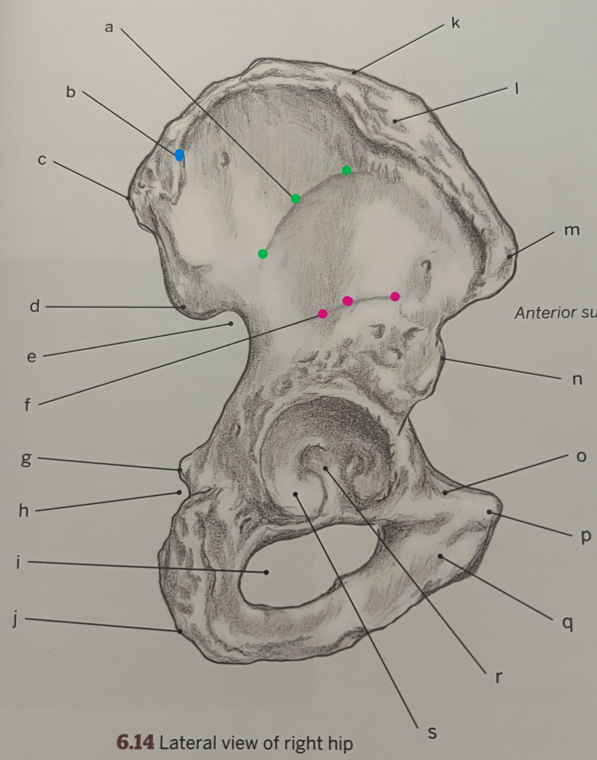
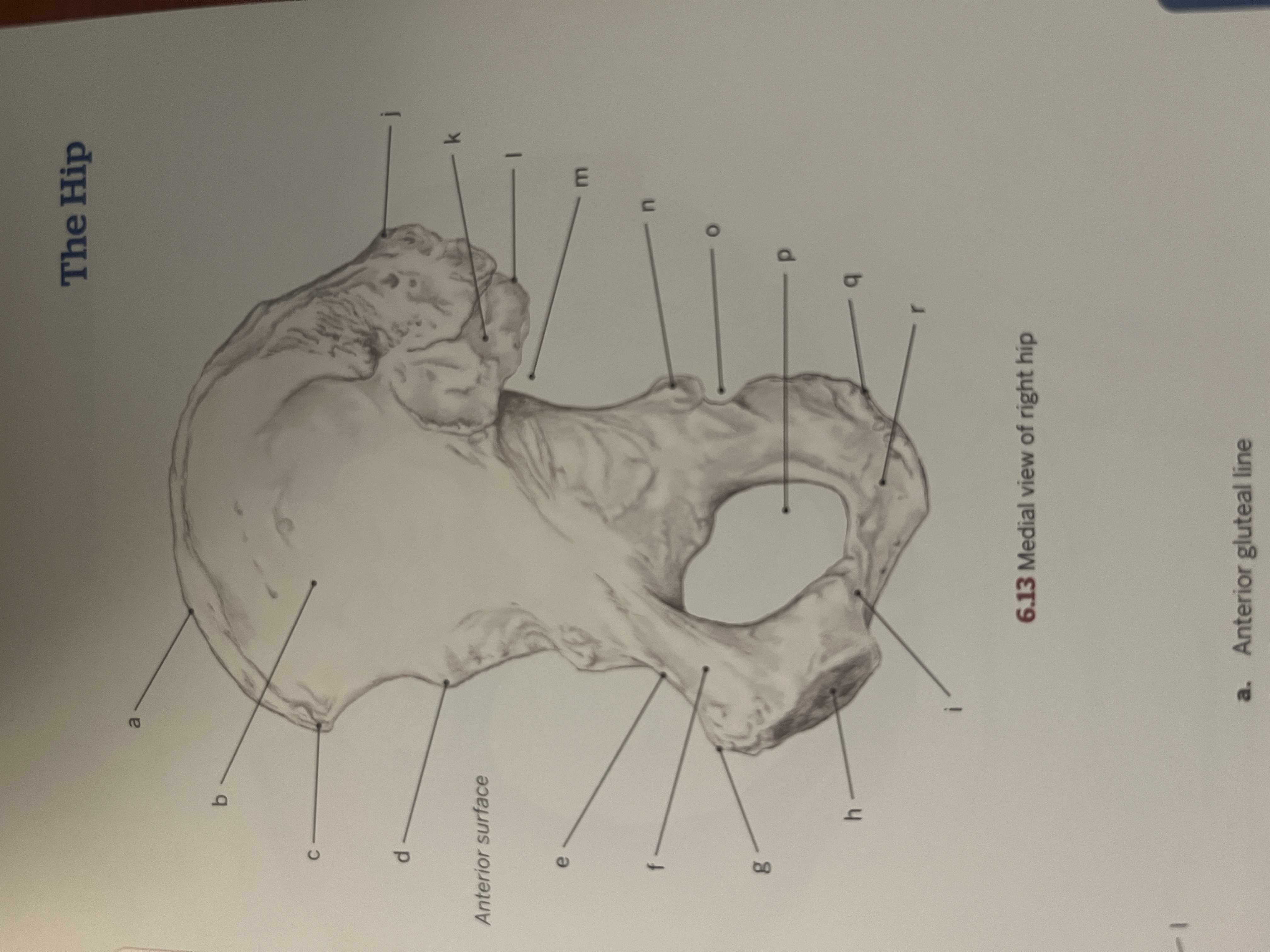
Anterior Inferior Iliac Spine (AIIS):d
Proximal Attachment: Rectus femoris (origin)
Function: Assists in hip flexion and knee extension
ADL Example: Walking upstairs
Concentric Exercise: Leg raises
Anterior Superior Iliac Spine (ASIS):c
Proximal Attachment: Sartorius (origin), Inguinal ligament (attachment)
Function: Assists in hip flexion, abduction, and external rotation
ADL Example: Sitting cross-legged
Concentric Exercise: Leg swings (abduction)
Gluteal Lines: Mark attachment sites for gluteal muscles
Posterior Gluteal Line:blue
Proximal Attachment: Gluteus medius (origin)
Function: Hip abduction and stabilization
ADL Example: Standing on one leg
Concentric Exercise: Side-lying leg lifts
Anterior Gluteal Line:green
Proximal Attachment: Gluteus minimus (origin)
Function: Assists in hip internal rotation and abduction
ADL Example: Balancing while walking
Concentric Exercise: Hip abduction with resistance bands
Inferior Gluteal Line: pink
Proximal Attachment: Gluteus minimus (origin)
Function: Hip stabilization
ADL Example: Controlling gait during walking
Concentric Exercise: Lateral step-ups
Iliac Fossa:b
Proximal Attachment: Iliacus (origin)
Function: Hip flexion
ADL Example: Bringing knee to chest
Concentric Exercise: Seated knee raises
Iliac Crest:a
Proximal Attachments: Abdominal muscles, TFL, gluteus maximus
Function: Core stabilization and hip abduction
ADL Example: Twisting torso while reaching
Concentric Exercise: Side bends
Posterior Inferior Iliac Spine (PIIS):K
Proximal Attachment: Sacrotuberous ligament
Function: Supports pelvis stabilization
ADL Example: Maintaining posture while sitting
Concentric Exercise: Hip bridges
Posterior Superior Iliac Spine (PSIS):J
Proximal Attachment: Gluteus maximus (partial origin)
Function: Hip extension
ADL Example: Standing from a seated position
Concentric Exercise: Squats
Ischium (Posterior-Inferior Portion of the Os Coxae)
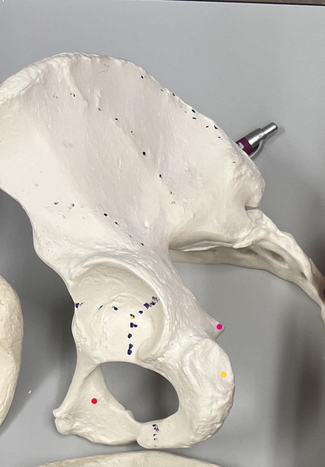
Ischial Spine: pink
Proximal Attachment: Sacrospinous ligament, gemellus superior (origin)
Function: Assists in lateral rotation of the hip
ADL Example: Turning while walking
Concentric Exercise: Clamshell exercise
Ischial Tuberosity: yellow
Proximal Attachments: Semitendinosus, semimembranosus, biceps femoris (long head), quadratus femoris, gemellus inferior (origins)
Function: Hip extension, knee flexion
ADL Example: Sitting down
Concentric Exercise: Deadlifts
Inferior Ramus of the Ischium: red
Proximal Attachments: Adductor brevis, adductor magnus, gracilis
Function: Hip adduction
ADL Example: Crossing legs while sitting
Concentric Exercise: Seated hip adduction
Pubis (Anterior-Inferior Portion of the Os Coxae)
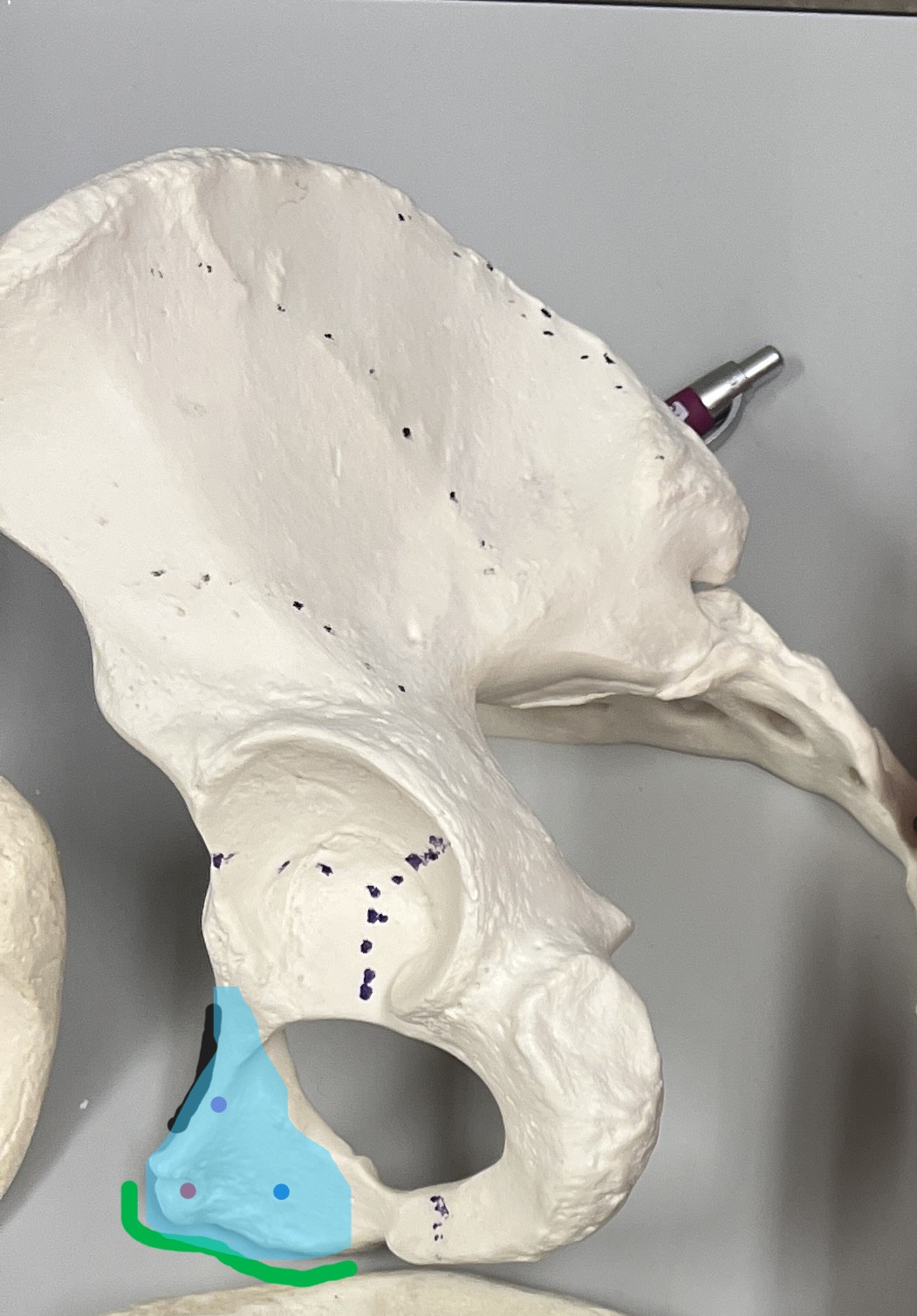
Inferior Ramus of the Pubis: blue
Proximal Attachments: Adductor brevis, adductor magnus, gracilis (origin)
Function: Hip adduction
ADL Example: Pulling legs together
Concentric Exercise: Hip adduction machine
Superior Ramus of Pubis: purple
Proximal Attachments: Adductor longus (pubic tubercle), pectineus (pectineal line), rectus abdominis (pubic crest/symphysis)
Function: Assists in hip adduction and core stabilization
ADL Example: Kicking a soccer ball
Concentric Exercise: Hanging leg raises
Pectineal Line: black
Proximal Attachment: Pectineus (origin)
Function: Hip flexion and adduction
ADL Example: Lifting leg to step onto a curb
Concentric Exercise: Standing hip flexion with resistance bands
Pubic Crest: blue highlight
Proximal Attachment: Rectus abdominis (origin)
Function: Trunk flexion
ADL Example: Bending forward to tie shoes
Concentric Exercise: Sit-ups
Pubic Symphysis: green line
Function: Provides stability between the two os coxae
ADL Example: Supporting pelvis during walking
Concentric Exercise: Pelvic tilts
Pubic Tubercle: red
Proximal Attachment: Adductor longus (origin)
Function: Hip adduction
ADL Example: Bringing legs together while standing
Concentric Exercise: Inner thigh squeezes
Joints
Knee (Tibiofemoral & Patellofemoral Articulations)
Proximal Attachment: Femur (Condyles)
Distal Attachment: Tibia (Condyles)
Function: Knee flexion and extension
Exercise: Squats
ADL: Walking
Hip Joint
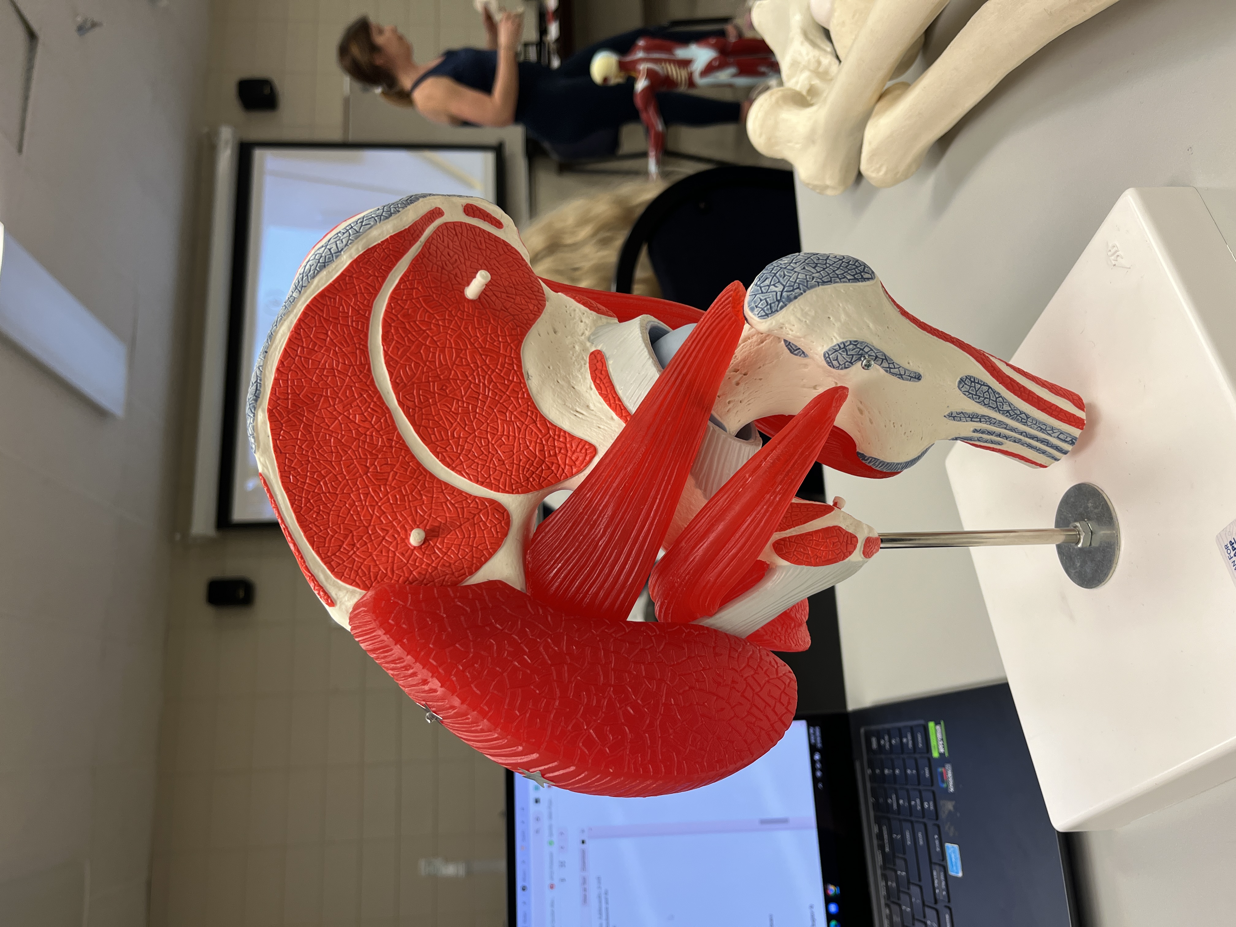
Proximal Attachment: Acetabulum
Distal Attachment: Head of Femur
Function: Hip movement
Exercise: Hip thrusts
ADL: Sitting down
Sacroiliac Joint
:background_color(FFFFFF):format(jpeg)/images/library/13208/ZzTBXyAOEp5TcstuOZbGqg_Sacroiliac_Joint_01.png)
Proximal Attachment: Sacrum
Distal Attachment: Ilium
Function: Transfers weight from spine to pelvis
Exercise: Bridge exercises
ADL: Standing balance
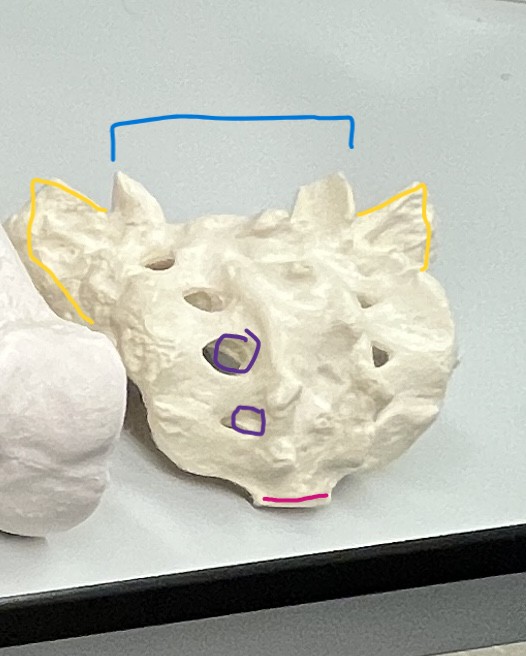
. Base of the Sacrum (blue)
Proximal Attachments:
Connects to the 5th lumbar vertebra (L5) through the Lumbosacral joint.
Distal Attachments:
The sacrum attaches to the ilium (pelvic bones) forming the sacroiliac joint.
Insertions:
No direct insertions.
Actions:
Primarily involved in the support and stability of the pelvis and the trunk.
Concentric Exercise Example: Squats (especially deep squats) help engage muscles around the sacral base by stabilizing the pelvis and spine.
ADL Example: Lifting an object from the ground with a neutral spine, activating muscles near the sacral base to protect the lower back.
2. Apex of the Sacrum(pink)
Proximal Attachments:
The apex faces downward and connects with the coccyx.
Distal Attachments:
The apex has no direct distal attachments to muscles, but it connects indirectly through ligaments and fascia.
Insertions:
No specific muscle insertions.
Actions:
Supports the weight transferred from the upper body and provides attachment points for various ligaments and muscles.
Concentric Exercise Example: Deadlifts, as they involve posterior chain activation that affects the apex region by helping stabilize the sacrum.
ADL Example: Sitting down and standing up from a chair, where the sacrum helps with posture and balance.
3. Sacral Foramina (anterior and posterior) purple
Proximal Attachments:
No direct proximal attachments, but the foramina are pathways for nerves and vessels.
Distal Attachments:
These openings allow the passage of sacral nerves and vessels that emerge from the spinal canal.
Insertions:
The foramina allow nerve roots to exit and insert into various areas of the pelvis and lower limbs.
Actions:
The sacral foramina help in nerve signaling for lower extremity movements and sensation.
Concentric Exercise Example: Leg raises or lunges. These exercises recruit the lower body muscles, which are affected by the sacral nerve signals.
ADL Example: Walking or cycling, which require nerve transmission through the sacral foramina for motor control of the legs.
4. Ala (Wing of the Sacrum) yellow
Proximal Attachments:
Connects to the lumbar spine and ilium (pelvic bones) through the sacroiliac joint.
Distal Attachments:
Supports various ligaments like the posterior sacroiliac ligaments and iliolumbar ligaments.
Insertions:
No specific muscle insertions, but the area provides attachment for ligaments and some muscles of the back.
Actions:
Plays a role in stabilizing the pelvis and transferring body weight to the lower limbs.
Concentric Exercise Example: Hip thrusts or glute bridges, as they activate the muscles attached to the sacrum, especially around the ala region.
ADL Example: Climbing stairs or standing from a seated position. The ala helps stabilize the pelvis and aids in weight transfer
Ligaments
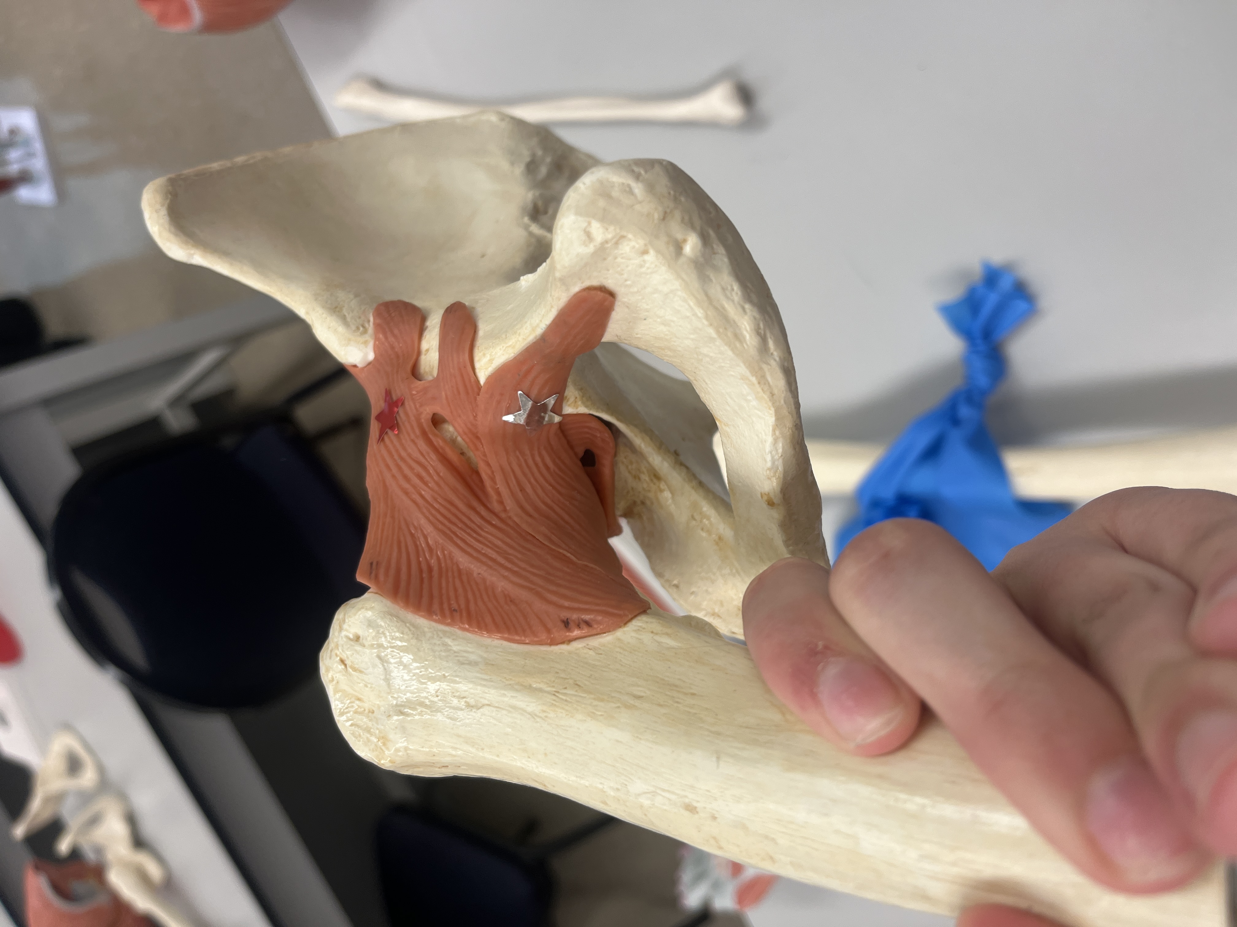
Iliofemoral Ligament (Y-Shaped Ligament) red
Attachments: From the ilium at the AIIS to the acetabular rim, extending down to the intertrochanteric line of the femur.
Function: Resists excessive hip extension, external rotation, and abduction.
ADL Example: Prevents overextension when walking or standing upright.
Concentric Exercise: Hip flexion exercises like leg raises.
Pubofemoral Ligament silver
Attachments: From the body of the pubis and adjacent superior pubic ramus, passing anterior to the femoral neck (intertrochanteric line).
Function: Resists excessive hip extension, abduction, and external rotation.
ADL Example: Stabilizes the hip when stepping sideways.
Concentric Exercise: Side leg lifts targeting hip adduction.
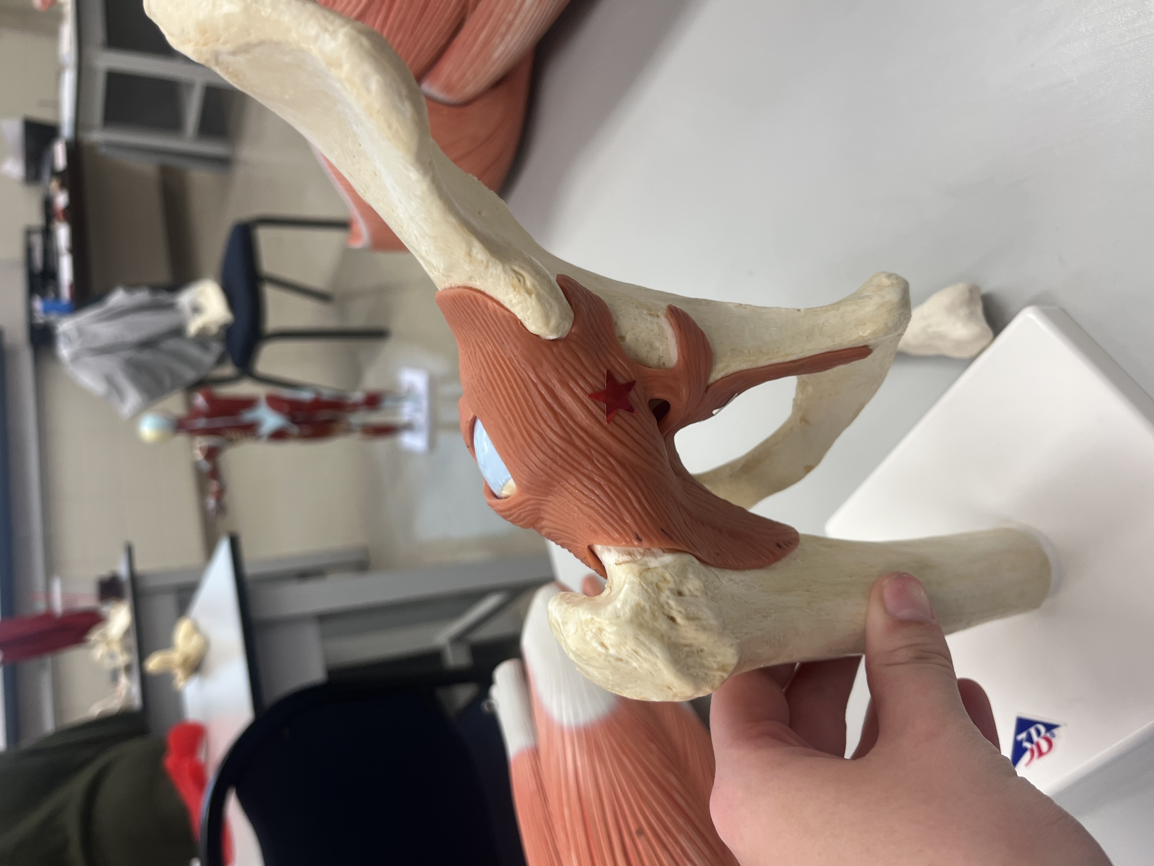
Ischiofemoral Ligament red
Attachments: From the body of the ischium behind the acetabulum to the femoral neck, with fibers spiraling up and out to the femur.
Function: Resists excessive hip extension, abduction, and internal rotation.
ADL Example: Provides stability during twisting movements.
Concentric Exercise: Hip internal rotation exercises.
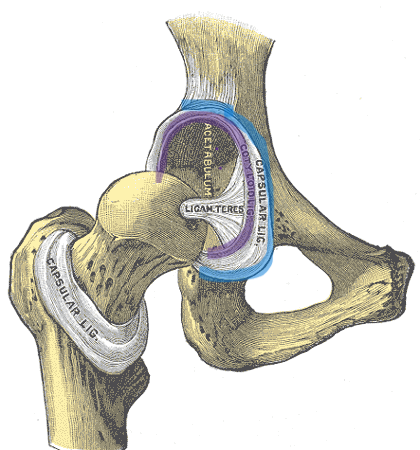
Acetabular Labrum- purple
Structure: Fibrocartilaginous rim that deepens the acetabular socket, increasing stability and protecting bone edges.
Function: Enhances joint congruency and reduces stress on the hip joint.
ADL Example: Helps maintain balance and stability while walking or running.
Concentric Exercise: Hip stabilization exercises, such as banded lateral walks.
Transverse Acetabular Ligament- blue
Attachments: Completes the acetabular labrum circle at the acetabular notch, spanning from end to end.
Function: Holds the femoral head in the acetabulum and bridges the notch, converting it into a foramen for vascular passage.
ADL Example: Supports the hip joint while transitioning between sitting and standing.
Concentric Exercise: Deep squats and step-ups.
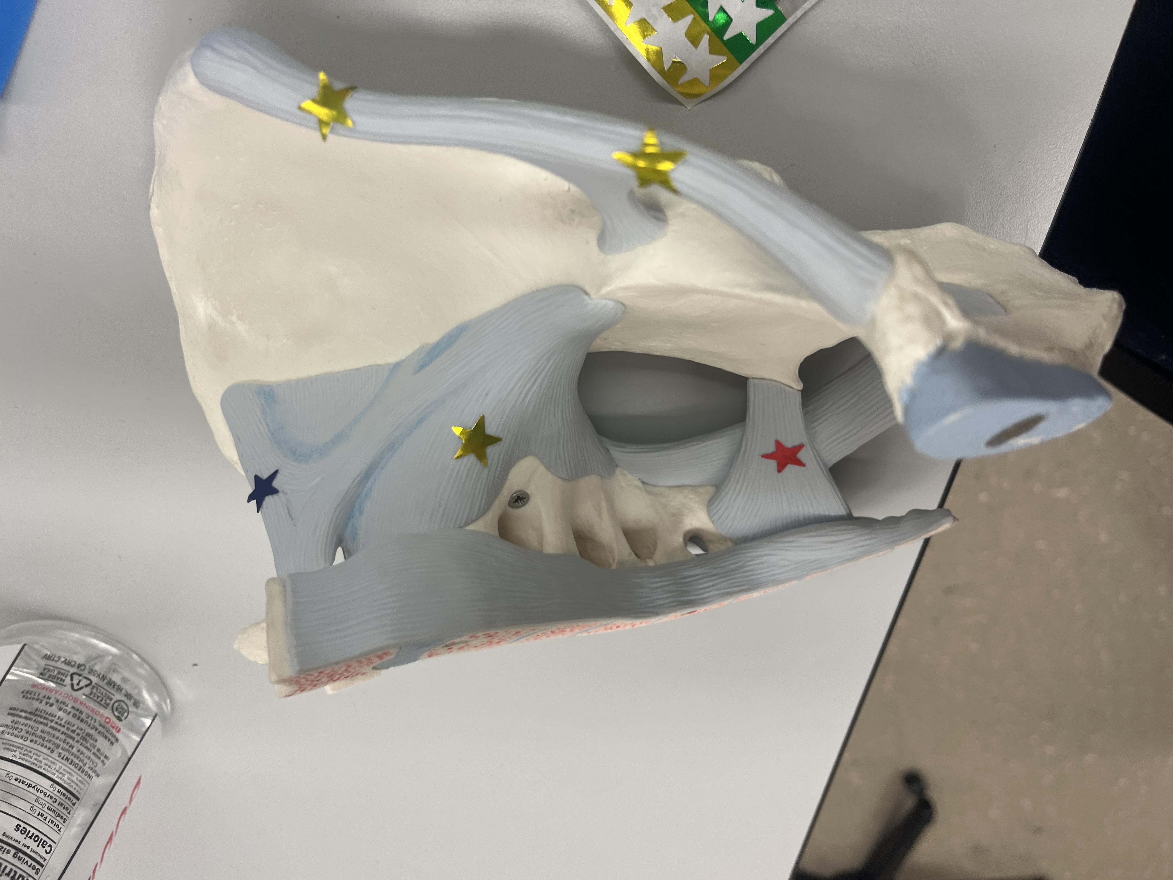
Inguinal Ligament- yellow star closer to bottom of photo
Attachments: Supports soft tissues of the groin; serves as an attachment site for the internal oblique and transverse abdominis.
Function: Stabilizes the anterior pelvis and lower abdominal wall.
ADL Example: Assists in core engagement when bending forward.
Concentric Exercise: Core stabilization exercises like planks.
Iliolumbar Ligament- blue star
Attachments: Superior and inferior bands extend from the transverse processes of L4 and L5 to the iliac crest.
Function: Holds L4, L5, and the sacrum together, maintaining pelvic stability.
ADL Example: Helps maintain posture and lower back support during standing or lifting.
Concentric Exercise: Deadlifts and back extensions.
Sacrotuberous Ligament- pink
Attachments: Extends from the ischial tuberosity to the posterior spines of the ilium and the lateral sacrum and coccyx.
Function: Limits anterior pelvic tilt, stabilizing the sacroiliac joint.
ADL Example: Maintains pelvic alignment when walking.
Concentric Exercise: Glute bridges and hip thrusts.
Sacrospinous Ligament- red
Attachments: Connects the ischial spine to the lateral border of the sacrum and coccyx.
Function: Limits anterior pelvic tilt, aiding in sacroiliac joint stability.
ADL Example: Provides support when transitioning from sitting to standing.
Concentric Exercise: Hip abduction exercises, such as clamshells.
Anterior & Posterior Sacroiliac Ligaments - gold start closer to blue star/ sacrum
Attachments: Both ligaments connect the sacrum to the pelvis.
Function: Provide strong stability for the sacroiliac joint, limiting excessive movement.
ADL Example: Assists in load distribution when carrying heavy objects.
Concentric Exercise: Weighted squats and lunges.