Ch. 2 - Brain Overview & Function
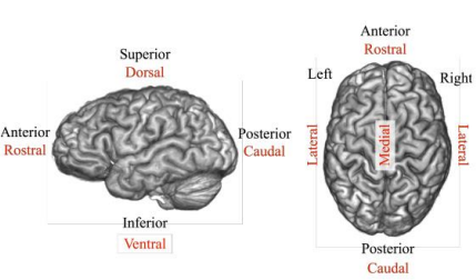
Brain Structure
Hindbrain (life support)
medulla oblongata (bottom of brain stem) - respiration, BP, coughing, sneezing, vomiting, heart rate
pons (middle of brain stem) - bridge between cerebrum and cerebellum; balance, sleep, arousal
reticular formation (also part of midbrain!) - main job is consciousness & wakefulness
cerebellum - muscle coordination, balance, general motor behavior
Midbrain (top of brain stem)
bridge between lower brain stem & forebrain; relays visual/auditory signals
has superior & inferior colliculi (4 bumps on the back of midbrain)
Forebrain
thalamus - relays info to cerebral cortex (w/ a little bit of preprocessing)
hypothalamus - controls pituitary gland (controls endocrine system) & homeostatic behaviors (e.g. temp. control, eating, drinking, sleep etc.)
hippocampus - long term memory
amygdala - emotional learning/memories
basal ganglia - voluntary motor behavior
cerebrum - brain’s largest part
white matter - inner layer of brain (at brain stem, it is outer); carries info between brain structures
cerebral cortex (grey matter) - outer layer of brain; processes information
frontal lobe
motor cortex - fine motor movements
prefrontal cortex (PFC) - planning, decision making, inhibitory control; last to develop in brain, first to decline in age
parietal lobe - has the somatosensory cortex which processes sensory info (e.g. pain, pressure, touch, temp.)
temporal lobe - in the area of the hippocampus & amygdala
occipital lobe - visual information processing
corpus callosum - major bridge between left & right hemisphere
anterior commissure - below and in front of corpus callosum; bridges temporal lobes
Localization of Function
early ideas: different brain areas control different mental abilities (franz gall), phrenology (now discredited) - bumps on skull = ability strength/personality traits (gall’s student spurzheim)
current idea: different brain regions have specialized functions !!
aphasias (disruption of expressive language)
language disorder caused by damage to language areas in left side of brain, affecting expression/understanding of language + reading/writing
broca’s area (non-fluent aphasia) - damage to left frontal lobe → impaired speech production
“walk dog” “book book two table”
wernicke’s area (fluent aphasia) - damage to superior posterior temporal lobe → impaired language understanding
“you know the smoodle pinkered, and that i want to get him round and take care of him like you want before”
homunculus (body map)
areas with more sensory input (e.g. hands, tongue, face)
kluver-bucy syndrome (KBS)
neurological/psychiatric disorder caused by damage to both temporal lobes, specifically in the amygdala
first described in 1939 after observing dramatic behavioral changes in monkeys following surgical removal of both temporal lobes
symptoms: hyperorality, hypersexuality, visual agnosia, hyperphagia, memory loss, emotional “flattening”, reduced fear and aggression
limits of localization -
not all brain functions can be neatly mapped to specific regions
ablation (removal of parts of the brain) showed that memory deficits depended on the amount, not location, of the cortex removed
brain = reorganizes, some regions can take over lost functions (equipotentiality), especially in young brains (more plasticity)
Lateralization of Function
referring to how 2 cerebral hemispheres play different roles in cognitive functioning
→ left - language specialization (about 95%) though a few can be bilateralized or right hemisphered dominant
→ right - spatial processing, musical ability & other cognitive activities
analytic-synthetic theory: 2 basic thinking modes; analytic in the left (computerlike info processing) & synthetic in the right (gestalts)
split-brain patients: those whose corpus callosum were surgically severed (happened in some epilepsy cases to prevent seizure spread)
resulted in:
left hand able to pick up objects but can’t describe/name it
information on the left could be identified by touch without seeing, while right hand can’t
Methods for Brain Study
computerized [axial] tomography (CAT/CT)
uses x-rays (radiation; invasive) from different angles to create images of the body
scans allow us to see “slices” of a living brain, which helps with diagnosing brain injury
magnetic resonance imaging (MRI)
uses strong magnetic field + radio waves to make images
radio waves make molecules in body line up, before returning to normal and sending signals, which show different tissues of body
better contrast resolution & no radiation
positron emission tomography (PET)
injects radioactively labeled compound, measuring blood flow to different regions of the brain
uses radiation (invasive) → scans show “slices” of living brain
single-photon emission computed tomography (SPECT)
nuclear medicine scan using a radioactive tracer, creating 3D images of organ function, involving radiation exposure (invasive)
commonly used for cardiac perfusion/stress testing & brain disorders
lower contrast/spatial resolution but cheaper than PET scan
functional magnetic resonance imaging (fMRI)
examines brain function by evaluating blood flow across different brain regions
occurs when a part of the brain becomes more active → more blood flow → more oxygen → more MRI signal in that region
electrical recording methods
electroencephalography (EEG)
measures brain activity using scalp electrodes, detecting different states of consciousness
excellent temporal resolution (timing accuracy)
limited spatial detail
magnetoencephalography (MEG)
measures magnetic field from brain activity
precise location of brain function + timing accuracy
better spatial resolution
often used with MRI (more expensive than EEG)
event-related potential (ERP)
measures brain responses to specific events via EEG before, during and after stimulus
transcranial magnetic stimulation (TMS)
studies specific brain circuit activity via magnetic coil near scalp to excite/inhibit targeted brain areas
used in research/clinic
Brain Training
neuroplasticity: brain’s ability to adapt by changing its structure and function
enriched environments → better brain development + dendritic fields
brain games → improved cognitive skills (perhaps in memory/processing speed) but limited to trained stacks, lack broad/lasting benefits (doesn’t prevent major declines like alzheimer’s), and no studies yet meet the highest research standards
physical exercise + social activities = stronger health benefits (than only brain training)