Coursework Support
Abstract = 200 words = 10%
Background of proteomic study
Introduce the aim
Results
No difference in samples in western blot (p53)
84 proteins studied in microarray (cancer vs control)
Some proteins overexpressed in cancer compared to control
Conclusion
Both methods good in studying cancer
Protein microarray is better - you can study different proteins (in comparison to western blot)
Overall microarray will be cheaper than doing western blot as you can study multiple at once.
Western blot is also time consuming.
Western blot needs more samples
Write abstract after the entire report is done.
NO REFERENCES IN ABSTRACT
Introduction = 350 words = 10%
Key words:
p53 → primary antibody in western blot
Incidence of cancer
Proteomic study
Signaling pathways
Cell lysate and Control cell underwent two different experiments.
Western Blot
Protein Microarray
AIM: Effectiveness of using different proteomic methods in the studying of cancer.
Include clear background of the problem using the key words.
Need good references → Papers (not websites)
Results = 600 words = 20%
Present the results as text, figures and tables.
Text: Start of the results.
Figures
Tables
Just write your findings → do not explain!
e.g Western blot experiment showing xyz
Cross reference: “As shown is figure 1”
Reference the figures in the text.
Figure legends: Clear and shows sufficient details of the experiment and the findings.
Talk about the methodology and statistical test used to analyze.
Above for a table
Below for a figure
Read the PDF for microarray methodology
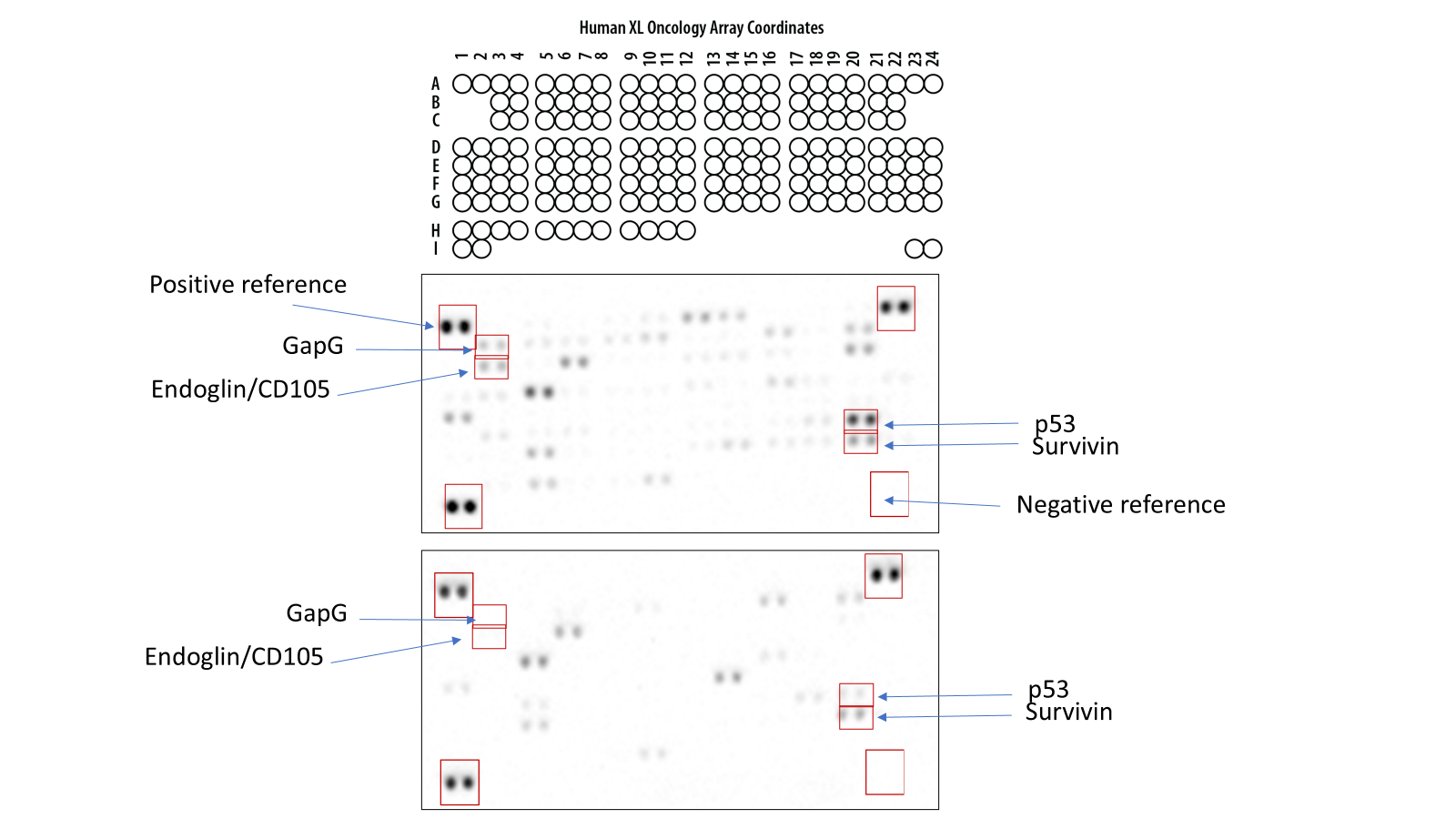
Image to be used in results section
Use PDF to learn how to explain the results of microarray
Negative reference: background to make the test reliable
If it comes up positive then it means a mistake was made in the results.
Excel sheet on Moodle with quantified results → Make a graph
Top one → cancer lysate
Bottom → Normal lysate
Western blot: Can use any image you want from the folders
No graph needed for western blot.
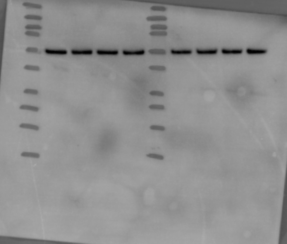
p53 revealed after addition of primary antibody (anti-p53).
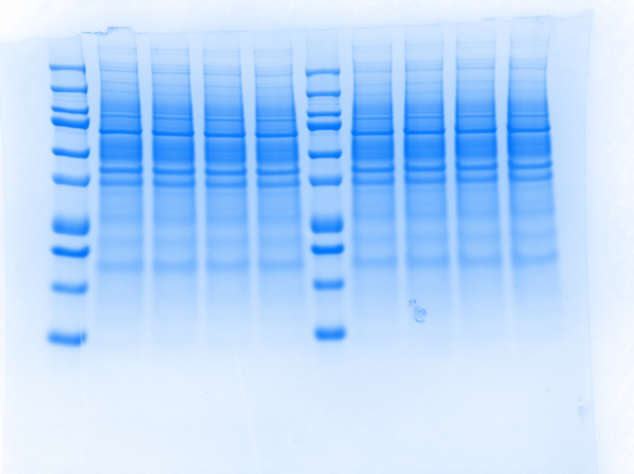
Coursework folder has the MR weight for each marker
Use the protocol from practical to see what sample was put in each well
Discussion = 850 words = 30%
Interpretation of the results and background of existing knowledge.
Publications which used your methodology.
First paragraph → Re-iterate your aim
Limitations
No repeats
Small sample size
Final paragraph → Conclusion