Muscular System Notes
I. The Muscular System
Muscles are responsible for all types of body movement
they contract or shorten and are the machine of the body
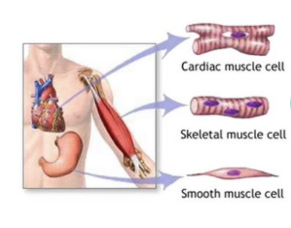
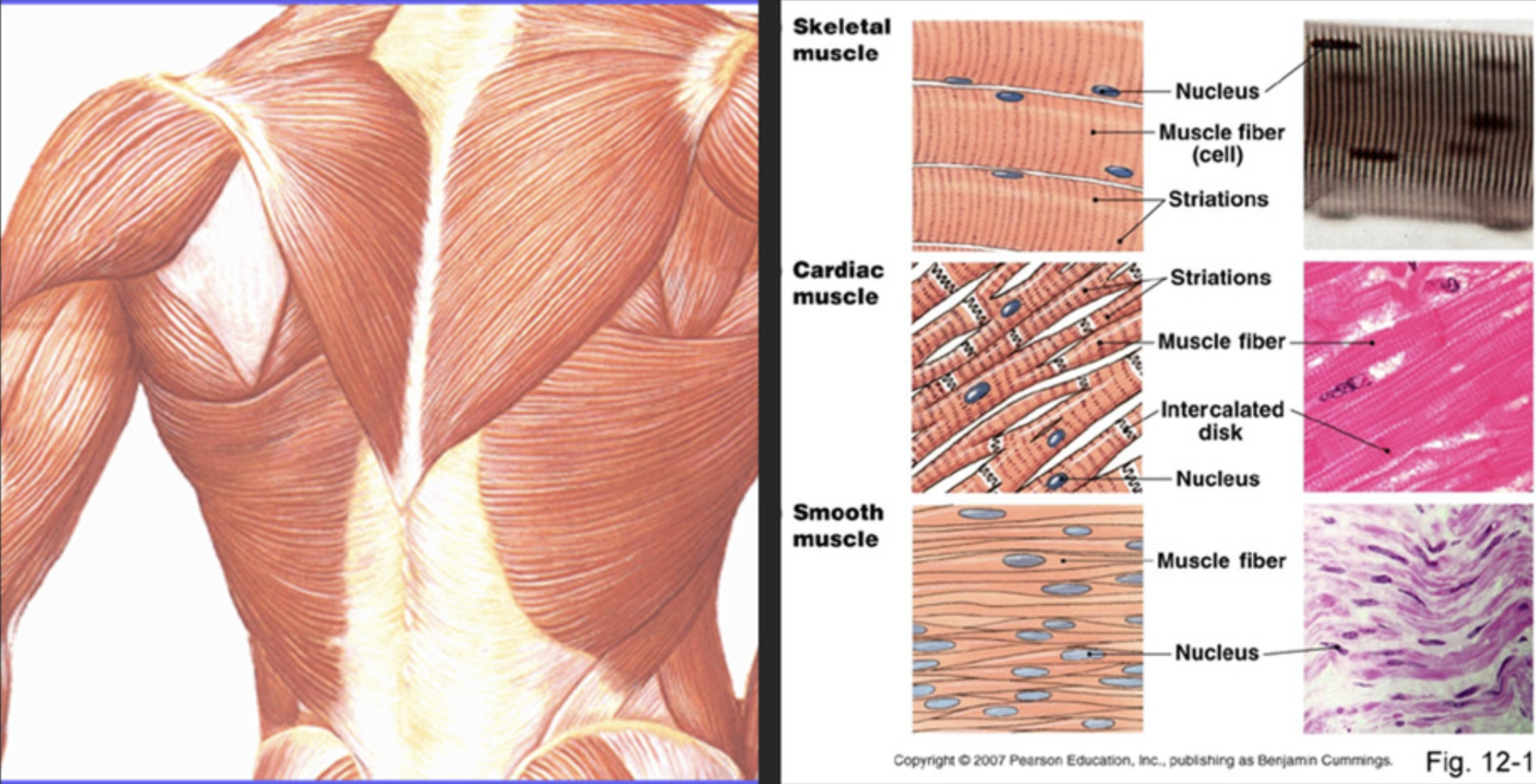
Three basic muscle types are found in the body
Skeletal muscle
Cardiac muscle
Smooth muscle
II. Function of Muscles
Support the body
Allow for movement by making bones and other body parts move
Maintain constant body temperature
Assist in movement of cardiovascular veins and lymph
Protect internal organs and stabilizes joints
III. Anatomy of a Skeletal Muscle
A. Organization of Skeletal Muscle
B. Coverings of Skeletal Muscle
Skeletal muscles are organs
They contain muscle fibers, nerves and blood vessels
Connective tissue membranes separate each muscle structure
Fascia - layer of fibrous tissue that separates muscles from each other and from the skin
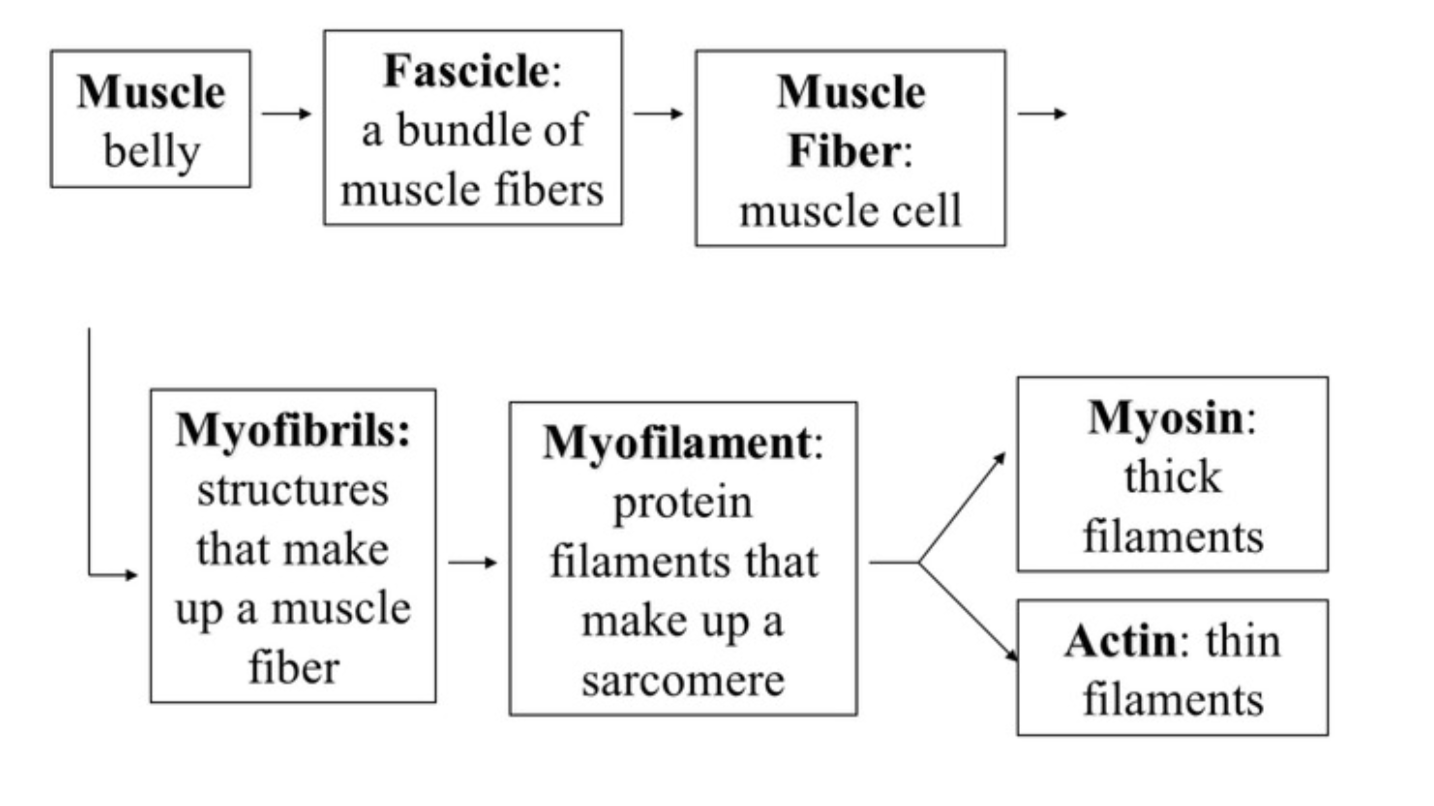
Coverings from largest to smallest:
Epimysium - covers the entire skeletal muscle
Perimysium - surrounds a bundle of muscle fibers (fascicle)
Endomysium - surrounds a single muscle fiber
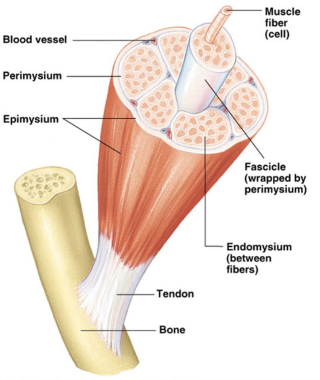
C. Skeletal Muscle Attachments
Epimysium blends into a connective tissue attachment, the tendon
Tendon - cord-like structure
Sites of muscle attachment
Bones, Cartilages, and connective tissue coverings
A. Microscopic Anatomy of Muscle Fiber (muscle cell)
Cells are multinucleate
Nuclei are just beneath the membrane
Sarcolemma - specialized plasma membrane
Sarcoplasmic reticulum - specialized smooth endoplasmic reticulum involved with muscle contraction
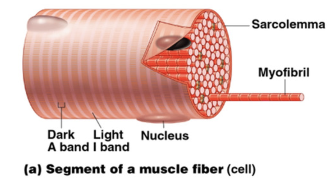
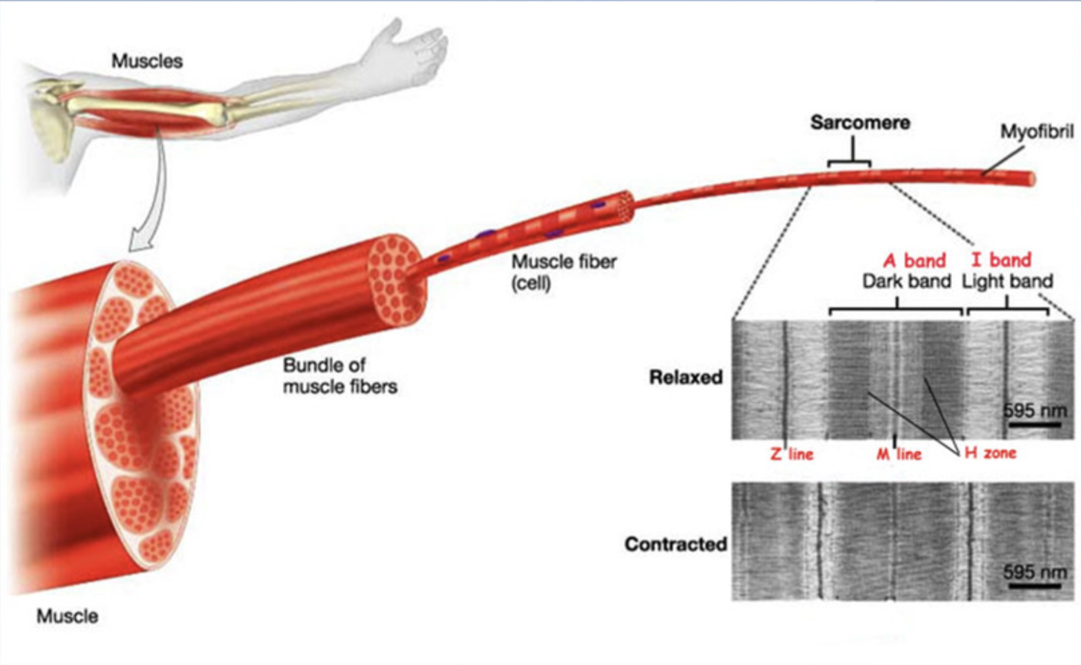
Myofibril
Bundles of myofilaments
Myofibrils are aligned to give distinct bands
Light band = I band
Dark band = A band
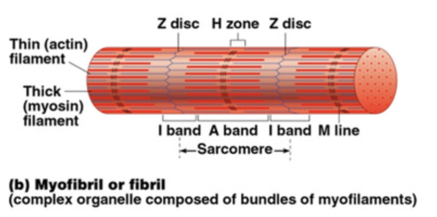
Sarcomere
Contractile unit of a muscle fiber
Organization of a sarcomere:
Thick filaments = myosin protein
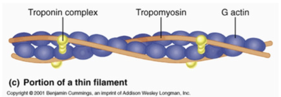
Thin filaments = actin protein
Myosin and actin overlap somewhat in the sarcomere
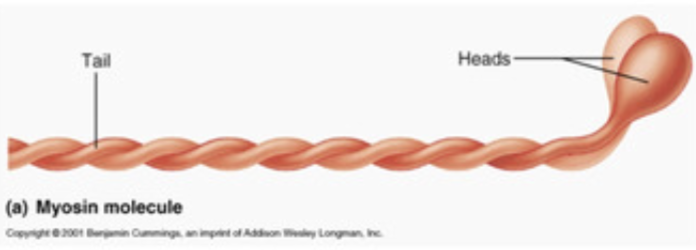
Myosin filaments have heads (extensions) that can “grab” onto actin forming a crossbridge
I. Physiology of Muscle Contraction
Skeletal muscles must be stimulated by a nerve (motor neuron) to contract
A. Transmission of Nerve Impulse to Muscle
Step 1: Nerve releases a neurotransmitter (acetycholine)
Step 2: Neurotransmitter causes muscle cell membrane gates to open
Step 3: Ions (Na+ and K+) exchange places causing the sarcoplasmic reticulum to release Ca2+
Step 4: This release of Ca2+ starts the muscle contraction as the actin filaments slide past the myosin filaments
B. The Sliding Filament Theory of Muscle Contraction
Sliding Filament Model - a muscle contracts when the thin filament in the muscle fiber slides over the thick filament
Activated by ATP and calcium (Ca2+) ions
Step 1: An influx of Ca2+ causes thick myosin filaments to form crossbridges with the thin actin filament by exposing the binding site on actin.
Step 2: The crossbridges change shape as it pulls on filaments which slides towards the center of the sarcomere in the power stroke
The distance between the Z line decreases, shortening the muscle
Step 3: The crossbridges detach from the actin filament when ATP bonds to myosin head
Step 4: The myosin head gets ready to bond to actin again using ATP energy
The cycle is repeated on another site of the actin filament
C. Contraction of a Skeletal Muscle
Muscle fiber contraction is “all or none”
Within a skeletal muscle, not all fibers may be stimulated during the same interval
Different combinations of muscle fiber contractions may give differing responses
Graded responses - different degrees of skeletal muscle shortening
Rapid stimulus = constant contraction or tetanus
D. Muscle Response to Strong Stimuli
Muscle force depends upon the number of fibers stimulated
More fibers contracting results in greater muscle tension
Muscle can continue to contract unless they run out of ATP or Ca2+
One molecule of ATP supplies enough energy for one actin and myosin cross-bridge
II. Energy for Muscle Contraction
Muscles use stored ATP for energy
Bonds of ATP are broken to release energy
Only 4-6 seconds worth of ATP is stored by muscles
Three ways for muscle to make energy (ATP)
Creatine Phosphate
Creatine phosphate is a high-energy compound and is the fastest way to make ATP available for muscles
Used for activities lasting less than 15 seconds
Anaerobic (no oxygen needed)
Reaction:
Creatine phosphate + ADP → creatine + ATP
Creatine phosphate is made when a muscle is at rest
Cellular Respiration
Mitochondria uses glucose molecules to make ATP in the presence of oxygen
Provides most of a muscle’s ATP
Aerobic (needs oxygen)
Used for activities lasting hours
Reaction;
C6H12O6 + 6O2 → 6CO2 + 6H20 + energy
1 glucose = 36 ATP
Anaerobic Respiration/Fermentation
Reaction that breaks down glucose without using oxygen
Used for activities lasting 30-60 seconds
Anaerobic (no oxygen needed)
Reaction:
Glucose → pyruvic acid + 2 ATP → lactic acid
Lactic acid is also produced causing pain in the muscle
Heavy breathing after exercise is a sign of oxygen deficiency
A marathon runner is exhausted after crossing the finish line because they have depleted not only their oxygen but their glucose as well
It takes up to two days to replace all of the glucose in the muscles and glycogen in the liver