Chapter 3: Anatomy and Research Methods (Chapter 13.1 Lateralization and language)
Learning Objectives
After studying this chapter, you should be able to:
1.Define the terms used to describe brain anatomy.
2.Describe the structure of the spinal cord.
3.Describe the structure and functions of the autonomic nervous system.
4.List the major components of the hindbrain, midbrain, and forebrain.
5.Distinguish between the laminae and columns of the cortex.
6.List the four lobes of the cerebral cortex and describe their principal functions.
7.Describe the binding problem and explain its theoretical importance.
8.Describe major methods for studying the relationship between brain activity and behavior.
9.Note the strengths and limitations of each method.
Introduction
Learning neuroanatomy (the anatomy of the nervous system) from a book is like learning geography from a road map. A map can tell you that Mystic, Georgia, is about 40 km north of Enigma, Georgia, and a book can tell you that the habenula is about 4.6 mm from the interpeduncular nucleus in a rat’s brain (proportionately farther in a human brain). But these little gems of information will seem mysterious and enigmatic unless you are interested in that part of Georgia or that area of the brain.
This chapter is more like a world globe than a road map, describing the larger structures and some distinctive features of each. Still, it will introduce a great many terms and facts. You cannot expect to learn them all at once, and you should review this chapter repeatedly.
The first module introduces key neuroanatomical terms and outlines overall structures of the nervous system. In the second module, we concentrate on the cerebral cortex, the largest part of the mammalian central nervous system. The third module outlines research methods that explore the functions of brain areas.
Module 3.1 Structure of the Vertebrate Nervous System
Learning Objectives
After studying this module, you should be able to:
1.Define the terms used to describe brain anatomy.
2.Describe the structure of the spinal cord.
3.Describe the structure and functions of the autonomic nervous system.
4.List the major components of the hindbrain, midbrain, and forebrain.
Your nervous system includes a huge number of neurons and an even huger number of synapses. How do they all work together to make one behaving unit? Does each neuron have a unique function? Does each brain area have a unique function? Or does the brain operate as an undifferentiated whole?
The answer is something between those extremes. Consider an analogy to human society: Each person has a specialty, such as teacher, farmer, or nurse, but no one performs any function without the cooperation of many others. Similarly, brain areas and neurons have specialized functions, but they perform those roles by connections with other areas.
Terminology to Describe the Nervous System
For vertebrates, we distinguish the central nervous system from the peripheral nervous system (Figure 3.1). The central nervous system (CNS) is the brain and the spinal cord. The peripheral nervous system (PNS) connects the brain and spinal cord to the rest of the body. Part of the PNS is the somatic nervous system, which consists of the axons conveying messages from the sense organs to the CNS and from the CNS to the muscles. Another part of the PNS, the autonomic nervous system, controls the body’s organs. A special part of the autonomic nervous system is the enteric nervous system, which controls the digestive system (Rao and Gershon, 2018).

To follow a map, you must understand north, south, east, and west. Because the nervous system is three-dimensional, we need more terms to describe it. As Figure 3.2 and Table 3.1 indicate, dorsal means toward the back and ventral means toward the stomach. (A ventriloquist is literally a “stomach talker.”) In a four-legged animal, the top of the brain and spinal cord is dorsal (the side of the animal’s back), and the bottom is ventral (the stomach side). The same would be true for you if you crawled on your hands and knees. However, when humans evolved upright posture, the position of the head changed relative to the spinal cord. For convenience, we still apply the terms dorsal and ventral to the same parts of human anatomy as in other vertebrates. Consequently, the dorsal–ventral axis of the human brain is at a right angle to the dorsal–ventral axis of the spinal cord. Figure 3.2 also illustrates the three ways of taking a plane through the brain, known as horizontal, sagittal, and coronal (or frontal).


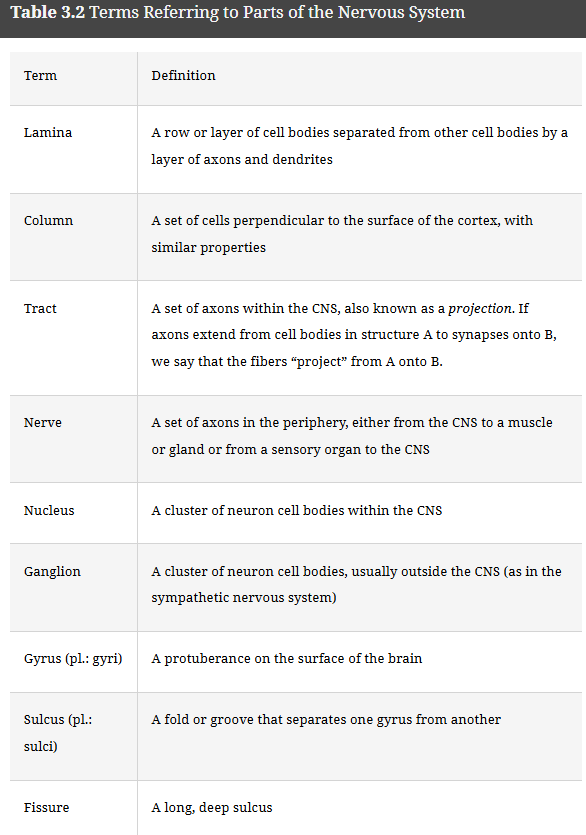
1.What does ventral mean, and what is its opposite? Ventral means toward the stomach side. Its opposite is dorsal.
2.What term means toward the midline, and what is its opposite? Medial; lateral
3.If two structures are both on the left side of the body, they are ____ each other. If one is on the left and the other is on the right, they are _____ to each other. Ipsilateral; contralateral
4.The bulges in the cerebral cortex are called ______. The grooves between them are called _____.
Gyri; sulci. To remember sulcus, think of the word sulk, meaning “to pout” (and therefore lie low).
The Spinal Cord
The spinal cord is the part of the CNS within the spinal column. The spinal cord communicates with all the sense organs and muscles except those of the head. It is a segmented structure, and each segment has on both its left and right sides a sensory nerve and a motor nerve, as Figure 3.3 illustrates. One of the earliest discoveries about the functions of the nervous system was that the entering dorsal roots (axon bundles) carry sensory information, and the exiting ventral roots carry motor information. The cell bodies of the sensory neurons are in clusters of neurons outside the spinal cord, called the dorsal root ganglia. (Ganglia is the plural of ganglion, a cluster of neurons. In most cases, a neuron cluster outside the CNS is called a ganglion, and a cluster inside the CNS is called a nucleus.) Cell bodies of the motor neurons are inside the spinal cord.

In the cross sections through the spinal cord depicted in Figures 3.3 and 3.4, the H-shaped gray matter in the center of the cord is densely packed with cell bodies and dendrites. Many neurons from the gray matter of the spinal cord send axons to the brain or to other parts of the spinal cord through the white matter, containing myelinated axons. Figure 3.5 depicts a close-up view of gray matter and white matter.


Each segment of the spinal cord sends sensory information to the brain and receives motor commands from the brain. All that information passes through tracts of axons in the spinal cord. If the spinal cord is cut at a given segment, the brain loses sensation from that segment and below. The brain also loses motor control over all parts of the body served by that segment and the lower ones.
The Autonomic Nervous System
The autonomic nervous system consists of neurons that receive information from and send commands to the heart, intestines, and other organs. Its two parts are the sympathetic and parasympathetic nervous systems (Figure 3.6). The sympathetic nervous system, a network of nerves that prepare the organs for a burst of vigorous activity, consists of chains of ganglia just to the left and right of the spinal cord’s central regions (the thoracic and lumbar areas). These ganglia have connections back and forth with the spinal cord. Sympathetic axons prepare the organs for “fight or flight,” such as by increasing breathing and heart rate and decreasing digestive activity. Because the sympathetic ganglia are closely linked, they often act as a single system “in sympathy” with one another, although certain events activate some parts more than others. The sweat glands, adrenal glands, muscles that constrict blood vessels, and muscles that erect the hairs of the skin have sympathetic input but no parasympathetic input.

The parasympathetic nervous system, sometimes called the “rest and digest” system, facilitates vegetative, nonemergency responses. The term para means “beside” or “related to,” and parasympathetic activities are related to, and generally the opposite of, sympathetic activities. For example, the sympathetic nervous system increases heart rate, and the parasympathetic nervous system decreases it. The parasympathetic nervous system increases digestive activity, whereas the sympathetic nervous system decreases it. The parasympathetic system also promotes sexual arousal, including erection in males. Although the sympathetic and parasympathetic systems produce contrary effects, both are constantly active to varying degrees, and many stimuli arouse parts of both systems.
The parasympathetic nervous system is also known as the craniosacral system because it consists of the cranial nerves and nerves from the sacral spinal cord (see Figure 3.6). Unlike the ganglia in the sympathetic system, the parasympathetic ganglia are not arranged in a chain near the spinal cord. Rather, long preganglionic axons extend from the spinal cord to parasympathetic ganglia close to each organ. Shorter postganglionic fibers then extend from the parasympathetic ganglia into the organs themselves. Because the parasympathetic ganglia are not linked to one another, they act more independently than the sympathetic ganglia do. Parasympathetic activity decreases heart rate, increases digestive rate, and, in general, conserves energy.
The parasympathetic nervous system’s axons release the neurotransmitter acetylcholine onto the organs. Most sympathetic nervous system axons release norepinephrine, although a few, such as those onto the sweat glands, use acetylcholine. Because the two systems use different transmitters, certain drugs excite or inhibit one system or the other. For example, over-the-counter cold remedies exert most of their effects by blocking parasympathetic activity or by increasing sympathetic activity. Because the flow of sinus fluids is a parasympathetic response, drugs that block the parasympathetic system inhibit sinus flow. The side effects of cold remedies stem from their pro-sympathetic, anti-parasympathetic activities: They increase heart rate, blood pressure, and arousal. They inhibit salivation and digestion. Certain decongestant pills containing pseudoephedrine have been withdrawn or restricted because of their potential for abuse.
5.Motor nerves leave from which side of the spinal cord, dorsal or ventral? Ventral.
6.Which functions are controlled by the sympathetic nervous system? Which are controlled by the parasympathetic nervous system? The sympathetic nervous system prepares the organs for vigorous fight-or-flight activity. The parasympathetic system increases vegetative responses such as digestion.
The Hindbrain
The brain has three major divisions: hindbrain, midbrain, and forebrain (Figure 3.7 and Table 3.3). Some neuroscientists prefer terms with Greek roots: rhombencephalon (hindbrain), mesencephalon (midbrain), and prosencephalon (forebrain). You may encounter these terms in other reading.


The hindbrain, the posterior part of the brain, consists of the medulla, the pons, and the cerebellum. The medulla and pons, the midbrain, and certain central structures of the forebrain constitute the brainstem (Figure 3.8).

The medulla, or medulla oblongata, can be regarded as an enlarged extension of the spinal cord. Just as the lower parts of the body connect to the spinal cord via sensory and motor nerves, the head and the organs connect to the medulla and adjacent areas by 12 pairs of cranial nerves (one of each pair on the right side and one on the left), as described in Table 3.4 and Figure 3.9. The size of each cranial nerve varies among species. In elephants, the trigeminal nerve that receives sensations from the face is huge because it innervates the trunk, a highly sensitive structure for elephants (Purkart, 2022). The elephant’s trigeminal nerve is thicker than its spinal cord!


Cranial nerves III, IV, and VI are coded in red to highlight their similarity: control of eye movements. Cranial nerves VII, IX, and XII are coded in green to highlight their similarity: taste and control of tongue and throat movements. Cranial nerve VII has other important functions as well. Nerve X (not highlighted) also contributes to throat movements, although it is primarily known for other functions.

The cranial nerves originating in the medulla control vital reflexes such as breathing, heart rate, vomiting, salivation, coughing, and sneezing. Because opiate receptors, which suppress activity, are abundant in the medulla, opiates can produce a dangerous decrease in breathing and heart rate.
The pons lies anterior and ventral to the medulla. Like the medulla, it contains nuclei for several cranial nerves. The term pons is Latin for “bridge,” reflecting the fact that in the pons, axons from each half of the brain cross to the opposite side of the spinal cord so that the left hemisphere controls the muscles of the right side of the body and the right hemisphere controls the left side.
The cerebellum is a large hindbrain structure with many deep folds. It is one of the first brain areas to start developing and one of the last to complete development (Sathyanesan et al., 2019). The cerebellum has long been known for its contributions to the control of movement, and many older textbooks describe the cerebellum as important for “balance and coordination.” True, people with cerebellar damage are clumsy and lose their balance, but the functions of the cerebellum extend far beyond balance and coordination. People with damage to the cerebellum have difficulty with timing, such as judging whether one rhythm is faster than another rhythm. Depending on the location and type of damage, they may have impairments of learning, memory, attention, visual-spatial processing, language, recognizing emotional expressions, or social behavior (De Zeeuw et al., 2021; Ferrari et al., 2018;
Koziol et al., 2014). One hypothesis is that the cerebellum’s primary function is to detect sequences of events (Van Overwalle et al., 2020; Vandervert, 2018).
7.Most of the cranial nerves connect to which brain area? The medulla and pons.
The Midbrain
As the name implies, the midbrain is between the hindbrain and the forebrain, although in adult mammals the forebrain surrounds it. The midbrain is more prominent in reptiles, amphibians, and fish. The roof of the midbrain is called the tectum. (Tectum is the Latin word for “roof.” The same root occurs in the geological term plate tectonics.) The swellings on each side of the tectum are the superior colliculus and the inferior colliculus (Figures 3.8 and 3.10). Both are important for sensory processing—the inferior colliculus for hearing and the superior colliculus for vision.

Under the tectum lies the tegmentum, the intermediate level of the midbrain. (In Latin, tegmentum means a “covering,” such as a rug on the floor. The tectum covers the tegmentum, but the tegmentum covers several other midbrain structures.) Another midbrain structure, the substantia nigra, gives rise to a dopamine-containing pathway that facilitates readiness for movement.
The Forebrain
The forebrain, the most prominent brain area for mammals and birds, consists of two cerebral hemispheres, left and right (Figure 3.11). Each hemisphere is organized to receive sensory information, mostly from the contralateral (opposite) side of the body. It controls muscles, mostly on the contralateral side, by way of axons to the spinal cord and the cranial nerve nuclei.

The outer portion is the cerebral cortex. (Cerebrum is a Latin word for “brain.” Cortex is a Latin word for “bark” or “shell.”) Under the cerebral cortex are other structures, including those illustrated in Figure 3.12. The thalamus is the main source of input to the cortex. The hypothalamus is essential for control of eating, drinking, temperature control, and reproductive behaviors. The amygdala is part of a circuit that is central for evaluating emotional information, especially with regard to fear. Figure 3.12 shows the positions of these structures in three-dimensional perspective. Figure 3.13 is a coronal (from the front) section through the human brain and a view of the ventral surface of the brain.


Thalamus
The thalamus is a pair of structures (left and right) in the center of the forebrain. It resembles two small avocados joined side by side. Most sensory information goes first to the thalamus, which processes it and sends output to the cerebral cortex. An exception to this rule is olfactory information, which goes from the olfactory receptors to the olfactory bulbs and then directly to the cerebral cortex.
Many nuclei of the thalamus receive their input from a sensory system, such as vision, process it in various ways, and transmit information to an area of the cerebral cortex, as in Figure 3.14. Another function of the thalamus relates to memory. When you hold information in working memory, activity bounces back and forth between the frontal cortex and the thalamus (Bolkan et al., 2017).

Hypothalamus and Pituitary Gland
The hypothalamus, a small area just ventral to the thalamus (Figures 3.10 and 3.12), has widespread connections with the rest of the brain. Damage in the hypothalamus leads to abnormalities in motivated behaviors, such as feeding, drinking, temperature regulation, sexual behavior, or fighting. Because of these important behavioral effects, the small hypothalamus attracts much research attention. Partly through nerves and partly by releasing hormones, the hypothalamus conveys messages to the pituitary gland, altering its release of hormones (Figure 3.10).
Basal Ganglia
The basal ganglia, a group of subcortical structures lateral to the thalamus, include the caudate nucleus, the putamen, and the globus pallidus (Figure 3.15). Many authorities include other structures as well, especially the nucleus accumbens.

It has long been known that damage to the basal ganglia impairs movement, as in conditions such as Parkinson’s disease and Huntington’s disease. The basal ganglia integrate motivational and emotional behavior to increase the vigor of selected actions. However, the role of the basal ganglia extends beyond movement. They are critical for gradual learning of skills and habits.
Basal Forebrain
A structure on the ventral surface of the forebrain, the nucleus basalis, receives input from the hypothalamus and basal ganglia and sends axons that release acetylcholine to widespread areas in the cerebral cortex (Figure 3.16). The nucleus basalis is a key system for arousal, wakefulness, and attention. Patients with Parkinson’s disease and Alzheimer’s disease have impairments of attention and intellect because of inactivity or deterioration in the nucleus basalis.

Hippocampus
The hippocampus (from the Latin word meaning “sea horse”) is a large structure between the thalamus and the cerebral cortex, mostly toward the posterior of the forebrain, as illustrated in Figure 3.12. We consider the hippocampus in more detail in the chapter on memory. The gist of that discussion is that the hippocampus is critical for memories, especially memories for individual events. It is also essential for monitoring where you are and where you are going.
Of the following, which are in the hindbrain, which in the midbrain, and which in the forebrain: basal ganglia, cerebellum, hippocampus, hypothalamus, medulla, pituitary gland, pons, substantia nigra, superior and inferior colliculi, tectum, tegmentum, thalamus? Hindbrain: cerebellum, medulla, and pons. Midbrain: substantia nigra, superior and inferior colliculi, tectum, and tegmentum. Forebrain: basal ganglia, hippocampus, hypothalamus, pituitary, and thalamus.
Which area provides most of the input to the cerebral cortex? Thalamus
The Ventricles
The nervous system begins its development as a tube surrounding a fluid canal. The canal persists into adulthood as the central canal in the center of the spinal cord, and as the ventricles, four fluid-filled cavities within the brain. Each hemisphere contains one of the two large lateral ventricles (Figure 3.17). Toward their posterior, they connect to the third ventricle, positioned at the midline, separating the left and right thalamus. The third ventricle connects to the fourth ventricle in the center of the medulla.
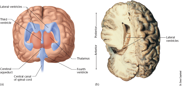
Cells called the choroid plexus along the walls of the four ventricles produce cerebrospinal fluid (CSF), a clear fluid similar to blood plasma. CSF flows from the lateral ventricles to the third and fourth ventricles, and then into the central canal of the spinal cord and the narrow spaces between the brain and the meninges, membranes that surround the brain and spinal cord.
Although the brain has no pain receptors, the meninges do, and meningitis—inflammation of the meninges—is painful. Swollen blood vessels in the meninges produce the pain of a migraine headache (Hargreaves, 2007).
Cerebrospinal fluid cushions the brain against mechanical shock when the head moves. It also provides buoyancy. Just as a person weighs less in water than on land, cerebrospinal fluid helps support the weight of the brain. It also provides a reservoir of hormones and nutrition for the brain and spinal cord.
If the flow of CSF is obstructed, it accumulates in the ventricles or in the space between brain and meninges, increasing pressure on the brain. When this occurs in infants, the skull bones spread, causing an overgrown head. This condition, known as hydrocephalus (HI-dro-SEFF-ah-luss), can impair cognitive development, although the results vary from one person to another.
Module 3.1 in closing
Summary
The vertebrate nervous system has two main divisions, the central nervous system and the peripheral nervous system.
Each segment of the spinal cord has a sensory nerve and a motor nerve on both the left and right sides. Spinal pathways convey information to the brain.
The sympathetic nervous system (one of the two divisions of the autonomic nervous system) activates the body's internal organs for vigorous activities. The parasympathetic system (the other division) promotes digestion and other nonemergency processes.
The central nervous system consists of the spinal cord, hindbrain, midbrain, and forebrain.
The hindbrain consists of the medulla, pons, and cerebellum.
The medulla and pons control breathing, heart rate, and other vital functions through the cranial nerves. The cerebellum contributes to movement, timing, certain types of learning and conditioning, and detecting sequences of events.The cerebral cortex receives its sensory information, except for olfaction, from the thalamus. Connections between the cortex and the thalamus maintain information in working memory.
The subcortical areas of the forebrain include the thalamus, hypothalamus, pituitary gland, basal ganglia, and hippocampus.
The cerebral ventricles contain fluid that provides buoyancy and cushioning for the brain.
Key Terms
amygdala
autonomic nervous system basal ganglia
brainstem
central nervous system (CNS)
cerebellum
cerebrospinal fluid (CSF)
cranial nerves
dorsal
dorsal root ganglia
enteric nervous system
forebrain
gray matter
hindbrain
hippocampus
hypothalamus inferior colliculus
medulla
meninges
midbrain
neuroanatomy
nucleus basalis
parasympathetic nervous system peripheral nervous system (PNS)
pituitary gland
pons
somatic nervous system
spinal cord
substantia nigra
superior colliculus
sympathetic nervous system
tectum
tegmentum
thalamus
ventral
ventricles
white matter
Thought Question
Being nervous may interfere with sexual arousal. Explain why, with reference to the sympathetic and parasympathetic nervous systems.
Module 3.1. End-of-Module Quiz
1. What does dorsal mean? b. Toward the back
2. If one structure is on the left and another is on the right,
what is their relationship? d. Contralateral
3. What is a gyrus in the brain? a. A bulge in the cortex
4. What is the function of the ventral roots of the spinal
cord? b. Thev control motor outout.