Acetylcholine Part 2
Overview and Classification of Muscarinic Acetylcholine Receptors (mAChRs)
Receptor Class and Structural Architecture:
Muscarinic acetylcholine receptors (mAChRs) belong to the superfamily of seven-transmembrane-domain G protein-coupled receptors (GPCRs), structurally configured as heptameric membrane-spanning proteins.
. They play a major role in regulating the central and peripheral nervous systems, particularly within the parasympathetic nervous system.
There are five distinct receptor subtypes designated as , and .
The receptors are named after muscarine, a natural trace alkaloid toxin found in the fly agaric fungus (Amanita muscaria), which selectively binds and activates these receptors.


Functional Bifurcation of Receptors:
Even-Numbered Receptors ( and ): Primarily couple to pertussis toxin (PTX)-sensitive proteins, inhibiting adenylyl cyclase to suppress intracellular cyclic adenosine monophosphate () levels.
Odd-Numbered Receptors (, and ): Primarily couple to proteins, stimulating phospholipase C () and activating the inositol phosphate pathway to mobilize intracellular calcium and activate protein kinase C ().
Even-Numbered Receptors ( and ): -Coupled Signalling Pathway
Sequential Activation Mechanism:


Acetylcholine (ACh) binds to the extracellular binding pocket of the or heptameric transmembrane receptor.
Ligand binding induces a conformational change in the receptor that is transmitted to the intracellular heterotrimeric protein complex.
This conformational transition promotes the dissociation and loss of guanosine diphosphate () from the -subunit ().
The nucleotide-free -subunit binds guanosine triphosphate (), generating active .
The active complex dissociates from the -subunit complex and directly inhibits the membrane-bound effector enzyme adenylyl cyclase ().
Inhibition of adenylyl cyclase prevents the conversion of adenosine triphosphate () into cyclic adenosine monophosphate (), halting downstream activation of Protein Kinase A ().
Intrinsic GTPase enzymatic activity within the -subunit eventually hydrolyzes bound back to , facilitating re-association with the -subunits and restoring the basal, resting conformation.
Effector Ion Channel Modulation:
In addition to adenylyl cyclase inhibition, signalling directly involves the activation of inwardly rectifying potassium () channels mediated by the dissociated subunit complex.
channel opening drives outward potassium efflux, producing membrane hyperpolarization.
Signalling through this pathway simultaneously suppresses voltage-gated calcium () channels, depressing inward calcium currents.
Odd-Numbered Receptors (, and ): -Coupled Signalling Pathway


Receptor Activation and Phospholipase Cascade:
Acetylcholine binds to the extracellular loops of the , or heptameric receptor.
Receptor activation triggers a conformational rearrangement leading to the loss of from the -subunit of the heterotrimeric protein.
The -subunit binds to form an active complex.
Active stimulates the effector enzyme phospholipase C ().
Active cleaves and hydrolyzes the membrane-bound phospholipid phosphatidylinositol-4,5-bisphosphate (noted as phosphatidylinositol-4,4-biphosphate, ), generating two distinct secondary messengers: diacylglycerol () and inositol-1,4,5-trisphosphate ().
Intracellular Secondary Messenger Actions:
Inositol-1,4,5-trisphosphate (): Diffuses through the cytoplasm and binds to ligand-gated calcium release channels/pumps on the membrane of the endoplasmic reticulum (ER), triggering rapid translocation of stored into the cytoplasm.
Diacylglycerol (): Remains situated within the plasma membrane where it activates Protein Kinase C () in concert with elevated cytosolic .
Protein Kinase C () Downstream Roles: Directly modulates receptor desensitization, alters membrane architecture, governs gene transcription, coordinates immune responses, controls cellular growth and division, and participates in synaptic plasticity associated with learning and memory.
Lipid and Eicosanoid Secondary Cascades: Receptor stimulation additionally drives activation of phospholipase D2 () and phospholipase A2 (), driving the liberation of arachidonic acid ().
Electrophysiological Outcome: Induces cellular depolarization and excitation, manifesting as an increase in the slow excitatory postsynaptic potential ().
Comprehensive Molecular, Anatomical, and Functional Profiles of mAChR Subtypes
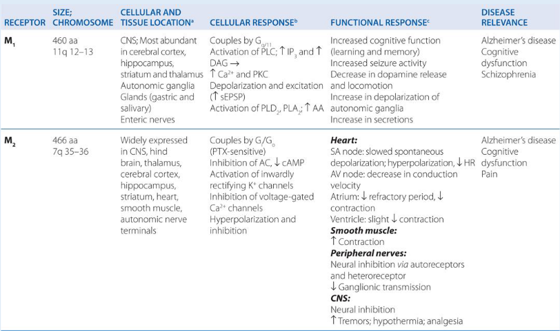

Pharmacology of mAChR Agonists
Clinical Spectrum of Direct Agonists:
Acetylcholine and synthetic choline esters (man-made drugs designed in a laboratory to mimic the exact structure and function of natural acetylcholine) exhibit potent agonist efficacy (it binds to a receptor and turns it "on" to trigger a cellular response) across all mAChR subtypes.
Due to rapid enzymatic degradation of endogenous acetylcholine by cholinesterases and lack of selectivity, only synthetic or alkaloid analogues—specifically bethanechol and pilocarpine—are utilized in clinical medicine.
Your body is packed with highly efficient enzymes called cholinesterases (primarily acetylcholinesterase). Their sole job is to clean up acetylcholine almost immediately after it is released. If a doctor injected you with pure, natural acetylcholine, these enzymes would destroy it in milliseconds before it could ever reach the target organ.
As noted previously, pure acetylcholine hits every single muscarinic and nicotinic receptor in the body simultaneously. Giving it as a drug would cause total systemic chaos—dropping your blood pressure, freezing your lungs, and emptying your bowels all at once
Agonist activity is directed predominantly at postsynaptic receptors located on autonomic effector organs (heart, lungs, stomach, intestines, bladder, and salivary glands)
Key Organ System Effects of Agonist Stimulation:
Cardiovascular System: Marked reduction in sinus rhythm (bradycardia, heart's natural pacemaker setting the tempo), diminished atrial contractile force (chambers are squeezing much weaker than normal), and peripheral vasodilation mediated by endothelial nitric oxide () release (When the drug hits the vessels, it forces this lining to release a gas called nitric oxide); these combined actions lead to a pronounced drop in systemic arterial blood pressure.
Visceral Smooth Muscle: Pronounced smooth muscle contraction; marked increase in gastrointestinal tract motility and peristaltic activity; stimulation of the detrusor muscle driving urinary bladder emptying.
Exocrine Glands: Generalized stimulation of fluid excretion, generating profuse diaphoresis (sweating), lacrimation (tears), sialorrhea (salivation), and hypersecretion of tracheobronchial mucus.
Ocular Apparatus: Contraction of the ciliary muscle causes the suspensory zonules to relax, enabling the crystalline lens to bulge for near-vision focus (accommodation); concurrent contraction of the iris sphincter (constrictor pupillae) produces pupillary constriction (miosis).
Pilocarpine Mechanism and Glaucoma Pathophysiology

Pathophysiology of Glaucoma:
Glaucomais a group of eye conditions that damage the optic nerve, which is vital for good vision. This damage is almost always caused by an abnormally high pressure inside your eye (intraocular pressure)
Aqueous humour is continuously synthesised and secreted by the specialised epithelial layer covering the ciliary body into the posterior chamber of the eye.
Fluid flows past the lens through the pupil into the anterior chamber, where it drains through the trabecular meshwork into the Canal of Schlemm to enter the episcleral venous circulation.
In glaucoma, congestion or anatomical closure of the anterior chamber angle impedes the entry of fluid into the Canal of Schlemm, particularly when the pupil is dilated.
Blockage leads to progressive accumulation of aqueous humour, producing a slowly rising intraocular pressure that damages the optic nerve head.
Mechanism of Action of Pilocarpine:
Pilocarpine is formulated as topical ophthalmic eyedrops.
Structurally, pilocarpine is a tertiary () amine, conferring adequate lipophilicity to cross the lipophilic conjunctival and corneal epithelial membranes.
Pilocarpine stimulates receptors on the constrictor pupillae muscle of the iris, triggering sustained contraction.
Pupillary constriction pulls the iris root away from the trabecular meshwork, widening the anterior chamber angle.
This mechanical opening relieves congestion at the entrance to the Canal of Schlemm, re-establishing continuous drainage of aqueous humor into the venous blood vessels and rapidly reducing intraocular pressure.
Pharmacology of mAChR Antagonists
Chemical Properties and Selectivity Limitations:
Antagonists act as competitive, reversible blockers that prevent acetylcholine binding.
Structurally, antagonists possess bulky, lipophilic aromatic or aliphatic sidechains in place of the compact acetyl group found in acetylcholine.
Prototypic agents include the naturally occurring belladonna alkaloids atropine and scopolamine.
Because mAChRs are ubiquitously distributed across all target tissues of the parasympathetic nervous system (PNS), non-selective antagonists display poor organ selectivity and produce widespread antimuscarinic side effects.
Pharmacological Spectrum of Atropine:
Exocrine Secretory Blockade: Profound suppression of salivary, bronchial, sweat, and lacrimal gland output occurs at extremely low doses, causing dry mouth (xerostomia).
Cardiovascular Modifications: Modest sinus tachycardia due to blockade of inhibitory receptors on the sinoatrial node, eliminating vagal tone; systemic arterial blood pressure remains essentially unaffected at therapeutic doses.
Ophthalmic Modifications: Prolonged pupillary dilation (mydriasis) that is refractory to light stimuli; cycloplegia (paralysis of accommodation) caused by ciliary muscle paralysis, leading to loss of near-vision accommodation (employed clinically during ophthalmic surgical interventions).
Gastrointestinal Effects: Marked reduction in smooth muscle peristalsis, tone, and motility throughout the gastrointestinal tract, causing prolonged transit times.
Vascular and Cutaneous Responses: Cutaneous vasodilation and enhanced superficial blood flow to the skin ("atropine flush"); concurrent bronchodilation and relaxation of airway smooth muscle.
Classic Anticholinergic Toxicity Mnemonic: "Hot as a hare, blind as a bat, dry as a bone, red as a beet, and mad as a hatter".
Clinical Indications for Representative Muscarinic Antagonists:
Atropine: Administered preoperatively to reduce salivary and respiratory secretions; prevents vagally mediated reflex bronchospasm and bradycardia; acts as the primary antidote against organophosphate and acetylcholinesterase inhibitor toxicity.
Scopolamine: Exhibits central and peripheral anticholinergic properties identical to atropine; uniquely crosses the blood-brain barrier to depress vestibular pathways, suppressing motion sickness.
Tropicamide: Administered topically to achieve rapid, short-duration mydriasis and cycloplegia for diagnostic ophthalmic examinations of the retina.
Ipratropium: Quaternary ammonium derivative utilized as a secondary () inhalation therapy in asthma and COPD; blocks airway receptors to prevent reflex bronchoconstriction and suppress excessive mucus production.
Pirenzepine: Relative selective blocker that suppresses -mediated gastric acid secretions; utilized historically for the medical management of peptic ulcer disease.
Presynaptic Modulation of Cholinergic Transmission
Inhibition of Acetylcholine Biosynthesis and Vesicular Storage:
Hemicholinium: Competitively blocks the high-affinity sodium-dependent choline transporter situated on the presynaptic terminal membrane, starving the terminal of intracellular choline and arresting de novo ACh synthesis (lacks clinical therapeutic utility; strictly an experimental pharmacological tool).
Vesamicol: Selectively inhibits the vesicular acetylcholine transporter (VAChT) residing on storage vesicle membranes, preventing the active translocation of newly synthesized ACh into synaptic storage vesicles.
Inhibition of Exocytotic Acetylcholine Release:
Magnesium Ions (): Competitively antagonizes extracellular calcium () influx through presynaptic voltage-gated channels, suppressing calcium-dependent vesicle fusion and neurotransmitter release.
Aminoglycoside Antibiotics: Specific antibacterial compounds, notably streptomycin and neomycin, exert secondary presynaptic neuromuscular blocking activity by antagonizing presynaptic calcium entry, blunting ACh release.
Potent Neurotoxins:
Botulinum Toxin: Produced by Clostridium botulinum; cleaves SNARE proteins responsible for vesicle docking, completely arresting exocytotic release of ACh at the neuromuscular junction and autonomic terminals.
-Bungarotoxin: Component of elapid snake venom; possesses phospholipase activity that irreversibly destroys the structural integrity of presynaptic motor nerve terminals, preventing ACh release.
Post-Synaptic Modulation: Cholinesterase Inhibitors and Enzyme Reactivation
Reversible Acetylcholinesterase (AChE) Inhibitors:
Compete reversibly with endogenous ACh for the active catalytic site of acetylcholinesterase, prolonging the residency time and concentration of ACh within the synaptic cleft.
Short-Acting Reversible Agents:
Edrophonium: Fast-acting, short-duration ionic binder utilized diagnostically for evaluating myasthenia gravis.
Donepezil: Centrally active, reversible cholinesterase inhibitor indicated for symptomatic cognitive management in Alzheimer's disease.
Medium-Duration Reversible Agents (Carbamates):
Neostigmine: Carbamylating inhibitor that forms an intermediate covalent adduct that hydrolyzes over hours; utilized to reverse non-depolarizing neuromuscular blockade and manage myasthenia gravis.
Pyridostigmine: Structurally related carbamate exhibiting a longer half-life; standard maintenance therapy for myasthenia gravis.
Irreversible Acetylcholinesterase Inhibitors (Organophosphates):
Compounds and Mechanism: Chemical warfare nerve agents (e.g., sarin) and agricultural insecticides/toxicants (e.g., dyflos / diisopropyl fluorophosphate).
Organophosphates covalently phosphorylate the serine hydroxyl residue situated within the catalytic esteratic triad of AChE.
The resulting phosphorylated enzyme complex does not undergo spontaneous hydrolysis, rendering the enzyme permanently inactivated.
Toxicity and Pathology: Uncontrolled accumulation of synaptic acetylcholine leads to continuous stimulation of nicotinic and muscarinic receptors, manifesting as autonomic hyperstimulation, flaccid skeletal muscle paralysis, and respiratory cessation.
Therapeutic Application: Historically employed in highly dilute topical formulations to treat refractory glaucoma; systemic therapeutic applications are strictly precluded due to extreme toxicity.
Enzymatic Reactivation via Pralidoxime:
Pralidoxime (2-PAM): A specific oxime antidote designed to reactivate phosphorylated acetylcholinesterase prior to chemical "aging" of the enzyme.
The positively charged quaternary nitrogen of pralidoxime anchors directly to the anionic subsite of the inactivated AChE enzyme.
This spatial alignment brings the nucleophilic oxime functional group () into contact with the covalently linked organophosphate group.
The oxime group executes a nucleophilic attack on the phosphorus atom, causing transfer of the phosphate moiety from the active-site serine to the oxime molecule.
The resulting phosphorylated oxime complex dissociates from the active site, restoring functional, active acetylcholinesterase.
