Divisions and Classifications of the Human Skeletal System
Overview of the Human Skeleton
The human skeleton is comprised of a large network of bones interconnected by joints.
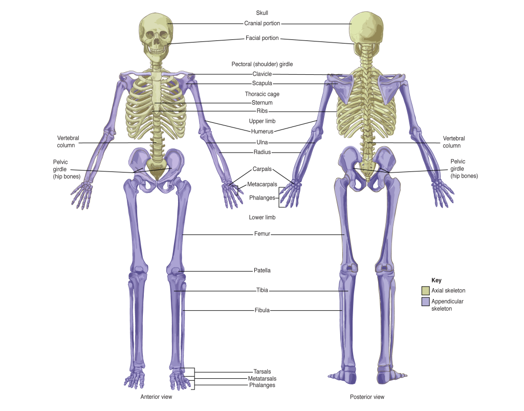
The skeletal system is organized into two primary divisions:
The Axial Skeleton.
The Appendicular Skeleton.
The Axial Skeleton
The term axial indicates that the bones lie along the midline of the body.
The midline refers to the vertical center of the body, running "right down the middle."
Identifying components of the Axial Skeleton:
The skull.
The vertebral column.
The sternum.
The laryngeal skeleton: Located in the neck region.
The thoracic cage.
The Appendicular Skeleton
The appendicular skeleton consists of all bones that are attached to the axial skeleton.
Identifying components of the Appendicular Skeleton:
The pectoral girdle: Described as a "fancy name for your shoulder."
The upper limbs: Located on both sides of the torso.
The pelvic girdle: Commonly known as the hips.
The lower limbs.
A simple rule for classification: if a bone is not part of the axial skeleton, it is attached to it and thus part of the appendicular skeleton.
Classification of Bones by Structure and Shape
Bones in the body are classified based on two main criteria:
Their structure.
Their shape.
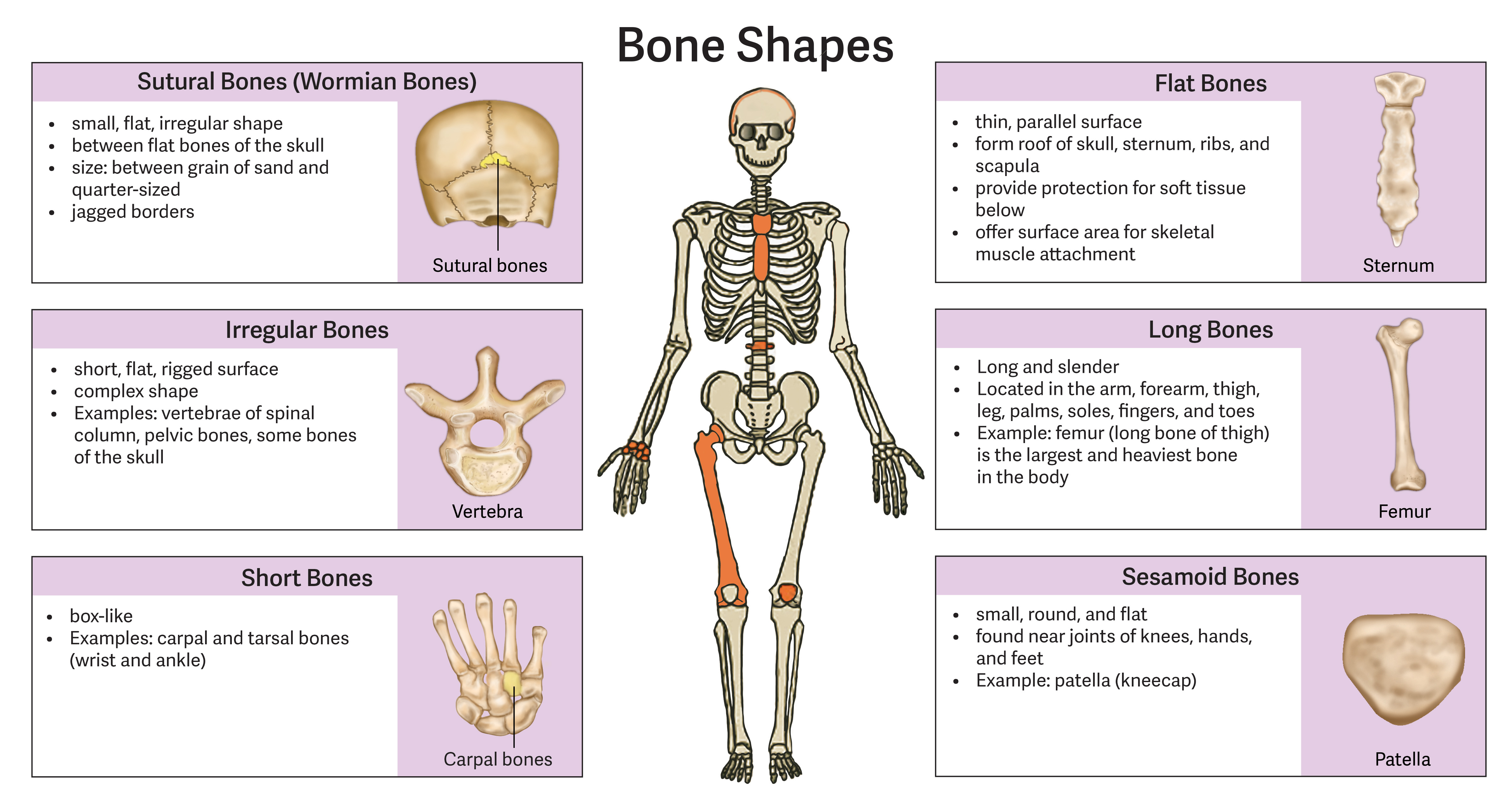
There are six primary categories of bones.
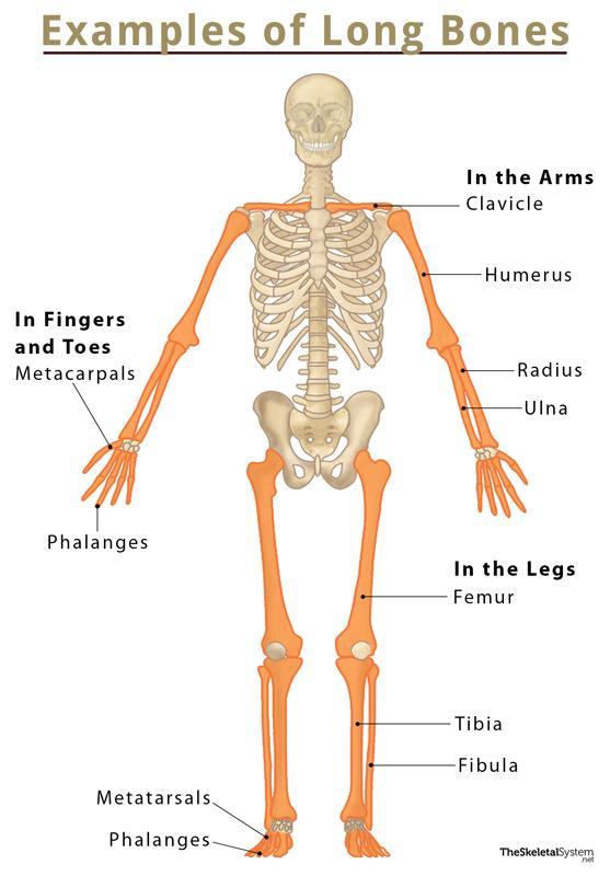
Long Bones
Characteristics: Long bones are defined as being more "long and thin."
Anatomical Features: They consist of a central shaft (diaphysis) and two distinct ends (epiphyses).
Function: Their structural design is specifically suited to support the weight of the body.
Named Examples:
Femur: A classic example of a long bone located in the thigh.
Radius and Ulna: The bones of the forearm.
Humerus: The bone of the upper arm.
Flat Bones
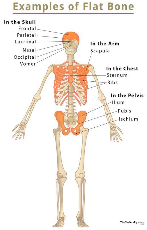
Characteristics: These bones are thin and flat.
Anatomical Features: They possess two parallel surfaces.
Named Examples:
Scapula: The shoulder blade; when viewed sideways, its thinness and parallel surfaces are evident.
Cranium: The bones that make up the skull.
Sternum.
Ribs.
Sutural Bones
Characteristics: These are small, flat bones.
Location: They are found lying in between the flat bones of the skull.
Variability: The specific number of sutural bones varies from person to person.
Representation in Models: Because the number varies individually, sutural bones are typically not included in general anatomical skeletal models.
Short Bones
Characteristics: Short bones are small and more "cube shaped."
Named Examples:
Carpals: The small bones located in the wrist region.
Tarsals: The short bones found in the ankle that provide stability and support during movement.
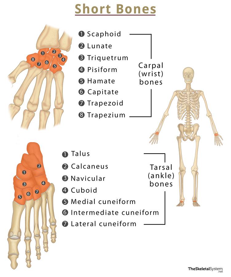
Irregular Bones
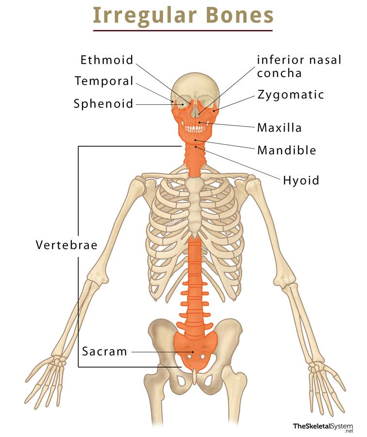
Characteristics: This is a "catch all category" for bone shapes that do not cleanly fit into other classifications.
Anatomical Features: These bones often have complex shapes with various ridges.
Named Examples:
Vertebra: A single unit of the spine is a prime example of an irregular bone due to its complex structure.
Sesamoid Bones
Characteristics: These bones are small and round in shape.
Development: Sesamoid bones uniquely develop inside of tendons.
Function: They serve to reinforce the tendon and provide it with greater strength and stability.
Named Examples:
Patella: The kneecap, located in the anterior (front) portion of the knee joint.
Hand Tendons: Sesamoid bones can also be found within some tendons in the hand.
Variability: While every person possess a patella, the presence of sesamoid bones in other tendons, such as those in the hands, varies from person to person.