Xrays
Chest xray
Black area is lucency
White area is opacity
Before reading xray, confirm these 4
Confirm patient name?
When taken?
Why requested to xray?
What view it is?
AP or PA
How to differentiate
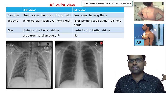
*for cardiomegaly PA is better(no magnification)
Erect or Supine
Quality of xray
Rotation/centering
distance between the clavicle and spinous process L and R are equal
Exposure
lower thoracic vertabrae is barely visible (not hyperexpose or hypoexposed)
Inspiration
How to know?
Q- where does origin of rib comes from
Criteria(above the diaphragm)
- visible 5-6 anterior end of ribs
- visible 8-10 posterior end of ribs
Normal Chest Xray interpretation
Inside out Method
Trachea
slightly tilted to Right because of aortic arch
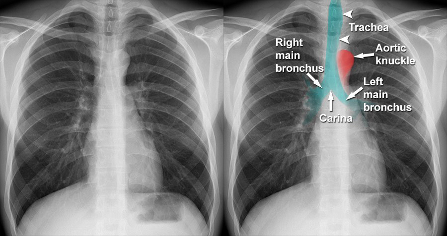
Hilum
Right Hilum is lower or equal than left hilum
concave shape
3.Heart size
CTR <0.5 = normal
PA view > 0.55 abnormal
AP view = False cardiomegaly (<0.6 is normal)
Mediastinum marking
Right
SVC, RA, IVC
Left
Aortic knuckle, pulmonary Artery trunk, LA appendage, LV
Diaphragm
Left dome is lower than right dome because heart is resting on left dome
Costophrenic and cardiophrenic angle
chest walls compare both side
Compare lungs, any pathology?
Bones- rib, scapula, sternum, clavicle