Urologic System Notes (Chapters 41 & 42)
Anatomy of the Urinary System
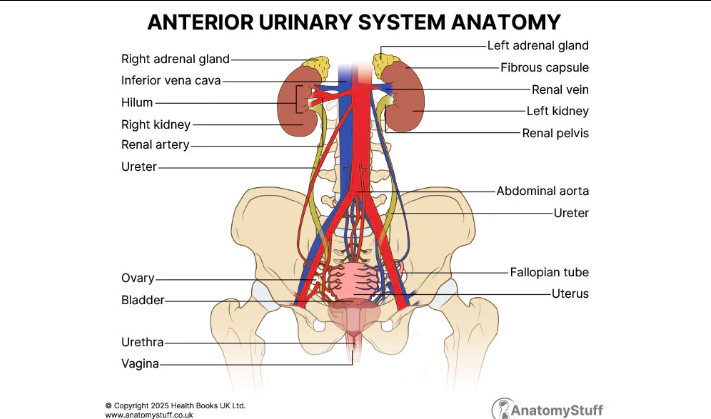
Major components: kidneys, ureters, urinary bladder, urethra
Supporting structures and relations:
Adrenal gland (near the kidney)
Kidneys located retroperitoneally; protected by fat pad and peritoneum
Relationship to vertebrae and vessels (twelfth rib, aorta, inferior vena cava)
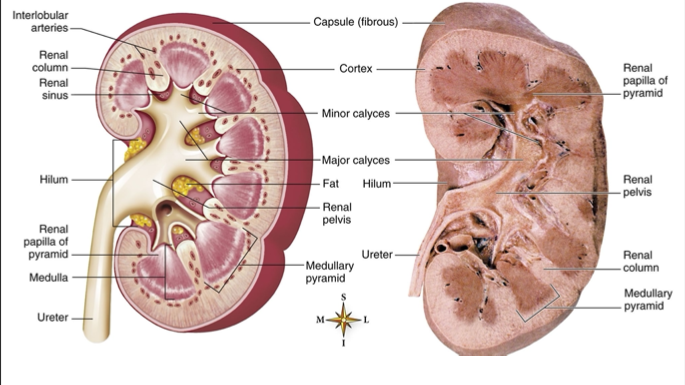
Renal hilum where vessels and ureter enter/exit
Cross-sectional anatomy includes renal capsule (fibrous), cortex (nephron is located), medulla, helim, renal columns, renal pyramids (medullary pyramids), minor and major calyces, renal pelvis
Nephron: the functional unit of the kidney, responsible for filtering blood and producing urine, consisting of a renal corpuscle and renal tubule.
Bowmen’s capsule : A double-walled epithelial structure surrounding the glomerulus, playing a crucial role in the filtration of blood to form urine.
Proximal tubular lumen: A segment of the nephron where reabsorption of water, ions, and nutrients occurs, significantly contributing to the concentration of urine.
Distal Tubular lumen: The final segment of the nephron involved in the regulation of electrolyte balance, where additional reabsorption and secretion of substances such as sodium, potassium, and hydrogen ions take place, ultimately influencing urine volume and composition.
Renal pelvis connects to ureter; ureter carries urine to the bladder
Peritoneal cavity and peritoneum separate intraperitoneal organs from retroperitoneal kidneys
Vascular and urinary drainage:
Renal artery and renal vein; interlobular arteries; peritubular capillaries
Fat, renal capsule, and the sinus containing calyces and pelvis
Ureter, urinary bladder, urethra as part of the excretory conduit system
Imaging references:
Radiographic visualization includes KUB (Kidneys, Ureters, Bladder)
Other imaging: intravenous pyelogram/urogram, renal arteriogram, cystogram, renal scan, CT/MRI, ultrasound
Key concepts: anatomy supports filtration, reabsorption, secretion, storage, and elimination of urine
Metabolic acidosis is for diabetes mellitus patients due to the increased production of keto acids, which can overwhelm the body's buffering capacity and lead to a decrease in blood pH.
Nephron and Renal Microanatomy
Renal corpuscle: glomerulus + glomerular capsule (Bowman’s capsule)
Afferent and efferent arterioles regulate glomerular blood flow and filtration pressure
Juxtaglomerular (JG) apparatus: regulates renin release and tubuloglomerular feedback
Tubular components:
Proximal convoluted tubule (PCT)
Loop of Henle with descending and ascending limbs
Distal convoluted tubule (DCT)
Collecting duct (CD)
Peritubular capillaries surround the nephrons and participate in reabsorption and secretion
Medullary structures:
Medullary pyramids and medullary columns
Renal papillae drain into minor calyces; multiple minor calyces converge to form major calyces then renal pelvis
Overall function: filtration at glomerulus, selective reabsorption and secretion along the tubules, concentration of urine in collecting ducts
Physiology of the Urinary System
Urine production involves three main processes:
Glomerular filtration
Tubular reabsorption
Tubular secretion
Urine elimination is the final excretion step
Regulation mechanisms:
Regulation of serum calcium and phosphate = opposite when calcium goes up phosphate goes down , and vice versa. This balance is crucial for maintaining bone health and various physiological functions in the body.
Regulation of blood pressure is also significantly influenced by renal function through the renin-angiotensin-aldosterone system (RAAS), which plays a key role in controlling fluid balance and systemic vascular resistance. When BP drops , the kidneys release renin, which initiates the conversion of angiotensinogen to angiotensin I,then goes to angiotension 2 leading to a cascade of events that ultimately results in increased blood pressure.
Kidneys secret renin then goes to angiotensin 1 then angiotensin 2 then aldosterone which leads to Na+ retention and potassium excretion, thus contributing to the elevation of blood volume and blood pressure.
ADH → decreased BP then Increased BP ; ADH (antidiuretic hormone) is released when blood pressure decreases, promoting water reabsorption in the kidneys. This increase in water retention helps to raise blood volume, which ultimately leads to an increase in blood pressure.
Hormonal regulation:
Erythropoietin production by the kidneys stimulates red blood cell production and bone marrow , which enhances oxygen delivery to tissues and supports overall blood health. This hormone is crucial for maintaining adequate hemoglobin levels in response to hypoxia.
RBC production is enhanced by erythropoietin, ensuring that the body can efficiently transport oxygen and prevent anemia, especially during situations of reduced oxygen availability.
Key terms to remember:
Glomerular filtration rate as a measure of kidney function
Distinct nephron segments contribute to selective reabsorption/secretion
Tubular Secretion: The process by which substances are transported from the blood into the tubular fluid, allowing for the removal of waste products and the regulation of electrolyte balance. K+ & H+ ph:4.6-8.0 average 6
Urine end product consists of water, urea, creatinine, uric acid, and potassium, reflecting the body's metabolic waste and maintaining homeostasis.
Urine: protiens and glucose (-) (5-6) void normal
ADH (Antidiuretic Hormone) plays a crucial role in regulating water balance by promoting water reabsorption in the kidneys, leading to more concentrated urine.
Aldosterone is a hormone that regulates sodium and potassium levels, facilitating water retention and influencing blood pressure, thus impacting overall fluid balance in the body.
PTH (Parathyroid Hormone) is involved in regulating calcium and phosphate levels in the blood, which is essential for maintaining bone health and metabolic functions.
Age-Related Changes in the Urinary System
Nephron loss and thickening of membranes; sclerosis of renal vessels
Creatinine clearance declines with age , leading to a decreased ability of the kidneys to filter waste products effectively.
Nocturia (awakening at night to void) common; bladder muscles weaken; connective tissue increases , contributing to urinary incontinence and bladder capacity reduction, which can lead to increased frequency of urination. Additionally, hormone levels such as estrogen and testosterone may decrease, further impacting the regulation of urinary function and promoting changes in bladder sensitivity. Loss of nephrons generally results in a reduced glomerular filtration rate (GFR), which contributes to the accumulation of metabolic waste in the bloodstream. Membrane gets thicken // Sclerosis renal artries , leading to decreased renal blood flow and impaired renal function, which can exacerbate the age-related decline in the efficiency of waste elimination from the body.
Incontinence is not a normal consequence of aging, but is common
In men, urethral obstruction frequently a problem due to benign prostatic hyperplasia (BPH), which can result in urinary retention, increased frequency, and urgency. This condition may necessitate interventions such as medication or surgical options to relieve the obstruction and improve urinary flow.
Assessment and History
Health history components:
Reason for seeking care includes any urinary symptoms experienced by the patient, such as difficulty starting urination, weak stream, or nocturia, as well as any previous diagnoses of BPH or other urological conditions. Change in skin color or urine color // Uremic frost can be white crystals in the skin that occur in severe cases of kidney dysfunction, indicating high levels of urea in the blood. It is crucial to assess these changes as they may suggest underlying renal pathology or complications related to urinary obstruction.
Present health status includes an evaluation of current urinary habits, any instances of incontinence, or hematuria, as well as the presence of flank pain, which may indicate renal stones or infections. It is essential to gather information regarding the patient's recent laboratory results, including serum creatinine and electrolyte levels, to further assess kidney function. Polyuria, oliguria, anuria, hematuria, dysuria
polyuria refers to excessive urination, often defined as the production of more than 3 liters of urine per day, and may be associated with conditions such as diabetes mellitus or diabetes insipidus.
oliguria refers to reduced urine output, typically defined as less than 400 milliliters per day, and can be indicative of acute kidney injury, dehydration, or urinary tract obstruction.
anuria refers to a severe reduction in urine output, defined as less than 100 milliliters per day, which may suggest complete kidney failure or severe dehydration and often requires immediate medical intervention.
hematuria refers to the presence of blood in the urine, which can be a sign of various underlying issues such as urinary tract infections, kidney stones, or tumors. It is essential to evaluate the cause of hematuria promptly to determine appropriate treatment and management.
dysuria refers to painful or difficult urination, which can result from infections, inflammation, or irritation of the urinary tract. Identifying the underlying cause is crucial for effective management and treatment.
30 ml per hour is typically considered the minimum acceptable urine output for proper kidney function. A sustained output below this level may indicate the need for further assessment and possible intervention.
Past health history should include any previous urinary tract infections, kidney stones, or surgeries related to the urologic system, as these factors can significantly impact current symptoms and treatment options.
Family history should document any hereditary conditions affecting the urologic system, such as polycystic kidney disease, bladder cancer, or prostate issues, which may predispose the patient to similar disorders.
Review of systems should encompass an examination of any changes in urinary frequency, urgency, incontinence, or hematuria, as well as any associated symptoms such as flank pain or dysuria, which can provide important clues to underlying urologic conditions. Skin, respiratory, fatigue, chills, fever
Functional assessment should evaluate the patient's ability to perform activities of daily living related to urologic health, including voiding habits, the presence of any difficulties during urination, and the impact of urinary symptoms on quality of life. Daily intake usual voiding patten
Physical examination focus:
Inspection for fluid status and edema, skin should be a priority, as these can indicate potential renal complications or fluid overload. Skin might turn ashen color (gray) , Uremic Frost may also be observed, indicating a severe accumulation of waste products due to renal failure. This should prompt further investigation into the patient's renal function and potential treatment options. Skin turgor , breath will smell like urine if there is kidney failure
Respiratory assessment , dyspnea , respiratory pattern, breath sounds
Abdominal examination should include assessment for any tenderness, distention, or masses, as these may signify underlying renal pathology or complications. Brutus whooshing sound when asscultate near belly button may indicate the presence of a possible renal artery stenosis or an abdominal aortic aneurysm, warranting further evaluation and diagnostic imaging.
Genitalia assessment should focus on inspecting for any abnormalities, such as lesions or swelling, which can also reflect underlying urologic issues. Palpation may be necessary to evaluate for tenderness or masses that could indicate infection or other pathological conditions. Prostate exam
Diagnostic Tests and Procedures
Urine tests:
Urinalysis is a common diagnostic test that examines the physical and chemical properties of urine, including its appearance, concentration, and content of cells or substances that could indicate disease. Specific Gravity of urine : 1.005- 1.030 Ph: 4-8
Urine culture & sensitivity (C&S) is another essential test that involves growing bacteria from a urine sample to identify any infections and determine the most effective antibiotics for treatment.
Creatinine clearance is a test that measures the rate at which creatinine, a waste product, is cleared from the blood by the kidneys. This test is significant for assessing kidney function and estimating the glomerular filtration rate (GFR).
Blood tests:
Blood urea nitrogen (BUN) is another important blood test that measures the amount of nitrogen in the blood that comes from urea, a waste product formed in the liver after protein metabolism, helping to evaluate kidney function and hydration status. 10-20 mg/dL is considered a normal range for BUN levels. An elevated BUN level may indicate kidney dysfunction, dehydration, or high protein intake, while low levels could suggest liver disease or malnutrition.
Serum creatinine is a waste product generated from muscle metabolism, and its concentration in the blood is used as a key indicator of kidney function. Normal serum creatinine levels typically range from 0.6 to 1.2 mg/dL, and elevated levels may suggest impaired kidney function or decreased renal perfusion.
Serum electrolytes (sodium, potassium, calcium) sodium 135-143 potassium 3.5-5 calcium 8-10
Uric acid is another waste product that is formed from the breakdown of purines, which are found in certain foods and are also produced by the body. Normal serum uric acid levels typically range from 3.5 to 7.2 mg/dL, and elevated levels may lead to conditions such as gout or kidney stones.
Radiographic tests and procedures:
KUB (Kidneys, Ureters, Bladder) is a common radiographic imaging technique used to assess the urinary tract and help identify abnormalities such as stones, tumors, or anatomical variations.
Intravenous pyelogram/urogram is another important diagnostic tool that involves injecting a contrast dye into the bloodstream, allowing for clear imaging of the kidneys, ureters, and bladder, thus providing valuable information about their structure and function.
Renal arteriogram is a specialized imaging technique that uses contrast dye and X-rays to visualize the renal arteries, helping to detect blockages, malformations, or other vascular conditions affecting kidney perfusion.
Cystogram is an imaging procedure that utilizes a contrast medium inserted into the bladder through a catheter, allowing for detailed visualization of the bladder's structure and function, particularly in diagnosing conditions such as bladder tumors, inflammation, or injury.
Renal scan is a nuclear medicine procedure that provides information about kidney function and structure by using radioactive material, enabling the assessment of blood flow, glomerular filtration rate, and the detection of abnormalities such as cysts or tumors.
CT/MRI
Ultrasound is a non-invasive imaging technique that uses sound waves to create images of the kidneys and bladder, offering real-time visualization to assess the presence of stones, masses, or structural abnormalities.
Invasive procedures:
Renal biopsy is performed to obtain kidney tissue for histological examination, allowing for the diagnosis of various renal diseases including glomerulonephritis and renal tumors.
Cystoscopy is a procedure that involves inserting a thin tube with a camera into the bladder through the urethra, providing direct visualization of the bladder and urethra to diagnose issues such as tumors, inflammation, and urinary tract obstructions.
Urodynamic studies (rate & flow):
Cystogram/voiding cystourethrogram (VCUG) is a specialized X-ray exam that highlights the bladder and urethra, allowing for assessment of bladder function and the detection of conditions such as vesicoureteral reflux and other structural abnormalities.
Cystometrogram (CMG) is a test that measures the pressure and volume in the bladder, helping to evaluate bladder capacity, compliance, and the function of the detrusor muscle during filling and voiding. This procedure is essential for diagnosing urinary incontinence and other bladder dysfunctions.
Postvoid residual measurement is another crucial aspect of urodynamic studies, assessing the amount of urine left in the bladder after voiding. This measurement helps to identify any obstructive uropathy or detrusor muscle weakness, providing insight into the efficiency of the bladder during the voiding process. >300 in bladder after voiding may indicate urinary retention, which can be caused by an obstruction in the urinary tract or inadequate detrusor muscle contractility. This finding necessitates further evaluation and may require interventions to improve urinary function.
Therapeutic Measures
Catheterization: insertion of urinary catheter
Reasons for placement
Types: size, retaining vs nonretaining, number of lumens
Ureteral catheters: Used to drain urine directly from the renal pelvis, often indicated in cases of obstruction or to facilitate urinary drainage during surgery.
Nephrostomy tube : A tube placed through the skin into the kidney to drain urine when the ureters are obstructed, often used in cases where ureteral catheters are not feasible.
Urinary stent : A thin, flexible tube inserted into the urinary tract to help keep it open, often used to relieve obstructions and allow for proper urine flow.
Urologic surgery:
Removal of kidney, removal of calculi stones, bladder incision/removal , and prostatectomy are common procedures performed under this category, each addressing various urologic conditions and ensuring optimal urinary function.
Preoperative care is crucial for minimizing complications during surgery and enhancing recovery outcomes. This includes patient education, ensuring the correct fasting protocols, reviewing medications, and assessing any preexisting conditions that may impact the surgical procedure.
Postoperative care: assessment, complications management, pain control, and monitoring for signs of infection are essential components of postoperative care to support recovery and prevent adverse outcomes.
Lithotripsy (SWL) as a noninvasive stone-disintegration option utilizes sound waves to break up kidney stones into smaller fragments, promoting easier passage through the urinary tract while minimizing risks associated with invasive procedures. Vibration to break stones
Drug therapy:
Diuretics: water pills, fuerosimide
Antihypertensives
Vitamins/supplements folic acid
Hormones arythroporiten
Antibiotics
Urinary antiseptics: nitrofurantone, trimethoprim, methenamine, and phenazopyridine are commonly used to treat urinary tract infections by targeting bacterial growth and providing symptomatic relief.
Antispasmodics: oxybutynin and tolterodine are utilized to alleviate bladder spasms and reduce urgency in patients with overactive bladder symptoms.
Analgesics Pyridium
Dialysis:
Hemodialysis and peritoneal dialysis serve different purposes in managing kidney failure, with hemodialysis filtering blood through a dialysis machine and peritoneal dialysis using the lining of the abdominal cavity to remove waste.
Chapter 42: Urologic Disorders
Risk factors for UTIs
Use of indwelling or intermittent catheters
Urinary or kidney stones
Compromised immunity
Female anatomy
Vaginal infections
Bubble baths and vaginal deodorant sprays
Dehydration
Tight-fitting, synthetic undergarments
Infrequent voiding
Pregnancy
Menopause
Urethritis
Signs and symptoms : dysuria, frequency, urgency, bladder spasms, pus discharge, Lower abdominal discomfort, Possible fever, Cloudy or foul-smelling urine
Medical diagnosis typically involves urinalysis to identify infection, imaging studies if anatomical abnormalities are suspected, and a thorough patient history to rule out other potential causes.
Medical treatment often includes antibiotics to address bacterial infections, analgesics to relieve pain, and sometimes antispasmodics to reduce bladder spasms, along with patient education on hydration and proper hygiene practices.
Nursing care: sit baths, patient education on medication adherence, and monitoring of fluid intake to ensure adequate hydration. Additionally, providing emotional support and reassurance to patients experiencing discomfort can enhance their overall care experience.
Cystitis
Risk factors for developing cystitis include female gender, sexual activity, a history of urinary tract infections, diabetes, and urinary retention. calculi, immboility, urinary diversion, indwelling catheters, and any anatomical abnormalities can also contribute to the development of cystitis.
Signs and symptoms of cystitis commonly include frequent urination, a burning sensation during urination, urinary urgency, cloudy or strong-smelling urine,Dark color urine and pelvic discomfort. In some cases, patients may also experience lower abdominal pain and fever. fever, nausea, and chills may indicate a more severe infection, necessitating further evaluation and possible intervention.
Medical diagnosis: urinalysis, culture & sensitivity
Medical treatment: antibiotics; analgesics; possible phenazopyridine (Pyridium) ; increased fluid intake to help flush the bladder; and in some cases, urinary antiseptics or antispasmodics may be prescribed to alleviate discomfort and promote recovery.
Antispasmodics (oxybutynin) may be utilized to help alleviate bladder spasms and provide additional relief from discomfort associated with cystitis.
Nursing care should include monitoring vital signs, assessing urinary output, and providing education on medication administration and potential side effects.
Interstitial Cystitis
Pathophysiology: chronic inflammatory disease; scar tissue formation causing bladder stiffness and decreased capacity of bladder
Diagnosis: cystoscopy and bladder biopsy to rule out other conditions and confirm the presence of inflammation.
Signs/Symptoms: moderate to severe bladder/pelvic pain with urinary frequency and urgency
Medical treatment: address possible causes; supportive and symptom-directed treatments; cystectomy last resort removal of bladder
Nursing care: patient education about the importance of hydration, medication adherence, and recognizing early signs of complications is essential to improve outcomes. Food diet modifications may include avoiding irritants such as caffeine, alcohol, and spicy foods, while encouraging a balanced intake of fruits and vegetables to support overall urinary health.
Pyelonephritis
Inflammation of the renal pelvis due to ascending bacteria going up(can be bloodborne) can lead to significant complications if not treated promptly, including renal scarring and chronic kidney disease.
Can lead to chronic pyelonephritis with scarring and kidney atrophy; HTN (hypotension) and renal ischemia(low blood supply); risk of ESRD (end-stage renal disease)
Signs: acute vs chronic
Acute pyelonephritis: symptoms include fever, chills, flank pain, dysuria, and increased frequency of urination, sever pain.
Chronic pyelonephritis: symptoms often appear less pronounced, including recurrent urinary tract infections, persistent flank pain, and gradual loss of kidney function, bladder irritation, chronic fatigue, slight pain.
Medical treatment: antibiotics, urinary tract antiseptics, analgesics, antispasmodics; antihypertensives; IV fluids; dietary sodium/protein restrictions
High Blood pressure effect of kidneys high blood pressure can lead to renal damage, contributing to the progression of chronic kidney disease and worsening overall kidney function.
Nursing care: manage acute pain; assess activity tolerance; fluid volume status and nutrition; education for self-care restict sodium and protien why protein intake is important as excessive protein can strain the kidneys and exacerbate the progression of kidney disease by increasing the buildup of nitrogenous waste. amonia build up
Polycystic Kidney Disease
Slow, progressive hereditary disorder with grape-like cysts replacing normal tissue
Signs and symptoms: (as listed in context) lose ability to urinate, htn, heart failure, lower back pain or flank pain, hematuria, and recurrent urinary tract infections.
Medical treatment: supportive care to preserve kidney function; treat UTI; control hypertension; sodium/fluid restrictions
Nursing care should focus on monitoring vital signs, assessing fluid balance, and educating patients about the importance of adhering to dietary restrictions and prescribed medications. Dialysis may be required in advanced stages of kidney disease to perform the filtering functions that the kidneys can no longer handle. or transplant
Acute Glomerulonephritis
Pathophysiology: immunologic inflammation of glomerular capillary loops; often post-streptococcal antigen-antibody reaction problem in the glomerulus
Consequences: inflammation leads to scar tissue; increased glomerular permeability with protein loss; decreased GFR; rise in BUN and serum creatinine, A-negative hemolytic streptococcus antibody titer may also be present, indicating an ongoing immune response. This condition may result in presenting symptoms such as hematuria, hypertension, and edema, which can complicate the patient's overall health status. Increased permeability of glomeruli can lead to significant proteinuria, as proteins that are normally retained in the bloodstream leak into the urine, further exacerbating the patient's condition and contributing to edema.
Signs: tea-colored urine; edema (peripheral and periorbital); HTN; hypervolemia
Medical diagnosis: urinalysis (protein and RBC casts); blood tests (BUN, creatinine, albumin); streptococcal antibody tests , protein in the urine
Medical treatment: diuretics; antibiotics if strep; bedrest/activity restrictions during active urine/blood/protein elevation or high BP; dialysis if renal failure
Nursing care: assess; manage fluid volume excess; activity tolerance; self-care; anxiety, monitor BP
Renal Calculi (Urolithiasis)
Pathophysiology and contributing factors (stasis, crystallization, supersaturation) stones in the kidneys can form due to a variety of factors including metabolic conditions, dietary choices, and genetic predisposition, leading to potential obstruction and varied symptoms. lots of vitamin D, B, C, antiacids can lead to renal calculi due to their role in increasing calcium or oxalate levels in the urine.
Signs and symptoms depend on stone location and size, but commonly include severe pain (renal colic), hematuria, nausea, vomiting, and urinary frequency.
Medical diagnosis and treatment: medications; IV fluids; stone-disintegration options such as SWL shock wave lthotripsy, ureteroscopic lithotripsy, laparoscopic stone removal, percutaneous nephrolithotomy
Prevention strategies
Nursing care: assessment; interventions for acute pain; monitor urinary elimination and electrolyte balance; post-op care; monitor for infection; assess breathing and circulation; patient education
Urologic Trauma
Penetrating injuries (knives, guns) and blunt trauma
Assessment for shock, pain, abdominal mass
Imaging: KUB, urography, CT, ultrasound as needed
Hematuria common; severe injuries may require surgery
Renal Cancer
Risk factors include smoking, obesity, hypertension, and family history of kidney cancer, long term acetaminophen use, and exposure to certain chemicals like aniline dyes and asbestos.
Signs and symptoms: early vs advanced
Early signs may include hematuria, flank pain, and weight loss.
Advanced symptoms can manifest as kidney mass, fatigue, and significant weight loss, along with possible metastasis-related symptoms such as bone pain or respiratory issues.
Medical diagnosis and treatment: nephrectomy removal of kidney; radiation/chemotherapy; immunotherapy; antiangiogenic therapy; thermal ablation for metastasis, CT scan and MRI for imaging studies. Comprehensive evaluation and management strategies are essential for optimizing patient outcomes.
Nursing care: preoperative and postoperative; pain management; monitor for fluid volume deficit, ventilation integrity, infection risk, coping and self-care , check vital signs, bleeding, fluid volume deficit, inadequate deficit
Native americans, alaskan natives, and african americans are at a higher risk for developing urologic conditions, highlighting the importance of tailored screening and prevention strategies within these populations.
Bladder Cancer
Risk factors for bladder cancer include exposure to carcinogens such as tobacco smoke and chemical solvents, chronic bladder irritation from urinary infections or catheters, and a family history of the disease. Causes smoking tabacco tryptophan exposure and genetic predispositions, which can significantly elevate the likelihood of developing malignant cell growth in the bladder.
Signs and symptoms painless and intermitten hematuria (blood in urine), frequent urination, urgency, and pelvic pain, which may indicate the presence of tumors or other bladder abnormalities. dysuria low stream urinary flow
Medical diagnosis and treatment: surgery with urinary diversion, cystoscopy, urinaliysis, cystology , and imaging studies such as ultrasound or CT scans play crucial roles in confirming the diagnosis and determining the extent of the disease. Treatment options often include chemotherapy, immunotherapy, or radiation therapy, depending on the stage and grade of the cancer.
Nursing care: preoperative and postoperative; assessments and interventions; focus on pain, urinary elimination, skin integrity, infection risk, and self-care capacity, vital signs, respiratory rate , monitoring for signs of bleeding, and educating the patient about recovery expectations and the importance of follow-up appointments.
Acute Kidney Injury (AKI)
Causes: prerenal, intrarenal, postrenal causes should be evaluated to identify the underlying issue affecting kidney function. Prerenal causes often involve decreased blood flow to the kidneys, intrarenal causes are related to direct damage to kidney tissue, and postrenal causes result from obstruction of urine flow.
increased BUN and creatinine levels may indicate a decline in kidney function, necessitating close monitoring and possible adjustments in treatment protocols.
Stages: onset, oliguric (anuric), diuretic (↑ urine production), recovery
Medical treatment: medications, diet, fluids, dialysis options (hemodialysis, peritoneal dialysis, CRRT continuous renal replacement therapy )
Nursing care: assess and intervene for fluid balance, cardiac output, anxiety, immobility complications, and self-care capabilities
Chronic Kidney Disease (CKD)
Signs and symptoms; medical treatment; nursing care
Stages of kidney function (based on GFR):
Stage 1: No manifestations
Stage 2: GFR decreases without manifestations
Stage 3: Moderate decline in GFR, can advance to ESRD with infection or nephrotoxicity
Stage 4: Chronic manifestations of uremia
Stage 5: End stage kidney disease (ESRD)
Renal Transplantation
Kidney donation considerations:
Preoperative nursing care of recipient
Surgical procedure specifics
Postoperative nursing care of recipient
Care of kidney donor
Connections to foundational principles:
Structure-function relationships in the nephron drive filtration, reabsorption, and excretion
Homeostasis of fluids, electrolytes, and acid-base balance hinges on intact renal function
Pathophysiology of infection/inflammation affects glomerular permeability, urine composition, and systemic symptoms
Chronic disease progression (AKI → CKD → ESRD) highlights the importance of early detection, prevention, and multidisciplinary management
Practical implications for nursing care:
Thorough assessment and documentation of urinary symptoms, intake/output, and fluid status
Education on catheter use, infection prevention, medication adherence, and lifestyle modifications (dietary/sodium restrictions, hydration)
Monitoring for complications post-procedure or post-surgery (infection, bleeding, thrombosis, electrolyte disturbances, respiratory issues, pain, mobility)
Coordination of care with physicians, dietitians, and social workers for transplantation and long-term management
Key abbreviations and terms to recall:
$KUB$, IVP, Urogram, SWL, CBC, BUN, Cr, $GFR$, PYRidium (phenazopyridine), PCT, DCT, CD, JG apparatus
Quick references from the content:
Diagnostic tests span urine, blood, radiographic, invasive, and functional studies
Therapeutic measures include catheterization, stents, nephrostomy, lithotripsy, and dialysis modalities
Common urologic cancers involve nephrectomy and urinary diversions with relevant nursing considerations for preop/postop care