Unit 4
Microscopes
Light
Uses light as an energy source
Mirrors direct the light to your eyes
Lenses are used to enlarge and focus image
Electron
Uses electricity as energy source
Magnets direct flow of electrons for viewing
Scanning
e- reflect off the specimen for 3d viewing
Transmission
e- flow through providing an internal structure
Structure of Cell membrane
Fluid mosaic model
Fluid
Phospholipids: create flexible bilayers act as water barrier
Cholesterol: regulates the fluidity of the cell membrane
Mosaic
Proteins: carry out various functions
Transport proteins move substances from one side of the membrane to another.
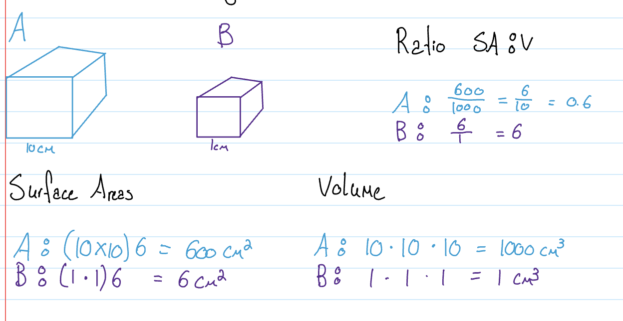
Why are cells small in size
Prokaryotic Cell
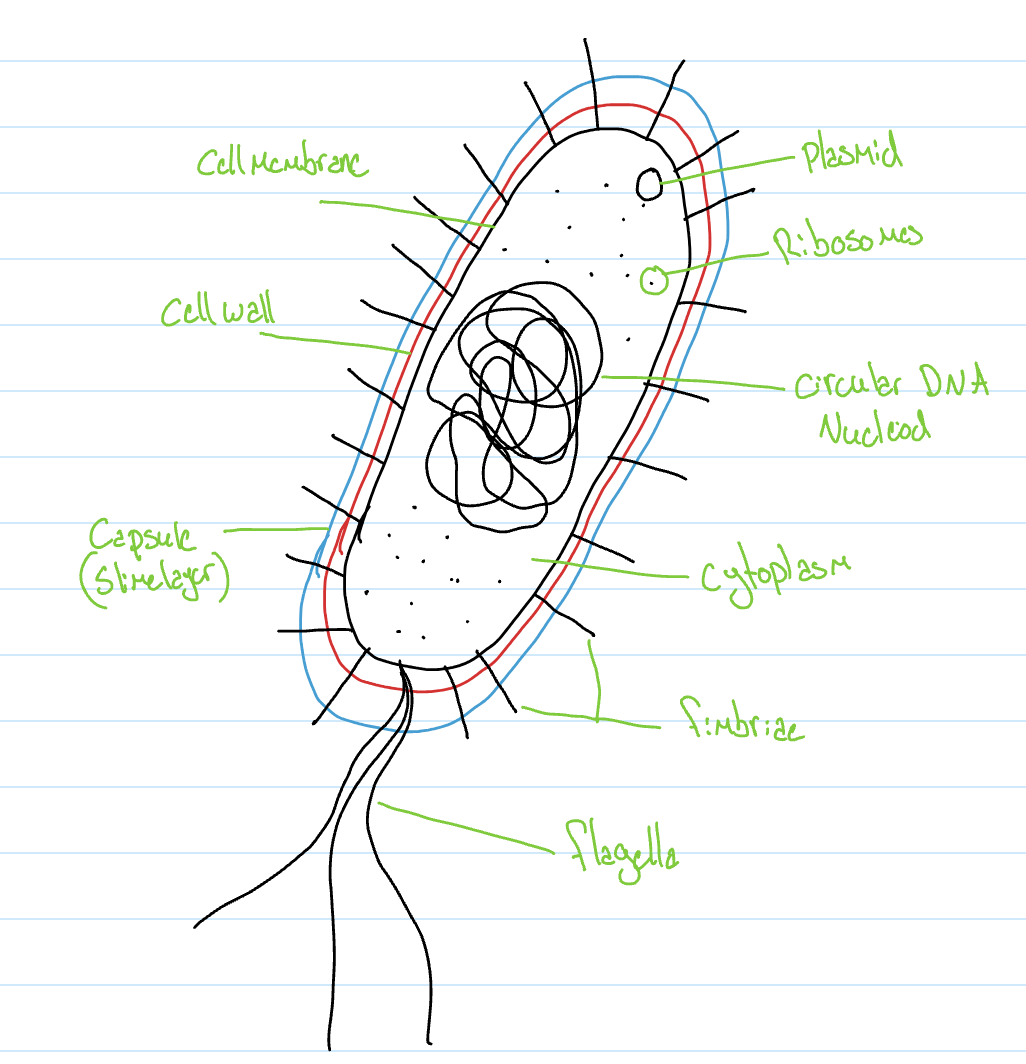
Cytoplasm: Gel-like fluid (cytosol) that contains chemicals (proteins) and cell structures (ribosomes) inner region of the cell contained by cell membrane
Bacterial chromosome: circular DNA that contains genetic information
Nucleoiod: region of the cytoplasm where the bacterial chromosome is located
Ribsomoes: Proton synthesis, creation of proteins for the cell
Plasmid: extra small pieces of DNA that provide superpowers to cells
Antibiotic resistance: cant be fought off by antibodies & neither will its offsprings
Motility: mobility by itself & spreads easily
Production of a capsule: proteins itself & moves around & sticks to surfaces
Fertility: ability to reproduce sexually, creation of new bacteria
Pathogenic: ability to cause infection or disease
Cell membrane: made up of phospholipids, cholesterol proteins & carbohydrates, it creates the boundaries of the cell it acts as a barrier controlling what comes in & out
Cell well: made up of a mixture of carbohydrates and proteins called peptidoglycan provies structure & shape
Capsule: may or may not be present, attaches to surfaces to induce infections, strong enough to resist digestion by white blood cells
Flagella: made up of protons, extending from the cell membrane, allow cells to move, moves by rotation.
Fimbriae: made up of protons that extend from the cell membrane for attachment ot surfaces.
Extracellular fluid: fluid on the outside of the cell
Eukaryotic Cells
Have compartmentalisation while prokaryotic don’t
The process of creating compartments closed off within the cell
Vacuole
Large vesicles with specialised functions
Food vacuole
Part of phagocytosis brings in large substances
Central vacuole
Found in plants; usually the largest structure
Stores water to create ‘pressure’ that helps with structure & shape of the cell & plant
Stores chemicals
Stores waste
Contractile vacuole
Found in freshwater protists; like the paramecium
Removes excess water from the cell
Water always moves to regions that have more solutes.
Paramecium
Water rushes in to fill up the vacuole when it reaches a certain point it opens a trap dopr and push it out to maintain a certain pressure
Endomembranal System
Nucleus
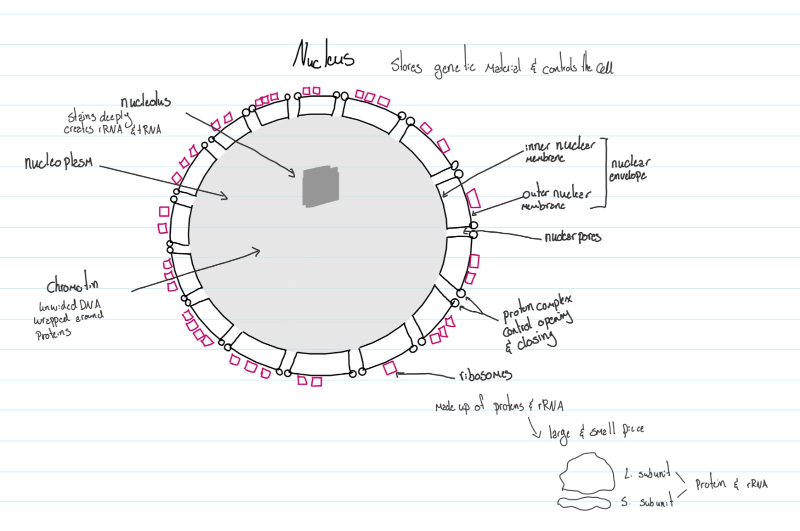
Controls the function of the cell & stores genetic material
Nucleolus: stains deeply create rRNA & tRNA
Chromatin: unwinded DNA wrapped around proteins
Nucleoplasm: the cytoplasm of a nucleus
Inner nuclear membrane: inner wall
Outer nuclear membrane: outer wall
Nuclear pores: openings between the membranes
Proton complex control the opening and closing of the pores
Ribosomes: made up of proteins & rRNA on outer membrane
Nuclear envelope: where the nucleus is held
Ribosomes
Proteins synthesis
Made up of two parts; large subunit and small subunit
Made with proteins & rRNA
Bound ribosomes: attached to the membrane
Free ribosomes: located in the cytoplasm
Bound ribosome creates proteins for:
Organisms they are attached to
Other organelles
Cell membrane
Exporting out the cell
Endoplasmic reticulum (ER)
Endo: in
Plasmic: thick fluid
Reticulum: network
Smooth ER
Tubular network of membrane
Liquid synthesis stores Ca^2+ detoxification of harmful chemicals
Rough ER
Flattened sack-like membrane covered with in bound ribosomes for protein synthesis
Functions
Bound ribosomes release the polypeptide chain into the lumen of the RER
Lumen: space contained within a structure
Polypeptide chain twists & folds into 2 or 3-degree structure % sugar chains are attached to create glycoprotein.
Sugar chains act as tags for sorting & delivering.
Glycoproteins are sorted into the ends of the RER for delivery when the transport vesicle is created by building
Trans vesicle
Budding: a process that creates vesicles by pinching off segments from a membrane
Newly created transport vehicle packages with protons is delivered to the next destination.
Vesicles: membrane-bound structures that have a spherical appearance and store or transport substances
Big ones are called vacuoles
Golgi Apparatus
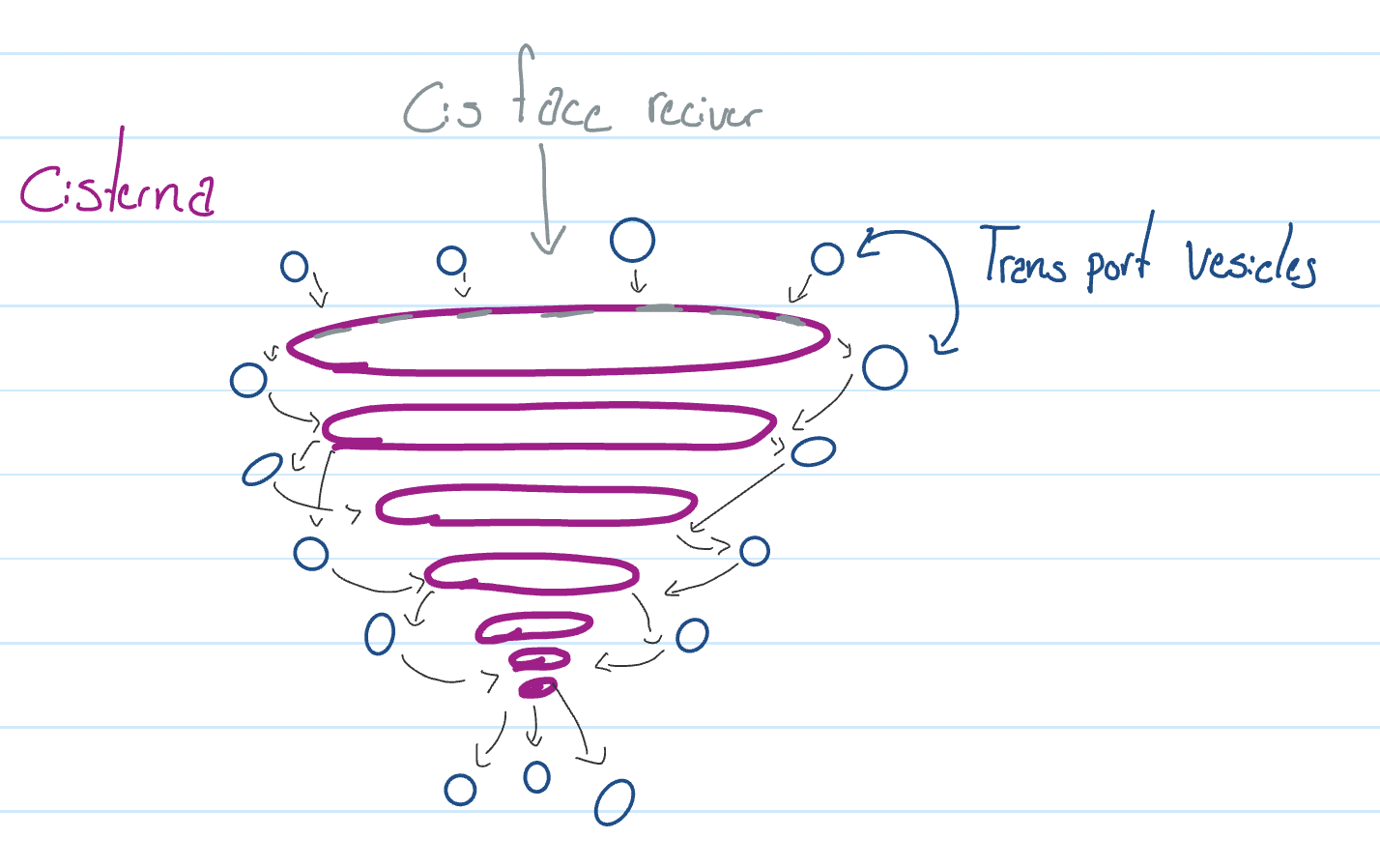
Functions
Receives chemicals from other organelles
Modifies the chemicals
Sorts & packages the chemicals
Ships the chemicals to their final destination
Possible destination
Other organelles
Cell membrane
Export out of the cell
They become new organelles
Exp: Lysosome
Lysosome
Specialised vesicle filled with hydrolytic enzymes
Breaking down… using water
Protons
Carbohydrates
Lipids
Nucleic acid
Major functions
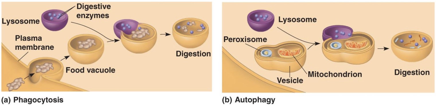
Phagocytosis
A cell process that involves lysosomes which breaks down large structures
Autophagy
A cell process where the cell digests…
Old worn-out organelles
Damages organelles
Uneeded organelles
End of endomembranal system
Energy organelles
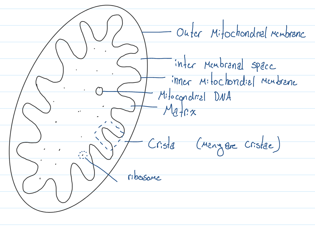
Mitochondrion
Generates energy in the form of ATP
Found in all eukaryotic cell
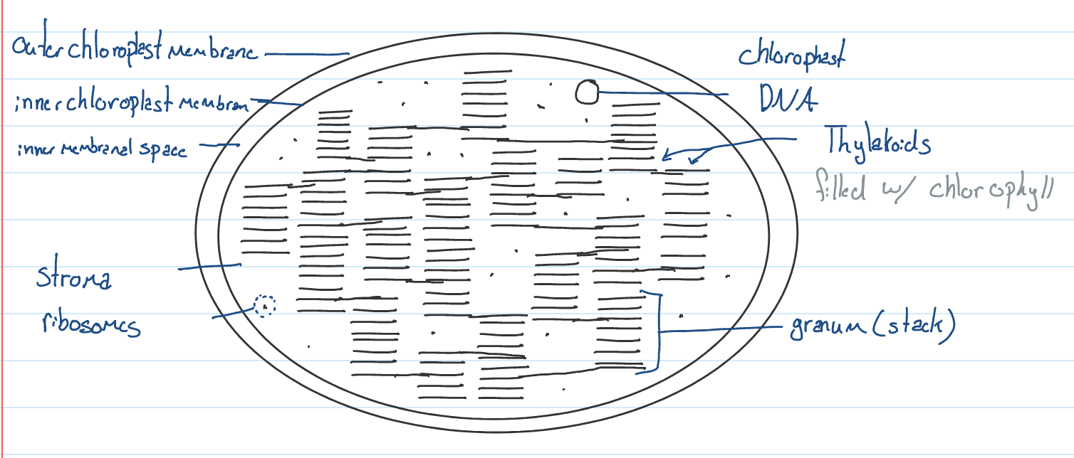
Chloroplast
Harvest light to produce food by photosynthesis found in plants & algae
Chlorophyll is the chemical that absorbs light to provide energy to the chloroplast
Cytoskeleton
Skeleton of the cell located in the cytosol
Cytoplasm = cytosol + organelles
Components
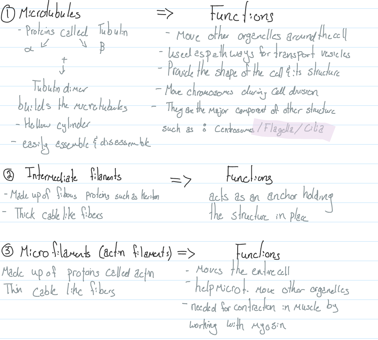
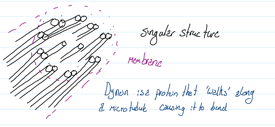
Flagells & cilia
Flagella
Few and very long
Sperm cell
Cilia
Many & very short
Inside the throat
Flagells & cilia have the microtubles in a 9 (doubles) and 2 (singles) arrangement
Extra Cellular Matrix
Located on the outside of the cell made up of a network of proteins and carbohydrates found in the extracellular fluid & attached to the cell membrane
Fibronectin: holds cell matric to the membrane.
Functions
Holds cells together, creating tissues using collagen
Communication between cells
Identification of self using immunoglobulins (glycoproteins)
Cell junction
Attach cells together that are adjacent to each other.
Functions:
Tight junction
Holds cells tightly, preventing any substance from passing between them
Important for the skin & intestine
Made up of intermediate filaments (fibrous)
Desmosome (anchoring J.)
Holds cells together in place but not tightly
Important for lung cells & cells in capillaries
Made up of intermediate filimates
Gap junction
Membrane protein (globular) that connects two adjacent cells to create tunnels
which allows the cytoplasm of one cell to mix with the cytoplasm of another
Such as cardiac muscle
Cell Wall Plants
Made up of cellulose (B Glu)
Provides the shape of a structure of a plant cell
Resits pressure created by cells to become rigid
All plants have a primary cell wall, but some woody plants will also have a secondary cell wall
Cell walls are always built on the outside of the cell membrane.
Plasmodesmata
Plant cell junctions create opening which connect the cytoplasm of adjacent cells
Permit rapid movement & communication between plant cells
Peroxisomes
Specialised vesicles filled with hydrogen peroxide (H2O2)
Mainly found in plants (found only in our liver)
Oxidation of amino acids & fatty acids
Detoxify poison
* not produced by Golgi apparatus
Anatomy of a microscope
Handling
Hold with 2 hands
Don’t slide the microscope
Rotate the head if someone wants to see
Parts
Eyepiece (ocular lens)
Magnifies specimen 10x
Diopter adjustment
Zooms in & out
Head
Tube w/ mirrors direct light to the eye
Nose piece
Changes objective lens
Objective lens
Scanning objective lens (4x)
Low-power objective lens (10x)
High-power objective lens (40x)
Oil immersion lens (100x)
Arm
Holds upper structure
Course adjustment
Moves stage up & down quickly
Fine adjustment
Moves stage slightly up & down
Stage
Where you place your specimen/ must be over the hole
Stage clip
Holds specimen in place
Stage control
Moves stage towards or away
Moves stage left or right
Condensor
Controls how much light is coming in (pupil)
Brightness adjustment
Dimmer switch
Illumination
Light source
Light switch
Base
Supports the microscope, keeps it sturdy
How to draw your specimen
Use a pencil & unlined paper.
Take up most of the paper
Draw on the left side
Boundary structures of the specimen is more important than the secondary
Stipple method to show dark regions
Draw clear, continuous lines, no sketches
Draw part by part → look & draw
Label parts you can identify; lines need to be drawn by a ruler
Do not cross label lines; must touch what they point to
No arrowheads
Include descriptors
Include magnification used
Possible specimen descriptors
Cross section
Longitudinal section
Dry mount
Water mount
Stained
Include the type of magnification bottom right