HF Pathophysiology
Lecture Objectives
Define Heart Failure and the classification of different kinds of heart failure
Distinguish between left-sided heart failure vs. right-sided heart failure, systolic vs diastolic heart failure, chronic heart failure vs. acute decompensated heart failure
Explain the normal processes of cardiac contraction and its regulation
Describe the role of compensatory mechanisms including pathophysiological and beneficial physiological responses activated in heart failure
Describe the pathophysiological progression of heart failure
Describe the role of neurohumoral responses (sympathetic nervous system, renin-angiotensin system, natriuretic peptides) play in heart failure
List the signs and symptoms of heart failure
Identify the blood markers used in the diagnosis of heart failure
Classify heart failure based on ACC/AHA and NYHA staging and classification systems
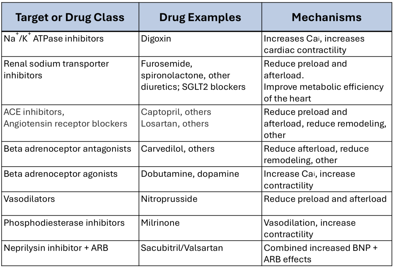
Describe the pharmacology of drugs used in heart failure – rationale for their use, mechanism of action, pharmacological effects, effects on preload, afterload, ventricular modeling, nonhumoral modulation, contractility, heart rate, and cardiac energy metabolism, adverse effects, and contraindications.
Definitions
Heart Failure (HF): clinical syndrome caused by inability of the heart to pump sufficient blood to meet the metabolic needs of the body
Most commonly because of low CO due to cardiac dysfunction → Low-output failure
High Output HF: heart is functionally normal but there is an excessive increase in the body’s metabolic demands that outpaces the increase in CO
May occur in presence or absence of fluid overload
Classification of HF
Ejection fraction (EF)
Heart Failure with Reduced LV Ejection Fraction (HFrEF): EF < 40%, systolic (contraction) dysfunction
Heart Failure with Preserved LVEF (HFpEF): EF > 50%, diastolic (filling) dysfunction)
HF with mid-range EF → Symptoms of HF and LVEF between 41-49%
HF with recovered EF → LVEF greater than 40% but at one time had LVEF less than 40%
Left or right ventricle
Left Ventricle Failure: Pulmonary congestion → SOB and fatigue
Right Ventricle Failure: Systemic congestion → Peripheral and abdominal fluid accumulation
Left-sided failure
Left-to-right cardiac shunt due to atrial or ventricular septal defect
Cor pulmonale
Failure of one ventricle leads to failure of other ventricle
Chronic vs. Acute-Decompensated HF
Etiology of HF
Most Common
CAD
HTN
Less Common
Valvular disease
Excessive alcohol intake
Illicit drugs
Cardiotoxic drugs
Chest radiation
Viral infections
Diabetes
HFrEF Etiology
CAD (i.e. MI, ischemia) - Major cause (70%)
Dilated cardiomyopathies
Pressure overload
HTN, aortic or pulmonic valve stenosis
Volume overload
Valvular regurgitation, shunts, high-output states
Rate and rhythm disorders
Chronic bradyarrhythmias, chronic tachyarrhythmias
HFpEF Etiology
Increased ventricular stiffness → Impaired ventricular relaxation
Ventricular hypertrophy
Hypertrophic cardiomyopathy, HTN
Infiltrative myocardial disease
Amyloidosis, sarcoidosis, endomyocardial fibrosis
Myocardial infarction or ischemia
Mitral or tricuspid valve stenosis
Pericardial disease
Neurohumoral Activation in HF
In response to decrease in cardiac output → Body activated compensatory neurohumoral responses
Pathophysiological Compensatory Responses
RAAS
Angiotensin II
Aldosterone
Vasopressin
SNS
Norepinephrine
Epinephrine
Helpful short-term, harmful long-term
Beneficial Compensatory Responses
Natriuretic peptides

Pathophysiological Compensatory Responses - RAAS + SNS

Pathophysiological Responses: Short-Term Benefit vs. Long-Term Harm

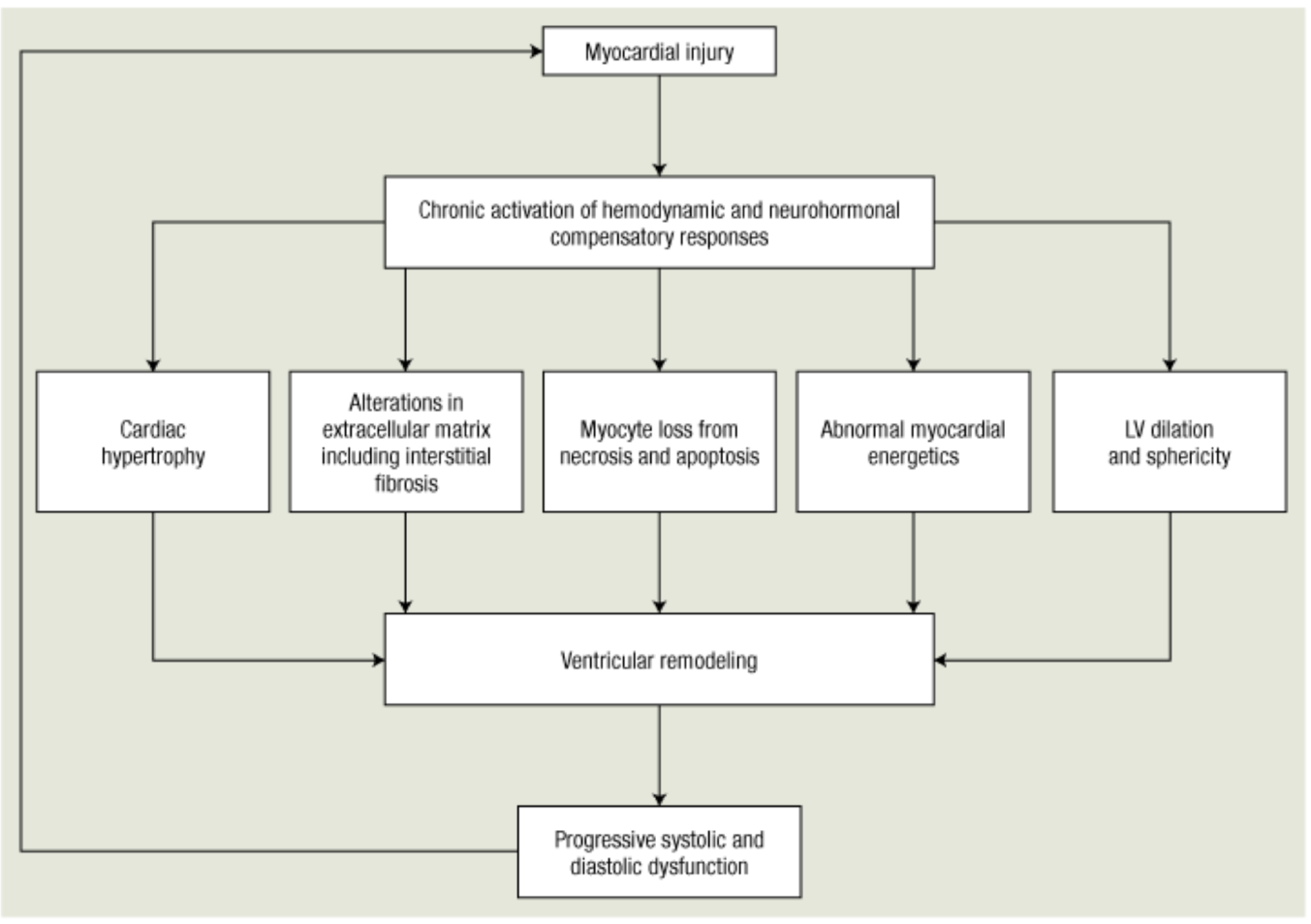
Ventricular Remodeling and Hypertrophy
Most important compensatory mechanism is myocardial hypertrophy → Increase in muscle mass helps maintain cardiac performance
Hypertrophy can lead to ischemic changes → Impairment of diastolic filling and alterations in ventricular geometry
Remodeling refers to:
Dilation of cardiac wall
Slow structural changes that occur in stressed myocardium
Proliferation of connective tissue cells as well as myocardial cells with abnormal biochemical characteristics
Neurohormones + factors that drive ventricular remodeling → Angiotensin II, norepinephrine, aldosterone, vasopressin
Pathophysiological Cardiac Remodeling
Beneficial Compensatory Response - Natriuretic Peptides
ANP is synthesized and stored in atria and to some extent in ventricles → Released into circulation during atrial stretching (filling)
BNP is synthesized by ventricles and released in LV dysfunction or early HF
Act via guanylate cyclase receptors to stimulate
Vasodilation: results in lowering BP
Natriuresis: results in Na+ loss
Antagonize
Sympathetic nervous system
RAAS activity (angiotensin II, aldosterone)
Growth or hypertrophy of cells → Antifibrotic/anti-remodeling effects
Natriuretic Peptides: Triggers for Release
Physiological triggers
Atrial wall stretch (volume overload)
Changing from standing to supine position
Exercise
Pathological triggers
HF
Primary aldosteronism
Chronic renal failure
Inappropriate ADH secretion syndrome
Natriuretic Peptides: Degradation
ANP/BNP are degraded by an enzyme called neprilysin → Inactive metabolites
Neprilysin also breaks down angiotensin II
Progression of HF
Progressive decline in cardiac performance with HF progression
Reduced cardiac output → Increase in vasoconstriction (norepinephrine, angiotensin II, endothelin)
Increased afterload and further decreases EF and cardiac output
Cycle continues and performance keeps decreasing
HF Signs + Symptoms
LV Failure (Pulmonary Congestion)
Dyspnea on exertion, orthopnea, paroxysmal nocturnal dyspnea, exercise intolerance, tachypnea, cough
Pulmonary rales, pulmonary edema
RV Failure (Systemic Congestion)
Abdominal pain, anorexia, nausea, bloating, poor appetite with early satiety, ascites
Peripheral edema, jugular vein distention, hepatojugular reflux, hepatomegaly
Diagnosis of HF
Laboratory tests
ECG, thyroid function tests, serum creatinine, echocardiogram
BNP and NT-proBNP levles in blood
Cannot be used in isolation to diagnose or exclude
Conditions that increase BNP: older age, renal dysfunction, pulmonary embolism, chronic pulmonary disease
Treatment with valsartan/sacubitril increases BNP
NYHA HF Functional Classification and ACC/AHA HF Staging

Acute Decompensated HF (ADHF)
NYHA class III or IV
III: comfortable at rest, less than ordinary physical activity causes fatigue, dyspnea, palpitation, angina
IV: symptoms at rest, any physical activity increases discomfort
Sudden worsening of HF symptoms → Requiring urgent medical attention
Symptoms upon minimal exertion or at rest
Causes of decompensation
40-50% have no cause
Noncompliance with medications or dietary restrictions, uncontrolled HTN, ischemia, arrhythmias, exacerbation of COPD, renal dysfunction, infections, DM, anemia, side effects of medications (NSAIDs, CCBs, thiazolidinediones)
Features: systemic volume overload (congestion), low cardiac output (hypoperfusion), pulmonary edema, renal failure, hypotension
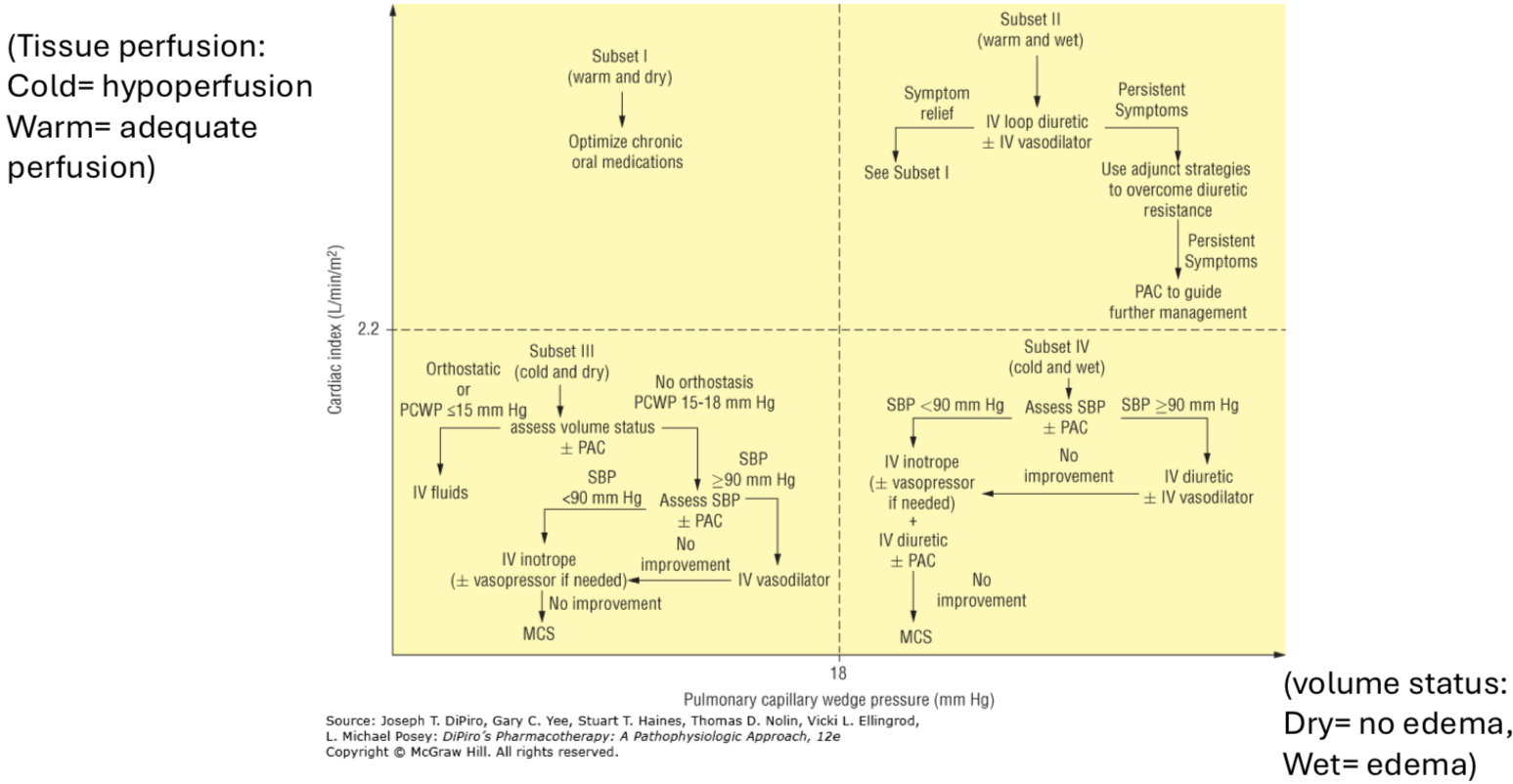
Management:
Cardiac Index: normalized value of cardiac output to body surface area → Determines tissue perfusion
Pulmonary Capillary Wedge Pressure (PCWP): indirect estimate of left atrial pressure, used to assess left ventricular filling and mitral valve function. Marker for congestion and volume status
High PCWP → High congestion or edema
Treatment of HF
Short-Term Goals
Improving symptoms of hemodynamics, avoid hypokalemia, renal dysfunction, symptomatic hypotension, correcting neurohumoral activation
Long-Term Goals
Correcting HTN, preventing MI and atherosclerosis, reducing hospitalizations, improving survival and quality of life
Treatment involves: dietary/lifestyle changes, drugs, surgery

Neurohumoral Blocking Agents
Improve symptoms, slow disease progression, improve survival in chronic HF
ACEi, ARBs, ARNI, beta-blockers, aldosterone receptor antagonists
ACEi/ARBs

Decrease levels (ACEi) or antagonize effects (ARB) of angiotensin II attenuating effects that drive initiation and progression
ACEi prevents breakdown of bradykinin → Enhances release of vasodilatory prostaglandins and histamines → Arterial and venous dilation
Reduce preload and afterload → Reducing myocardial work → Preventing ventricular remodeling
Concerns: Renal dysfunction and hyperkalemia
Cough and angioedema with ACEi
ARNI
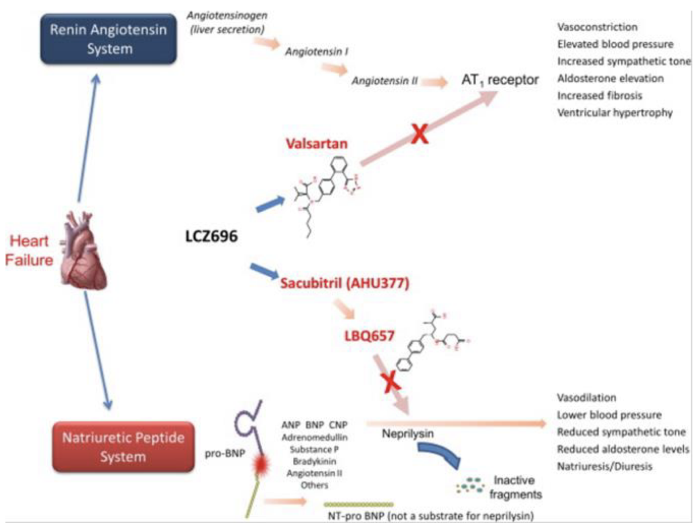
Angiotensin Receptor Neprilysin Inhibitor - Entresto
Valsartan: inhibits harmful effects of angiotensin II
Sacubitril: inhibits neprilysin mediated breakdown of BNP → Increase in BNP and beneficial effects
Neprilysin breaks down ANP, BNP, substance P, bradykinin, angiotensin II
Inhibition will increase BNP + angiotensin II levels → Need for an ARB
Reduces CV death and hospitalization
ADR: hypotension
Contraindications
ACEi use → 36 hour washout period
Pregnancy
Beta-Blockers (BB)
Antagonizes harmful effects of sympathetic nervous system in HF and slows disease progression
Antiarrhythmic effects
Blockade or reversal of ventricular remodeling
Reduction of myocyte death from catecholamine-induced necrosis or apoptosis
Improvement in LV systolic function
Reductions in HR and ventricular wall stress → Reduces myocardial oxygen demand
Inhibition of plasma renin release
Improves symptoms over time → May require a few months
Negative inotropic effects (reduction of CO) → Some symptomatic worsening may occur initially
Aldosterone Antagonists
Spironolactone, Eplerenone
Aldosterone causes fluid retention, cardiac fibrosis, electrolyte abnormalities
Aldosterone antagonists inhibit aldosterone effects on cardiac extracellular matrix and collagen deposition → Reduces cardiac fibrosis and ventricular remodeling
Decreases proinflammatory state, atherogenesis, oxidative stress
ADR: hyperkalemia
Eplerenone is selective for MR and does not cause endocrine ADR (gynecomastia, breast tenderness, menstrual irregularities)
Diuretics
Compensatory mechanisms in HF stimulate excessive sodium and water retention → Systemic and pulmonary congestion
Diuretics used for relief of acute symptoms of congestion and maintenance of euvolemia → Reduce preload and fluid retention
Thiazides
Weak, infrequently used alone
Preferred in those with mild fluid retention and elevated BP
Limited efficacy in renal insufficiency
Loops
Necessary to restore and maintain euvolemia
Retain efficacy in impaired renal function
Sodium Nitroprusside
MOA
Nitric oxide donor that causes venous and arterial vasodilation by activating guanylate cyclase which increase cGMP production in vascular smooth muscle cells
Effects
Reduces preload and afterload
Causes venous pooling and reduced arterial resistance
In patients with severely impaired LV function → Increase CO
Modest increase in HR and overall reduction in O2 demand
ADR
Hypotension, thiocyanate, cyanide toxicity, methemoglobinemia
PK
Onset is immediate
Duration: 1-10 min, continuous infusion required
Extremely potent, rebound HTN can occur upon discontinuation
Monitor: BP, HR, cyanide toxicity
Hydralazine + Isosorbide Dinitrate (ISDN)
Hydralazine
Activates voltage-gated K+ channels → Efflux of K+ and hyperpolarization of cell
Prevents calcium-mediated constriction of smooth muscle → Vasodilation
Acts mainly on arterial smooth muscle to reduce SVR (afterload) and BP
Increase stroke volume and cardiac output
Isosorbide Dinitrate
Releases NO and activated guanylyl cyclase resulting in an increase cGMP and venodilation
Venous pooling and reduction in preload
Beneficial effects
Increase in NO bioavailability secondary to NO donation and hydralazine-mediated reduction in oxidative stress
Reduces development of nitrate tolerance upon long-term use
Survival benefit in african-american
ADR
ISDN requires frequent dosing
Hypotension, headache, dizziness, GI distress, tachycardia
Hydralazine associated with lupus risk
Nitroglycerin
MOA
Releases NO → Increase cGMP → Vascular smooth muscle relaxation
Venodilation → Decrease preload
Mild arterial dilation at higher doses → Decrease afterload
Effects
Decrease LV end-diastolic pressure, pulmonary congestion, MVO2, rapid symptom relief
Useful when elevated preload is the issue
Tolerance develops with continuous exposure → Nitrate-free period
ADR
Hypotension, reflex tachycardia, headache, avoid with PDE-5 inhibitors
Guanylyl Cyclase
Family of enzymes that catalyze formation of cGMP
Membrane-bound (mGC)
Single extracellular domain, transmembrane, intracellular domain
Receptor for atrial natriuretic peptide
Soluble guanylate cyclase (sGC)
Heme and not bound to membrane
Receptor for NO → Activated by NO
Vericiguat
MOA: directly stimulates sGC independent of NO → Increases cGMP → Vasodilation
ADR: hypotension, syncope, anemia, fetal toxicity
Endothelial dysfunction and ROS → Reduced NO bioavailability→ Decrease in cGMP levels
Digoxin
Cardiac glycoside that acts as inotropic agent
Binds and inhibits Na+, K+, ATPase on cardiac myocytes → Decreases Na+ removal and increasing cytosolic Na+
Decreases Na+-Ca2+ exchange → Less Ca2+ removed and more Ca2+ accumulated in SR
Increase in Ca2+ → Enhanced myocardial contractility or positive inotropy
Benefits
Increased stroke volume
Increased EF
Decrease HR
Electrical Effects
Effects in HF
Reduces excessive sympathetic nervous system activation
Increases parasympathetic activity
Decreases HR and enhances diastolic filling
Positive inotropic and negative chronotropic
Toxicity
Non-cardiac ADR:
Anorexia, N/V, abdominal pain
Blurred vision, visual disturbances
Halos, photophobia, problems with color perception, scotomata
Fatigue, weakness, dizziness, headache, confusion, delirium
Cardiac ADR:
Ventricular arrhythmias
Premature ventricular depolarizations, bigeminy, trigeminy, ventricular tachycardia, ventricular fibrillation
AV block
Arrhythmias
Paroxysmal atrial tachycardia with AV block
Sinus bradycardia
Antidote
Narrow TI, long-half life
Digibind
K+ supplementation

Milrinone
Increases myocardial contractility and is also a vasodilator (inodilator)
MOA: inhibits PDE-3 → Increase in cAMP → Increase in intracellular Ca2+ → Increase force of contraction
Increase in SV, CO, HR
Lowers preload
ADR: hypotension, arrhythmias, headache, angina pectoris, thrombocytopenia
Dopamine
Low systolic BP and approaching cardiogenic shock
MOA: stimulates adrenergic receptors and releases NE from adrenergic nerve terminals
Higher doses → Chronotropy (increase HR) and vasoconstriction
Increase CO, BP, MVO2, cardiac work, and preload at high concentrations
ADR: arrhythmias
Dobutamine
Inotropic agent
MOA
B1/B2 receptor agonist with some A1 agonist effects
Does not result in release of NE
Positive inotropic effects due to B1
Vasodilation due to B2 effect
Increase in CO and decrease in preload
ADR
Tachycardia and increase in MVO2
Potential for producing angina or arrhythmias in patients with CAD
Tachyphylaxis

Ivabradine
MOA: inhibits funny current in SA node → Slows spontaneous depolarization of SA node → Dose-dependent slowing of HR
Does not affect BP or contractility
Reduced hospitalization and death
ADR: bradycardia, sinus arrest, heart block, a-fib, visual disturbances
Metabolized by CYP3A4
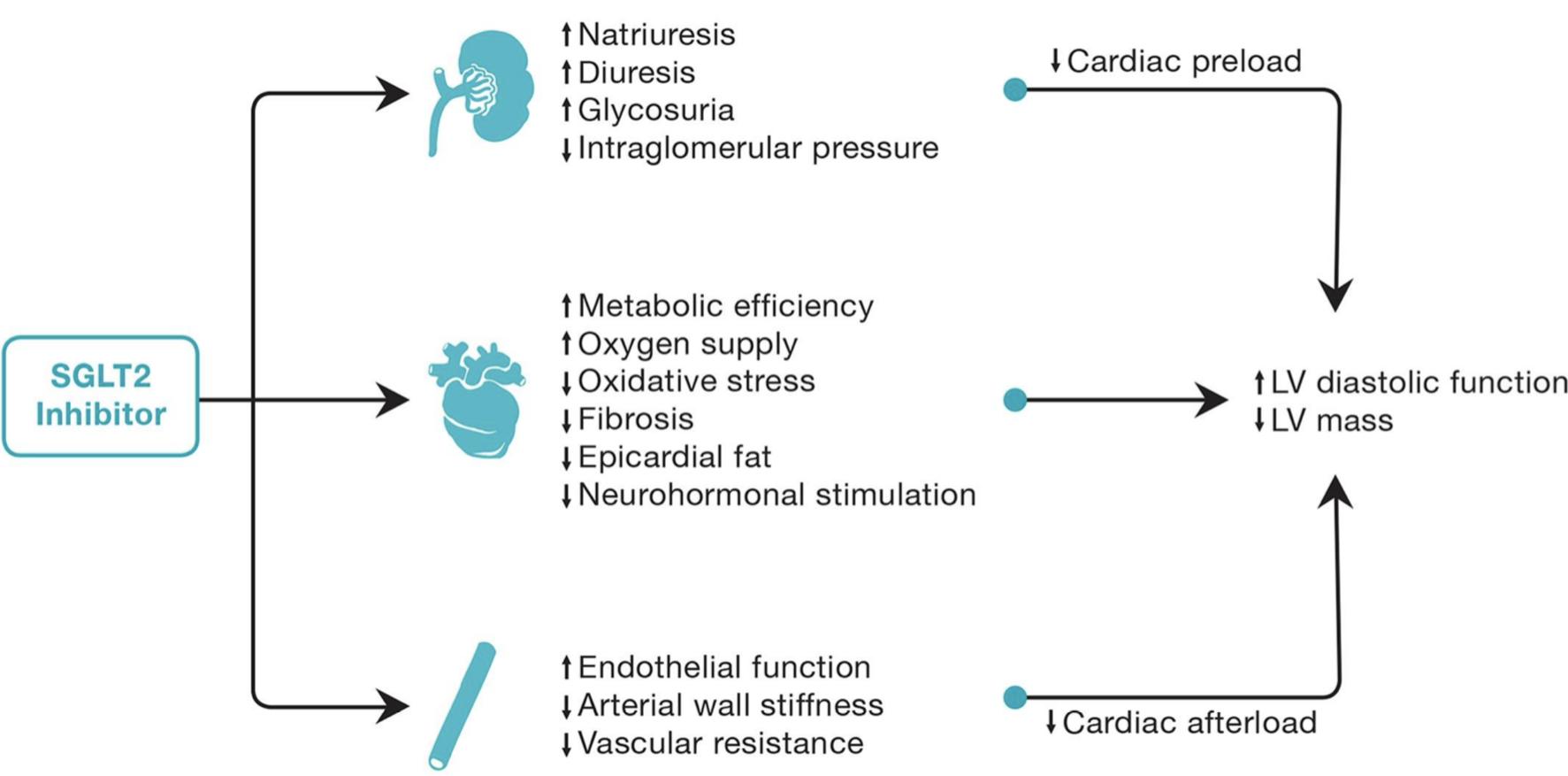
SGLT2 Inhibitors
Dapagliflozin reduces risk of hospitalization for HF, CV deaths, and urgent HF visits
Emapgliflozin reduces hospitalizations for HF and CV deaths
HF benefit
Cardiac energy metabolism
Overall HF benefits
Glycosuria, natriuresis
Decrease in preload, afterload, BP
Reduction in arterial stiffness
Renal protection → Reduction in renal blood flow and GFR
Improved cardiac energy metabolism
ADR
Diuresis, hypotension, dehydration
Slight increase in LDL cholesterol
UTI
Fournier’s gangrene
Risk of bladder and breast cancer
Increased fracture risk
Diabetic ketoacidosis
Sotagliflozin
Dual inhibitor of SGLT1/2
SGLT1-i: reduces intestinal absorption of glucose and sodium → Diarrhea
ADR: UTI, volume depletion, hypoglycemia, dizziness, genital mycotic infection

Summary