Week 2
NEURONS
The main part of a neuron is the cell body, which is also known as the soma (soma = “body”). The cell body contains the nucleus and most of the major organelles. But what makes neurons special is that they have many extensions of their cell membranes, which are generally referred to as processes. Neurons are usually described as having one, and only one, axon, a fibre that emerges from the cell body and projects to target cells. That single axon can branch repeatedly to communicate with many target cells. It is the axon that propagates the nerve impulse, which is communicated to one or more cells. The other processes of the neuron are dendrites, which receive information from other neurons at specialised areas of contact called synapses. The dendrites are usually highly branched processes, providing locations for other neurons to communicate with the cell body. Information flows through a neuron from the dendrites, across the cell body, and down the axon. This gives the neuron a polarity, meaning that information flows in this one direction.
Many axons are wrapped by an insulating substance called myelin, which is actually made from glial cells. Myelin acts as insulation much like the plastic or rubber that is used to insulate electrical wires. A key difference between myelin and the insulation on a wire is that there are gaps in the myelin covering of an axon. Each gap is called a node of Ranvier and is important to the way that electrical signals travel down the axon. At the end of the axon several branches extend toward the target cell, each of which ends in an enlargement called a synaptic bulb. These bulbs are what make the connection with the target cell at the synapse.

There are many neurons in the nervous system, a number in the trillions. And there are many different types of neurons. They can be classified by many different criteria. The first way to classify them is by the number of processes attached to the cell body. Using the standard model of neurons, one of these processes is the axon, and the rest are dendrites. Because information flows through the neuron from dendrites or cell bodies toward the axon, these names are based on the neuron's polarity.
Unipolar neurons
Unipolar neurons have a single process extending from the cell body, which quickly splits into two branches: one leading to dendrites and the other forming synaptic connections with a target. These neurons are exclusively sensory and have two key features:
Their dendrites receive sensory input, sometimes directly from the stimulus.
Their cell bodies are located in ganglia, near the central nervous system but still within the peripheral nervous system.
The axon extends from the dendritic endings, passes the cell body in the ganglion, and enters the CNS.
Bipolar neurons
Bipolar neurons have two distinct processes—one axon and one dendrite—emerging from opposite ends of the cell body. Though rare, they are found in specialised sensory regions such as the olfactory epithelium (smell) and the retina (vision).
Multipolar neurons
Multipolar neurons have one axon and two or more dendrites, often many more. They represent the most common neuron type in the nervous system. With the exception of unipolar sensory neurons and the specific bipolar neurons mentioned above, all other neurons are multipolar. These include motor neurons and interneurons, which are involved in integrating and transmitting signals throughout the CNS.
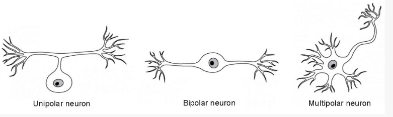
Neuron Classification by Shape Unipolar cells have one process that includes both the axon and dendrite. Bipolar cells have two processes, the axon and a dendrite. Multipolar cells have more than two processes, the axon and two or more dendrites.
Neurons can also be classified on the basis of the type of information they carry or direction of information flow. Neurons that carry information from the periphery to the central nervous system are called sensory neurons. Neurons that carry commands/instructions from the central nervous system to the periphery are called motor neurons. Motor neurons can then be divided into somatic motor for voluntary muscle commands or autonomic motor for involuntary glandular, cardiac or smooth muscle commands.
Structurally, a nerve is a bundle of axons, long, slender projections of neurons, wrapped in connective tissue layers that provide protection and support. Functionally, nerves carry electrical impulses that allow communication between sensory receptors, muscles, and glands, enabling processes such as sensation, movement, and reflexes. This rapid signaling system is vital for maintaining homeostasis and coordinating complex bodily functions.
Nerves
Bundles of axons in the PNS are referred to as nerves (referred to as tracts in the CNS). Nerves are composed of more than just nervous tissue. They have connective tissues invested in their structure, as well as blood vessels supplying the tissues with nourishment. Nerves are associated with the region of the CNS to which they are connected, either as cranial nerves connected to the brain or spinal nerves connected to the spinal cord.
Glial cells perform diverse functions in the nervous system. Ependymal cells line the ventricles of the brain, helping produce and circulate cerebrospinal fluid, astrocytes support neurons by maintaining the blood-brain barrier and regulating neurotransmitters, and microglia act as the immune defenders of the central nervous system.
Myelin
The insulation for axons in the nervous system is provided by glial cells called oligodendrocytes in the CNS and Schwann cells in the PNS. Myelin is a lipid-rich sheath that surrounds the axon and by doing so creates a myelin sheath that facilitates the transmission of electrical signals along the axon, ensuring the message is transmitted rapidly along each axon. Myelin is formed by the membrane of the glial cell.
Schwann cells wrap around a portion of only one axon segment and no others. The nucleus and cytoplasm of the Schwann cell are on the edge of the myelin sheath. The glial cell is wrapped around the axon several times creating several layers with little to no cytoplasm between the glial cell layers.
The blood-brain barrier is a physiological barrier that keeps many substances that circulate in the rest of the body from getting into the central nervous system, restricting what can cross from circulating blood into the CNS. Nutrient molecules, such as glucose or amino acids, can pass through the BBB, but other molecules cannot.

Action potentials and impulse transmission
The core functions of the nervous system: sensation, integration, and response, rely on the ability of neurons to communicate via electrical signals. This communication is driven by the action potential, a change in the membrane’s electrical state, which is dependent on an excitable membrane. Cells use ions to create electrical charges across the plasma (cell) membrane. This is essential for processes like muscle contraction, which depends on neuronal input. Both neurons and muscle cells regulate ion flow between the extracellular fluid and cytosol via the plasma membrane.
The membrane, a phospholipid bilayer, selectively allows substances to pass. Hydrophilic ions cannot cross unaided and require channel proteins or active transport pumps. A key player is the sodium-potassium pump, which moves Na⁺ out and K⁺ into the cell, maintaining ion balance and enabling the generation of action potentials.

Resting membrane potential describes the steady state of the cell, which is a dynamic process that is balanced by ion leakage and ion pumping. Without any outside influence, it will not change. To get an electrical signal started, the membrane potential has to change.
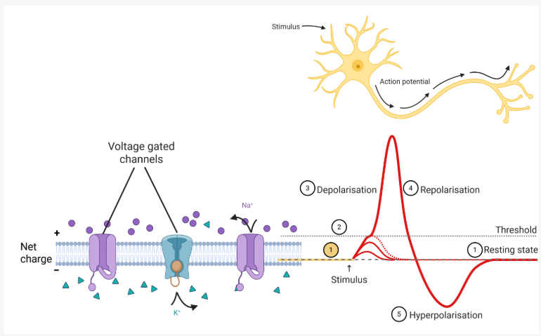
When a stimulus reaches the threshold potential, voltage-gated Na+ (sodium) channels in the cell membrane of the neuron open, allowing a rapid influx of Na+. This initiates an action potential.
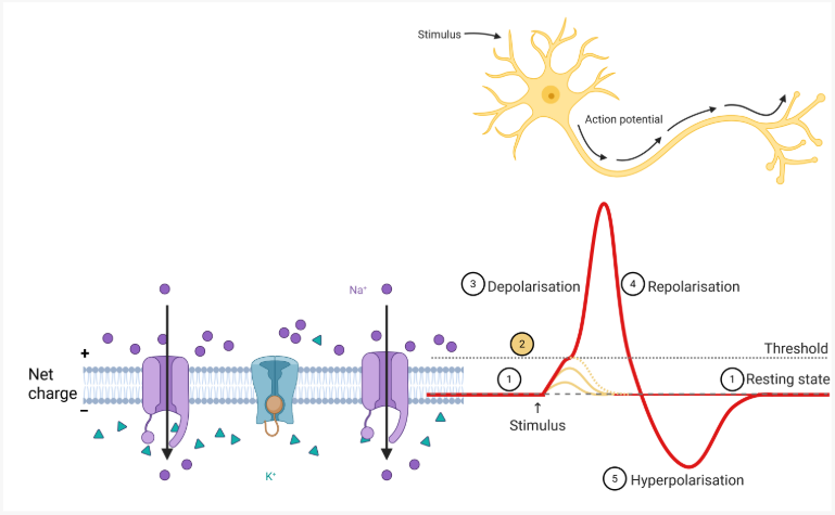
An action potential begins at the initial segment of the axon, where a high concentration of voltage-gated Na⁺ (sodium) channels allows rapid depolarisation. The resting potential is the state of the membrane at a voltage of -70 mV, so the sodium cation entering the cell will cause it to become less negative. As Na⁺ enters the cell, it spreads along the inner membrane, triggering nearby channels to open and continue the depolarisation. This wave-like process moves the signal toward the axon terminals.
Once peak depolarisation is reached, Na⁺ channels become temporarily inactivated, known as the absolute refractory period, preventing the signal from moving backward and ensuring one-way propagation.

As the membrane potential reaches approximately +30 mV, voltage-gated K⁺ (potassium) channels begin to open. These channels allow K⁺ ions to move out of the cell, driven by both the concentration gradient and the positive charge inside the membrane. As potassium exits, it carries positive charge away, causing the membrane potential to shift back toward its resting level. This process is called repolarisation, where the membrane voltage returns toward the resting value of about –70 mV.

Repolarisation returns the membrane potential back toward the resting level of –70 mV, but it briefly overshoots this value. This occurs because K⁺ (potassium) ions reach their equilibrium when the membrane voltage is slightly below –70 mV. As K⁺ channels remain open a little longer than necessary, additional potassium exits the cell, causing a short period of hyperpolarisation. This overshoot ends once the delayed K⁺ channels close and the membrane returns to its resting potential. The sodium-potassium pump then restores ionic gradients by actively transporting Na⁺ ions out and K⁺ ions into the cell, maintaining the resting potential and preparing the neuron for the next action potential.

Factors affecting neural impulse transmission speed |
Neural transmission speed refers to how quickly electrical impulses travel along neurons to transmit information throughout the nervous system. This speed is influenced by factors such as axon diameter and the presence of myelin, which acts as an insulating layer to accelerate signal conduction. Rapid transmission is essential for reflexes, sensory processing, and coordinated movement, ensuring the body responds efficiently to internal and external stimuli.
Conduction Types
Unmyelinated axons use continuous conduction, where Na⁺ channels open sequentially along the axon. This is slower due to constant ion influx.
Myelinated axons use saltatory conduction, where the action potential "jumps" between nodes of Ranvier. These gaps are spaced to maintain depolarisation above threshold, allowing faster signal transmission. Axon diameter also affects speed, wider axons conduct signals more quickly due to lower resistance.
Learning outcomes
Explain the structure and function of the synapse
Describe how neurotransmitters and receptors contribute to signal transmission
Structure and function of the synapse |
A synapse is a specialised junction mediating the transfer of electrical or chemical information between a neuron and its target effector cell. The synapse is comprised of the axon terminal membrane of the presynaptic neuron (the neuron transmitting the original message), the synaptic cleft (the gap between the two cells), and the cell membrane of the dendrite or cell body of the postsynaptic neuron or the target effector cell (the cell receiving the signal).

Signal transmission |
When an action potential reaches the axon terminal, synaptic vesicles fuse with the presynaptic membrane and release neurotransmitters into the synaptic cleft by exocytosis. These chemical messengers diffuse across the cleft and bind to specific receptors on the postsynaptic cell membrane, like a key fitting a lock. Receptor activation changes the electrical state or signaling pathways of the target cell, leading to a response such as generating a new action potential, muscle contraction, or hormone release. This process converts an electrical signal to a chemical signal and then back to electrical changes in the next cell.

Learning outcomes
Differentiate between the major anatomical components of the central and peripheral nervous systems
Describe the function of the nervous system
Structural divisions of the nervous system |
The nervous system can be divided into two major regions based on the location of nervous tissue: the central and peripheral nervous systems. The central nervous system (CNS) is the brain and spinal cord, and the peripheral nervous system (PNS) is everything else. The brain is contained within the cranial cavity of the skull, and the spinal cord is contained within the vertebral cavity of the vertebral column.

Central and peripheral nervous system The structures of the PNS are referred to as ganglia and nerves, which can be seen as distinct structures. The main organs of the CNS are the brain and spinal cord.
The nervous system can be divided on the basis of its functions, but anatomical divisions and functional divisions are different. The CNS and the PNS both contribute to the same functions, but those functions can be attributed to different regions of the brain or to different ganglia in the periphery.
Basic Functions
The nervous system is involved in receiving information about the environment around us (sensation) and generating responses to that information (motor responses). The nervous system can be divided into regions that are responsible for sensation (sensory functions) and for the response (motor functions). But there is a third function that needs to be included. Sensory input needs to be integrated with other sensations, as well as with memories, emotional state, or learning (cognition). Some regions of the nervous system are termed integration or association areas. The process of integration combines sensory perceptions and higher cognitive functions such as memories, learning, and emotion to produce a response.
Sensation
The first major function of the nervous system is sensation—receiving information about the environment to gain input about what is happening outside the body (or, sometimes, within the body). The sensory functions of the nervous system register the presence of a change from homeostasis or a particular event in the environment, known as a stimulus. The senses include taste, smell, touch, sight, balance, hearing and internal organ position and movement.
Response
The nervous system produces a response on the basis of the stimuli perceived by sensory structures. An obvious response would be the movement of muscles, such as withdrawing a hand from a hot stove, but there are broader uses of the term. The nervous system can cause the contraction of all three types of muscle tissue. For example, skeletal muscle contracts to move the skeleton, cardiac muscle is influenced as heart rate increases during exercise, and smooth muscle contracts as the digestive system moves food along the digestive tract. Responses also include the neural control of glands in the body as well, such as the production and secretion of sweat by the sweat glands found in the skin to lower body temperature.
Responses can be divided into those that are voluntary or conscious (contraction of skeletal muscle) and those that are involuntary (contraction of smooth muscles, regulation of cardiac muscle, activation of glands). Voluntary responses are governed by the somatic nervous system and involuntary responses are governed by the autonomic nervous system, which are discussed in the next section.
Integration
Stimuli that are received by sensory structures are communicated to the nervous system where that information is processed. This is called integration. Stimuli are compared with, or integrated with, other stimuli, memories of previous stimuli, or the state of a person at a particular time. This leads to the specific response that will be generated.
Controlling the Body
The nervous system can be divided into two parts mostly on the basis of a functional difference in responses.

Somatic nervous system
The somatic nervous system (SNS) is responsible for conscious perception and voluntary motor responses. Voluntary motor response means the contraction of skeletal muscle, but those contractions are not always voluntary in the sense that you have to want to perform them. Some somatic motor responses are reflexes, and often happen without a conscious decision to perform them. Other motor responses become automatic (in other words, unconscious) as a person learns motor skills (referred to as “habit learning” or “procedural memory”).
Autonomic nervous system
The autonomic nervous system (ANS) is responsible for involuntary control of the body, usually for the sake of homeostasis (regulation of the internal environment). Sensory input for autonomic functions can be from sensory structures tuned to external or internal environmental stimuli. The motor output extends to smooth and cardiac muscle as well as glandular tissue. The role of the autonomic system is to regulate the organ systems of the body, which usually means to control homeostasis. Sweat glands, for example, are controlled by the autonomic system. When you are hot, sweating helps cool your body down. That is a homeostatic mechanism. But when you are nervous, you might start sweating also. That is not homeostatic, it is the physiological response to an emotional state.
Learning outcome
Compare and contrast the organisation and functions of the grey and white matter in the brain and spinal cord
Grey matter = cell bodies and dendrite
White matter = axons
Looking at nervous tissue, there are regions that predominantly contain cell bodies and regions that are largely composed of just axons. These two regions within nervous system structures are often referred to as grey matter (the regions with many cell bodies and dendrites) or white matter (the regions with many axons). The colours ascribed to these regions are what would be seen in “fresh,” or unstained, nervous tissue. Grey matter is not necessarily grey. It can be pinkish because of blood content, or even slightly tan, depending on how long the tissue has been preserved. But white matter is white because axons are insulated by a lipid-rich substance called myelin. Actually, grey matter may have that color ascribed to it because next to the white matter, it is just darker, hence, grey.
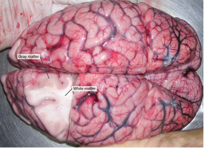
Naming grey and white matter in the CNS and PNS |
Regardless of whether tissue appears stained or unstained, neuronal cell bodies and axons are organised within distinct anatomical structures that require specific names. These names differ depending on whether the structure is part of the central or peripheral nervous system. In the CNS, a localised cluster of neuronal cell bodies is called a nucleus, whereas in the PNS, such a cluster is referred to as a ganglion.
The term nucleus has several meanings in anatomy and physiology:
it is the core of an atom, containing protons and neutrons.
it is the central part of a cell, housing DNA.
it is also a region of gray matter within the CNS.
Similarly, the term ganglion (plural: ganglia) has a historical usage that can cause confusion. Within the CNS, a group of interconnected nuclei was traditionally called the basal ganglia, even though “ganglion” now refers to peripheral structures. To avoid ambiguity, this group is currently referred to as the basal nuclei.
Terminology for bundles of axons also varies by location. In the CNS, a bundle of axons is called a tract, while in the PNS, it is called a nerve. Importantly, these terms can describe the same axons depending on their position. For example, axons projecting from the retina to the brain are called the optic nerve as they exit the eye, but once inside the cranium, they are referred to as the optic tract. The name changes at the optic chiasm, even though the axons themselves remain the same.

Learning outcome
Identify the location and function of major brain structures, including the brainstem, diencephalon, lobes of the cerebrum, and cerebellum
The four major parts of the brain are the:
Brainstem (midbrain, pons, medulla oblongata)
Diencephalon (thalamus, hypothalamus and epithalamus)
Cerebrum (including the cerebral cortex)
Cerebellum

The brainstem controls critical life-sustaining functions related to cardiovascular and respiratory function.
The cerebrum has many gyri (ridges) and sulci (shallow grooves), which increase the surface area of the brain. The cerebrum is separated into two halves called cerebral hemispheres.
The cerebrum contains four lobes on each side, named after the superficial bones:
Frontal lobe: location of higher executive functions including emotional regulation, planning, reasoning and problem solving.
Parietal lobe: location of sensory integration including touch, temperature, pressure and pain.
Temporal lobe: location of processing sensory information, particularly important for hearing, recognising language, and forming memories.
Occipital lobe: location of visual processing.
Brainstem |
The midbrain, pons and medulla oblongata are collectively referred to as the brainstem. The structure connects the brain to the spinal cord. Posteriorly attached to the brainstem, but considered a separate region of the brain, is the cerebellum. The midbrain coordinates sensory representations of the visual, auditory, and somatosensory perceptual spaces. The pons is the main connection with the cerebellum. The pons and the medulla oblongata regulate several crucial functions, including the cardiovascular and respiratory systems.
The cranial nerves connect through the brainstem and provide the brain with the sensory input and motor output associated with the head and neck, including most of the special senses. The major ascending and descending neuronal tracts between the spinal cord and brain, specifically the cerebrum, pass through the brainstem.
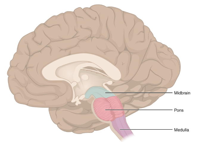
The Brainstem (Midsagittal view of brain) The brainstem comprises three regions: the midbrain (superior), the pons, and the medulla oblongata (inferior).
Diencephalon |
The majority of the brain, the spinal cord, and the PNS all send information to the cerebrum through the diencephalon. Output from the cerebrum passes through the diencephalon. The diencephalon is deep to the cerebrum and constitutes the walls of the third ventricle. The three major regions of the diencephalon are the thalamus centrally, the hypothalamus anteriorly and the epithalamus posteriorly. The epithalamus contains the pineal gland, which is an endocrine gland that produces the hormone melatonin important in sleep-wake cycles. The thalamus is a collection of nuclei (grey matter) that relay information between the cerebral cortex and the periphery, spinal cord or brainstem. Inferior and slightly anterior to the thalamus is the hypothalamus. The hypothalamus is a collection of nuclei (grey matter) that are largely involved in regulating homeostasis.
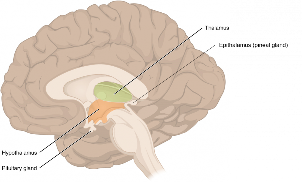
The Diencephalon (Midsagittal view of brain) The diencephalon is composed primarily of the thalamus, hypothalamus and epithalamus, which together define the walls of the third ventricle. The thalami are two elongated, ovoid structures on either side of the midline that make contact in the middle. The hypothalamus is inferior and anterior to the thalamus, culminating in a sharp angle to which the pituitary gland is attached. The epithalamus is the most posterior part of the diencephalon consisting largely of the pineal gland.
Cerebrum |
The part which appears to make up most of the mass of the brain, is the cerebrum. The wrinkled external portion composed of grey matter is the cerebral cortex, and the rest of the structure is deep to that external covering. There is a large separation between the two sides of the cerebrum called the longitudinal fissure. This fissure separates the cerebrum into two distinct halves, a right and left cerebral hemisphere. Deep within the cerebrum, the white matter of the corpus callosum provides the major pathway for axonal communication between the two hemispheres of the cerebral cortex.
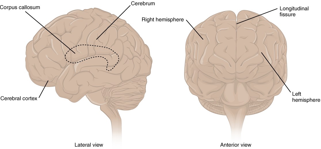
The Cerebrum The cerebrum is a large component of the CNS in humans, and the most obvious aspect of it is the folded surface called the cerebral cortex.
Many of the higher neurological functions, such as memory, emotion, and consciousness, are the result of cerebral function. The cerebrum comprises the outer grey matter that is the cortex (from the Latin word meaning “bark of a tree”) and several deep nuclei. The basal nuclei are responsible for cognitive processing, the most important function being that associated with planning movements.
Cerebral cortex
The cerebrum is covered by a continuous layer of grey matter called the cerebral cortex. This thin, extensive region of wrinkled grey matter is responsible for the higher functions of the nervous system. A gyrus (plural = gyri) is the ridge of one of those wrinkles, and a sulcus (plural = sulci) is the groove between two gyri. The pattern of these folds of tissue indicates specific regions of the cerebral cortex. Extensive folding in the cerebral cortex enables more grey matter to fit into this limited space. If the grey matter of the cortex were peeled off of the cerebrum and laid out flat, its surface area would be roughly equal to one square meter.
The cortex can be separated into four major regions, or lobes. The lateral sulcus separates the temporal lobe from the other regions. Superior to the lateral sulcus are the frontal lobe (anterior) and parietal lobe (more posterior), which are separated from each other by the central sulcus. The posterior region of the cortex is the occipital lobe, which has no obvious anatomical border between it and the parietal or temporal lobes on the lateral surface of the brain. From the medial surface, an obvious landmark separates the parietal and occipital lobes and is called the parieto-occipital sulcus.
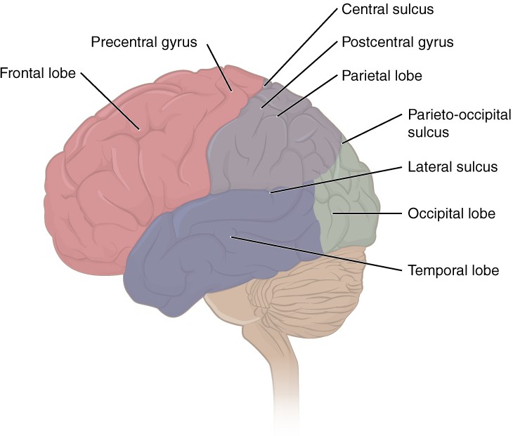
Lobes of the Cerebral Cortex The cerebral cortex is divided into four major lobes visible from the external view. Extensive folding increases the surface area available for cerebral functions. Note that the parieto-occipital sulcus is only visible from the medial aspect of the cerebral hemisphere within the longitudinal fissure.
The temporal lobe is associated with primary auditory sensation, memory and smell. The main sensation associated with the parietal lobe is somatosensation, meaning the general sensations associated with the body. Posterior to the central sulcus is the postcentral gyrus, the primary somatosensory cortex. All of the tactile senses are processed in this area, including touch, pressure, tickle, pain, itch and vibration, as well as more general senses of the body such as proprioception and kinesthesia, which are the senses of body position and movement, respectively.
Anterior to the central sulcus is the frontal lobe, which is primarily associated with motor functions. The precentral gyrus is the primary motor cortex. Cells from this region of the cerebral cortex are the upper motor neurons that instruct cells in the spinal cord to move skeletal muscles. Anterior to this region are a few areas that are associated with planned movements. The frontal lobe is also responsible for production of language and cognitive functions that can be the basis of personality, short-term memory and consciousness.
Subcortical structures
The basal nuclei are a set of nuclei (areas of grey matter in the CNS) in the cerebrum responsible for comparing cortical processing with the general state of activity in the nervous system to influence the likelihood of movement taking place. For example, while a student is sitting in a classroom listening to a lecture, the basal nuclei will keep the urge to jump up and scream from actually happening.
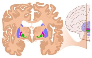
Coronal Section of Cerebral Cortex and Basal Nuclei The major components of the basal nuclei, shown in a coronal section of the brain, are coloured blue, purple and green, lying deep in the white matter of the cerebrum. The cerebral cortex can also be seen as the darker brown regions on the most superficial surface of the cerebrum
The cerebellum, as the name suggests, is the “little brain” and looks like a miniature version of that part of the brain. The cerebellum is largely responsible for comparing information from the cerebrum with sensory feedback from the periphery through the spinal cord. It accounts for approximately 10 percent of the mass of the brain.
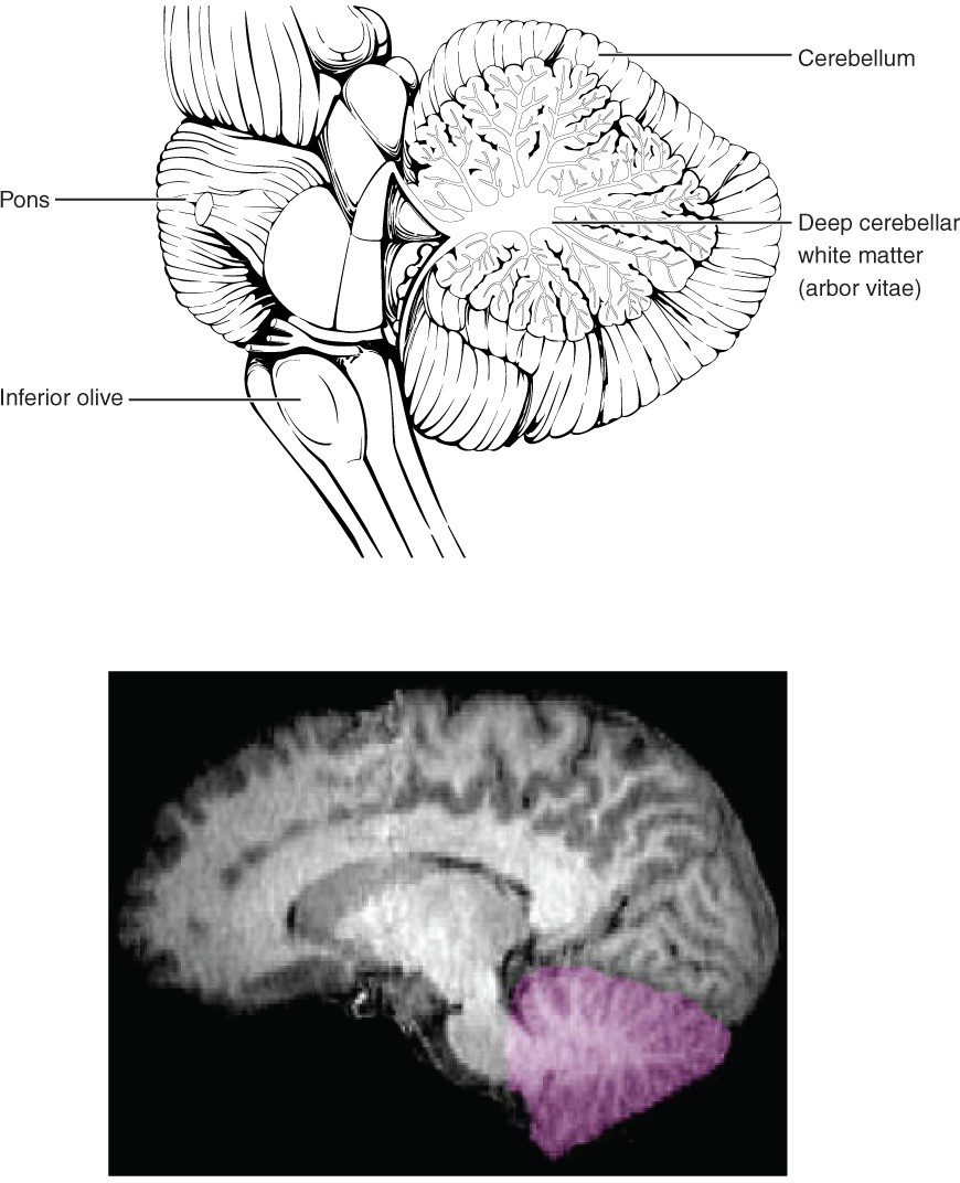
The Cerebellum The cerebellum is situated on the posterior surface of the brainstem.
If the primary motor cortex of the frontal lobe sends a command down to the spinal cord to initiate walking, a copy of that instruction is sent to the cerebellum. Sensory feedback from the muscles and joints, proprioceptive information about the movements of walking, and sensations of balance are sent to the cerebellum and the cerebellum compares them. If walking is not coordinated, perhaps because the ground is uneven or a strong wind is blowing, then the cerebellum sends out a corrective command to compensate for the difference between the original cortical command and the sensory feedback. The output of the cerebellum then sends signals into the spinal cord to correct the messages going to skeletal muscles.
Learning outcomes
Describe how ependymal cells, meninges, and peripheral nerve coverings contribute to the structure and function of the nervous system
Ependymal cells and the ventricular system |
Cerebrospinal fluid (CSF) is produced within the ventricles by a type of specialised membrane called a choroid plexus. Ependymal cells (a type of glial cell) surround blood capillaries and filter the blood to make CSF. The fluid is a clear solution with a limited amount of the constituents of blood. It is essentially water, small molecules, and electrolytes. Oxygen and carbon dioxide are dissolved into the CSF, as they are in blood, and can diffuse between the fluid and the nervous tissue. CSF circulates throughout and around the CNS. CSF functions to remove metabolic wastes from the interstitial fluids of nervous tissues and return them to the blood. It also acts as a liquid cushion for the brain and spinal cord. The ventricles are the open spaces within the brain where CSF circulates. CSF is continuously produced by the choroid plexuses each day and pulses through the ventricular system, keeping the fluid moving.
The ventricles
There are four ventricles within the brain. The first two are named the right and left lateral ventricles and are deep within each cerebral hemisphere. These ventricles are connected to the third ventricle by two openings called the interventricular foramina. The third ventricle is the space between the left and right sides of the diencephalon, which opens into the cerebral aqueduct that passes through the midbrain. The cerebral aqueduct opens into the fourth ventricle, which is the space between the cerebellum, and the pons and superior part of the medulla oblongata. The choroid plexuses are found in all four ventricles. The fourth ventricle is continuous with the thin passageway in the spinal cord called the central canal.

Cerebrospinal fluid circulation (Midsagittal view). The choroid plexus in the four ventricles produces CSF, which is circulated through the ventricular system and then enters the subarachnoid space through small apertures in the fourth ventricle. The CSF is then reabsorbed into the blood at the arachnoid granulations, where the arachnoid mater protrudes through the dura mater to emerge into the superior sagittal dural sinus.
Meninges of the brain and spinal cord |
The external surface of the CNS is covered by a series of membranes composed of connective tissue called the meninges (pl; meninx, singular), which protect the brain. The meninges are named from external to internal, the dura mater, arachnoid mater and pia mater.
Dura mater
Like a thick cap covering the brain, the dura mater is a tough external covering. The name comes from the Latin for “tough mother” to represent its physically protective role and durable tough texture. It encloses the entire CNS and the major blood vessels that enter the cranium and vertebral cavity. It is directly attached to the internal (periosteal) surface of the bones of the cranium and to the vertebral cavity. The dura also has an important role in forming the walls of the venous sinuses of the cranium, allowing blood to drain from the brain and into the internal jugular vein to return to the heart.
Arachnoid mater
The middle layer of the meninges is the arachnoid mater, named for the spider-web–like trabeculae between it and the pia mater. The trabeculae are found in the subarachnoid space, which is filled with circulating cerebrospinal fluid (CSF) and the cerebral arteries and veins. Small pockets of arachnoid protrude through the dura mater to enter the dural sinuses as the arachnoid granulations (also known as arachnoid villi when soft and flexible in young individuals), where the CSF empties into the venous blood circulation for recycling.
Pia mater
The surface of the brain and spinal cord is covered in the thin fibrous membrane of the pia mater, the deepest of the meningeal layers. The name pia mater comes from the Latin for “tender mother,” suggesting the thin membrane is a gentle covering for the brain. The pia extends into every convolution of the CNS, lining the inside of the sulci in the cerebral and cerebellar hemispheres.

Meningeal layers at the superior sagittal sinus (Coronal view) The layers of the meninges in the longitudinal fissure inferior to the superior sagittal sinus are shown, with the dura mater adjacent to the internal (periosteal) surface of the cranium, the pia mater adjacent to the surface of the brain, and the arachnoid and subarachnoid space between them. An arachnoid villus is shown emerging into the dural sinus to allow CSF to filter back into the blood for drainage.
The same meninges are found supporting the brain and spinal cord. In the spinal cord however the dura mater is not attached to the bony wall of the vertebral canal. Instead there is an adipose tissue filled space called the epidural space between the dura mater and the bone. This is an important location for venous blood flow and is the site for epidural blocks associated with anaesthesia of the body, such as when an expecting mother receives pain relief during childbirth.
Peripheral nerve coverings |
The outer surface of a nerve is a surrounding layer of connective tissue called the epineurium. Within the nerve, axons are further bundled into fascicles, which are each surrounded by their own layer of connective tissue called perineurium. Finally, individual axons are surrounded by connective tissue called the endoneurium.

Nerve structure The structure of a nerve is organised by the layers of connective tissue on the outside, around each fascicle, and surrounding the individual nerve fibres.