Brain and neuropsychology
The structure and function of the Nervous System
The nervous system is a complex network of nerve fibres and nerve cells.
This network helps to pass information around the body.
It’s divided into the central nervous system (CNS) and the peripheral nervous system (PNS)
The Central Nervous System (CNS)
It coordinates the information and makes the decisions about movement.
It consists of the brain and spinal cord.
Peripheral Nervous system (PNS)
The peripheral Nervous system (PNS) collects the information and helps to send it to different parts of the body.
The peripheral nervous system consists of two sections which are the somatic nervous system (SNS) and the autonomic nervous system (ANS)
Somatic Nervous System
Somatic Nervous System (SNS) is a network of nerve fibres that run throughout the body.
These nerve fibers pass information to and from the CNS using sensory and motor neurons which helps the messages travel faster.
Autonomic Nervous System
Autonomic Nervous System (ANS) is a division of the peripheral nervous system that controls involuntary bodily functions. It regulates processes such as heart rate, digestion, breathing, and glandular secretion.
It consists of two main branches: the Sympathetic nervous system, which prepares the body for "fight or flight" responses
The Parasympathetic nervous system promotes relaxation and "rest and digest" activities.
The fight or flight response allows you to call on energy and strength to deal with the situation regardless of whether you choose to run away or stay and fight
The James-Lange Theory of emotion
The James-Lange theory of emotion, proposed by psychologists William James and Carl Lange
According to this theory, our emotional experience is determined by the bodily changes we perceive.
For example, we feel fear because we experience a racing heart and trembling limbs.
This theory suggests that our emotions are not separate from our physical sensations, but rather, they are a direct result of them.
Neuron structure and function
The nervous system is made up of special cells which exchange chemicals to generate small electrical impulses and this is how information is passed around. These special cells are called neurons.
There are three types of neurons:
Sensory neurons: These neurons transmit sensory information from the sensory organs (such as the eyes, ears, and skin) to the central nervous system (brain and spinal cord).
Motor neurons: These neurons transmit signals from the central nervous system to the muscles and glands, enabling movement and bodily functions.
Relay neurons: These neurons are located within the central nervous system and facilitate communication between sensory and motor neurons. They process and interpret information, allowing for complex cognitive functions.
Synaptic Transmission
Synaptic transmission is the process by which information is transmitted between neurons in the nervous system.
It involves the release, diffusion, and binding of neurotransmitters across the synapse, which is the small gap between two neurons.
It is the process where neurons pass messages to other neurons or muscles by releasing special chemicals known as neurotransmitters into tiny gaps between dendrites.
These tiny gaps are called synapses.
The chemical is released from swellings at the end of each dendrite, called synaptic knobs.
These contain vesicles of neurotransmitters and when an electric impulse reaches them, the vesicles open and release the chemicals into the synapse.
These chemicals are then picked up at receptor sites on the next neuron, which is sensitive to that particular neurotransmitter.
This process allows for the transmission of signals and communication between neurons, enabling various functions such as sensory perception, motor control, and cognitive processes.
Donald Hebb’s Theory of Learning and Neuronal Growth
Donald Hebb's Theory of Learning and Neuronal Growth is a psychological theory that suggests that learning occurs through the strengthening of connections between neurons in the brain.
According to Hebb, when two neurons are repeatedly activated together, the connection between them becomes stronger, leading to more efficient communication.
This theory is often summarized by the phrase "neurons that fire together, wire together."
Hebb's theory has had a significant impact on our understanding of how learning and memory processes occur in the brain.
The structure and function of the brain
The brain consists of millions of relay neurons that are tightly packed together.
The cerebrum is the top layer of the brain.
The brain consists of two cerebral hemispheres, one on each side of the head and with each hemisphere divided into four areas known as lobes.
Following are the division
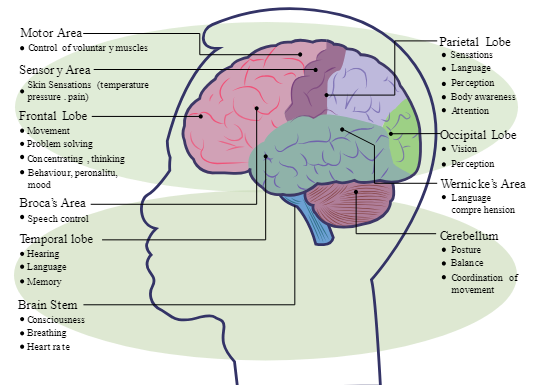
Frontal lobe: The frontal lobe is a region of the brain located at the front of each cerebral hemisphere.
It plays a crucial role in various cognitive functions, including decision-making, problem-solving, planning, and personality expression.
Parietal lobe: The parietal lobe is a region in the brain that is responsible for processing sensory information, such as touch, temperature, and pain.
It also plays a role in spatial awareness, perception, and language processing.
Occipital lobe: The occipital lobe is a region in the brain responsible for processing visual information. It is located at the back of the cerebral cortex.
Cerebellum: The cerebellum is a part of the brain that plays a crucial role in motor control, coordination, and balance.
It is located at the back of the brain, beneath the cerebral cortex.
The cerebellum receives information from various sensory systems and integrates it to fine-tune motor movements.
Localisation of function in the brain
Some brain functions are associated with particular areas on the folded outer layers of the cerebrum known as the cerebral cortex. These localized functions include movement, vision, hearing, language and touch.
The area which controls movement using motor neurons to send signals to our muscles is known as the motor area. Our fingers and thumbs have a larger share of the motor cortex than less active parts like the torso.
The area which is responsible for touch is known as the somatosensory area. The more sensitive a part of the body is, the larger the amount of the somatosensory cortex it will involve.
The two cerebral hemispheres of the brain control opposite sides of the body. For example, the right hemisphere’s sensory and motor strips deal with the left side of the body while those on the left hemisphere deal with the right side of the body.
The visual cortex receives information from both the eyes through the optic nerves while another area on the temporal lobe, the auditory cortex, serves the same job for hearing. The auditory cortex receives information from the ears so damage to this area of the brain can lead to hearing loss.
The angular gyrus is located at the back of the parietal lobe and receives information about written language from the visual cortex and interprets it as being similar to speech.
When people experience injury in this area, they develop a condition known as acquired dyslexia where they experience difficulties in reading.
Penfield’s study of the interpretive cortex
Penfield's study of the interpretive cortex refers to the research conducted by Canadian neurosurgeon Wilder Penfield on the functions of the cerebral cortex, specifically the areas involved in language and interpretation.
Penfield used electrical stimulation of the brain in awake patients undergoing surgery to map out the functions of different regions of the cortex.
His findings provided valuable insights into the organization and localization of various cognitive functions, including language processing and interpretation.
An Introduction to Neuropsychology
Cognitive Neuroscience
Cognitive neuroscience explores the relationship between the brain and cognitive processes.
It combines principles from neuroscience, psychology, and computer science.
The field investigates how the brain enables perception, attention, memory, language, and decision-making.
Researchers use techniques such as brain imaging, behavioural experiments, and computational modelling.
The interdisciplinary approach helps uncover insights into how the brain processes information and influences thoughts, behaviours, and experiences.
CT Scans
CT scans, also known as computed tomography scans, are medical imaging tests that use X-rays and computer technology to create detailed cross-sectional images of the body.
Bone is the densest however nerve cell bodies (grey matter), are less dense than myelinated nerve fibres (white matter), so they appear different too.
CT scans can be used to diagnose and monitor various conditions, including injuries, infections, tumours, and diseases of the organs.
Pet Scans
Pet scans (also known as positron emission tomography) work by monitoring a small amount of radioactive chemical which is put into the blood supply.
Active brain cells use more blood than passive brain cells which enables the scanner to see which parts of the brain are active and in use.
PET scans can highlight the brain pathways in use, as well as specific areas of activity or if there are blockages in blood flow around the brain.
Due to the slight risk from radioactivity, PET scans are not used as much but they do provide medical uses when required.
fMRI scans
fMRI stands for functional magnetic resonance imaging.
It is a neuroimaging technique used to measure brain activity.
fMRI scans detect changes in blood flow and oxygenation levels in the brain.
They can provide information about brain function and connectivity.
fMRI is commonly used in research and clinical settings due to the fewer health risks involved unlike x-rays or radioactive substances. g