Science
Ear Anatomy – Outer Ear

The outer ear comes in all types of shapes and sizes. This structure helps to give each of us our unique appearance. The medical term for the outer ear is the auricle or pinna.
The outer ear is made up of cartilage and skin. There are three different parts to the outer ear; the tragus, helix and the lobule.
EAR CANAL
The ear canal starts at the outer ear and ends at the ear drum. The canal is approximately an inch in length. The skin of the ear canal is very sensitive to pain and pressure. Under the skin the outer one third of the canal is cartilage and inner two thirds is bone.
EAR DRUM
The ear drum is about the size of a dime and is the same size in the new born baby as in the adult. The medical term for the ear drum is the tympanic membrane. The ear drum is a transparent gray membrane. Attached to the center part of the drum is the middle ear bone (the malleus).
MIDDLE EAR
The space inside the ear drum is called the middle ear. Three of the smallest bones of the body are found in the middle ear; they are called the malleus, the incus and the stapes. These bones are also known as the hammer, anvil and the stirrup. The medical term for all three bones together is the middle ear ossicles.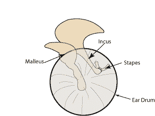
How the Ear Works
The Outer Ear
The auricle (pinna) is the visible portion of the outer ear. It collects sound waves and channels them into the ear canal (external auditory meatus), where the sound is amplified.
The sound waves then travel toward a flexible, oval membrane at the end of the ear canal called the eardrum, or tympanic membrane. Sound waves cause the eardrum to vibrate.
The Middle Ear
The vibrations from the eardrum set the ossicles into motion. The ossicles are actually tiny bones — the smallest in the human body. The three bones are named after their shapes: the malleus (hammer), incus (anvil) and stapes (stirrup). The ossicles further amplify the sound.
The tiny stapes bone attaches to the oval window that connects the middle ear to the inner ear. The Eustachian tube, which opens into the middle ear, is responsible for equalizing the pressure between the air outside the ear and that within the middle ear.
The Inner Ear
The sound waves enter the inner ear and then into the cochlea, a snail-shaped organ. The cochlea is filled with a fluid that moves in response to the vibrations from the oval window. As the fluid moves, 25,000 nerve endings are set into motion. These nerve endings transform the vibrations into electrical impulses that then travel along the eighth cranial nerve (auditory nerve) to the brain.
The brain then interprets these signals, and this is how we hear.
The inner ear also contains the vestibular organ that is responsible for balance.
Ultrasounds
A trained technician, called a sonographer, uses a small, hand-held device called a transducer. The technician presses the transducer against the area being studied and moves it as needed to capture the images. The transducer sends sound waves into your body and collects the ones that bounce back. The images appear on a computer.
Sometimes, ultrasounds are done inside the body. In this case, the transducer is attached to a probe that's inserted into a natural opening in the body. Examples include:
Transesophageal echocardiogram. A transducer, inserted into the esophagus, obtains heart images. It's usually done while under sedation.
Transrectal ultrasound. This test creates images of the prostate by placing a special transducer into the rectum.
Transvaginal ultrasound. A special transducer is inserted into the vagina to look at the uterus and ovaries.
What do we call sound waves we can’t hear?
Infrasound, sometimes referred to as low-frequency sound, describes sound waves with a frequency below the lower limit of human audibility (generally 20 Hz, as defined by the ANSI/ASA S1.
Why are ultrasound waves used to scan a mother’s abdomen?
Because ultrasound is not based on ionizing radiation, it is particularly useful for women of child-bearing age when CT or other imaging methods would otherwise result in exposure to radiation. Ultrasound is the most widely used medical imaging method for viewing the fetus during pregnancy.
Which parts of your body show up on X-rays?
X-rays can be used to examine most areas of the body. They're mainly used to look at the bones and joints, although they're sometimes used to detect problems affecting soft tissue, such as internal organs. Problems that may be detected during an X-ray include: bone fractures and breaks.
Which parts of a baby’s body show up on an ultrasound scan?
Abdomen and stomach
Arms, legs, and other body parts
Back of the neck
Head and brain
Heart chambers and valves
Kidneys
Placenta placement
Spine
Umbilical cord
Urinary bladder
Give two reasons why is it better to use ultrasound instead of an X-ray. Explain
your answers.
Because ultrasound images are captured in real-time, they can also show movement of the body's internal organs as well as blood flowing through the blood vessels. Unlike X-ray imaging, there is no ionizing radiation exposure associated with ultrasound imaging.
Why is a special gel used?
How could a doctor tell the sex of an unborn baby?
Doctors cannot always find out the sex of the baby. Why not?
Why is it useful to have a moving picture?
There are other medical uses of ultrasound. Find some examples.