Lab 2.5 to 2.8 and 3.1 - 3.2
2.5 Appendicular Skeleton
It contains the pectoral girdle and the pelvis girdle
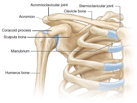
The pectoral girdle consists of the two bones that make up the shoulder scapula nd clavicle bones
Study t terms
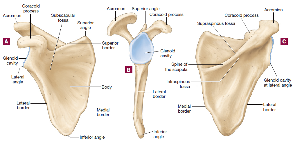
Fossa 2 on back of scapula and front of humorous
Fossa 1 on front of scapula and back of humorous
a before s
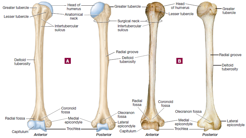
Humerus Bone
Only bone in the arm and forms the glenohumeral joint
Head of humerus - ball like structure and fits into the glenoid cavity
Greater tubercle - biggest “lump” on anterior proximal side of humorous
Lesser tubercle - smaller “lump” on the anterior proximal side of humorous
Intertubercular sulcus - grooves formed from the greater and lesser tubercles, on the anterior side
Deltoid tuberosity - middle “lump” on the humerus
Radial groove - just above the deltoid tuberosity on the posterior side of the humerus
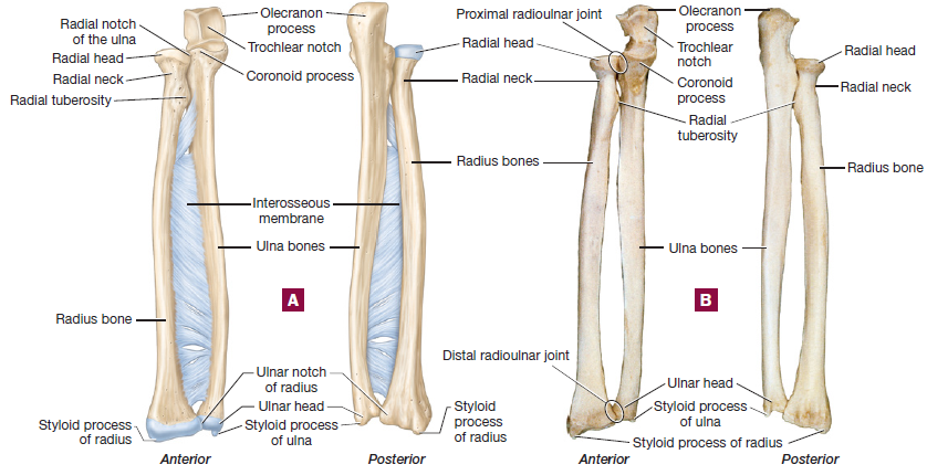
Ulna Bone
Olecranon process - the elbow bone, the most proximal part of ulna
Coroniod process - on the anterior part of ulna, smaller pointer part
Trochlear notch - deep curve below the olecranon process
Ulnar head - most distal pat of the ulna
Styloid process - pointy part distal of the ulna
Radius bone
Radial head - most proximal part of radius bone
Radial neck - just below the radial head
radial tuberosity - proximal “lump” on radius
Syoloid process of radius - most distal part of radius
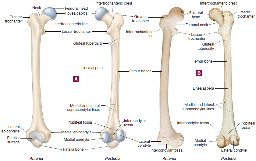
Gluteal tuberosity - rough lines on the posterior side of bone which allows for muscle connection
Linea aspera - line that runs on the posterior side of femur which allows for hamstring muscle to attach
Popliteal fossa - posterior distal side of femur
Intercondylar fossa - posterior side, middle hole in femur bone
Medial and Lateral supracondylar line - just below the Linea aspera
Medial and lateral epicondyle
Medial and lateral condyle - the blue ball looking parts of the femur
Patellar surface - where the knee joint goes U shaped at the distal part of femur
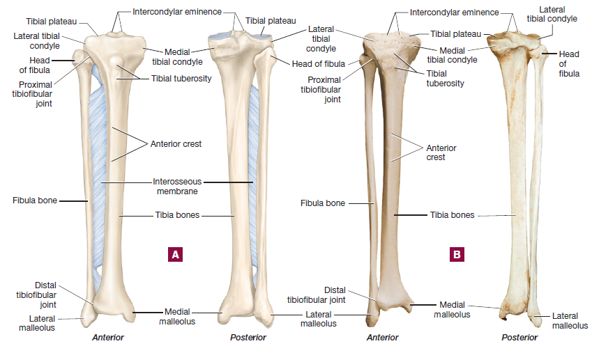
Tibia Bone
Tibial plateau - flat proximal part of tibia
Lateral tibial condyle - outside tip of tibia
Medial tibial condyle - inside tip of tibia
Intercondylar eminence - what forms the pointy parts of the proximal tibia
Tibial tuberosity - boney part that sticks out the tibia
Anterior crest (shin) - smooth anterior part of tibia
Anterior crest articulates with the talus bone (top part of ankle)
Medial malleolus - pointy distal part of the tibia
Fibula Bone
Lateral malleous - distal part of the fibula (pointy part)
2 tibiofibular joints - distal and proximal
Interosseous membrane - what the fibula and tibia are held together by
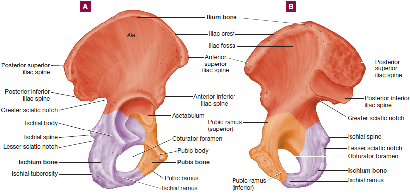
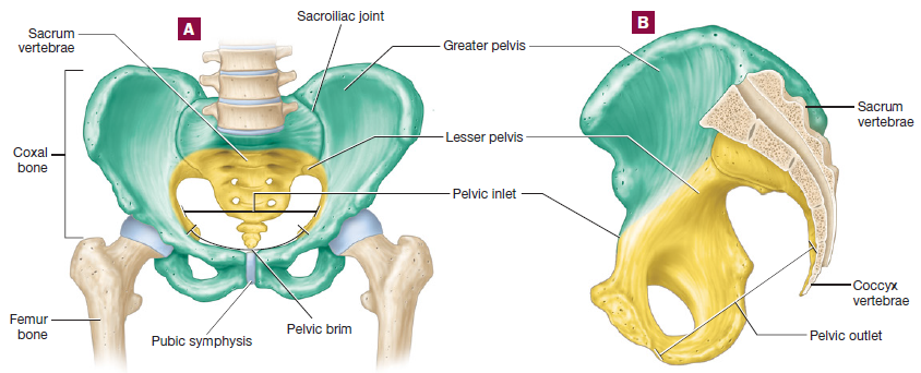
Three paired bones of the pelvic girdle
ilium bone
Ischium bone
Pubis Bone
The pelvis is made up of the sacrum vertebrae & pelvic girdle
While the pelvic girdle is made up of 2 (left & right) coxal bones
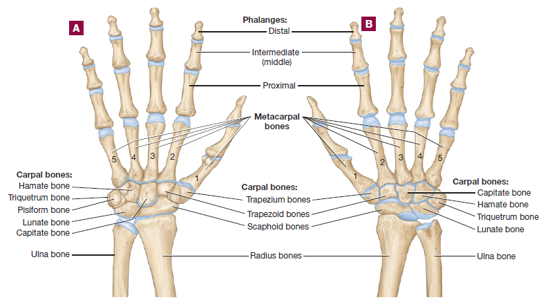
Scaphoid and Lunate bones attach to the radius
Lunate attaches to ulna
So → Scaphoid Attahces to Radius
Long → Lunate Attaches to Ulna
To → Triquetrum
Pinky → Pisform Think Pinky, and is only bone you can see anteriorly
Here → Hamate Ring finger think hooked for life
Comes → Capitate Think middle finger is a capital (capitate)
The → Trapezoid
Thumb → Trapezium Rymes with thumb (trapezium)
2.6 Synovial Joints
Features of a Synovial Joint
Synovial Joints
are freely movable joints
Hyaline Cartilage
provides smooth frictionless surface
Joint Capsule
dense irregular collagenous connective tissue
lined with connective and epithelial tissue called synovial membrane
Synovial Fluid
fills the joint cavity
reduces friction
exchanges oxygen nutrients, and waters with the cells of articular cartilage
Knee joint
Dense irregular collagenous connective tissue
Shoulder Joint
Dense regular collagenous connective tissue
Ligaments: the bones on a synovial joints are held together by ligaments
Extrinsic ligaments: are external to the joint capsule
Intrinsic ligaments: are embedded inside the joint capsule
Tendons: Synvovial joints typically are surrounded by tendons
Bursa: Fluid-filled sacs located between tendons and joints
Range Of Motion of Synovial Joints
Nonaxial Joints: A nonaxial joint is a type of joint where the bones can slide past each other but do not spin. An example of this is where a rib connects to the bone in your back
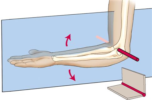
Unaxial Joint: Uniaxial joints are like door hinges that only move in one direction, back and forth. The elbow is a great example; it can bend and straighten but not twist.
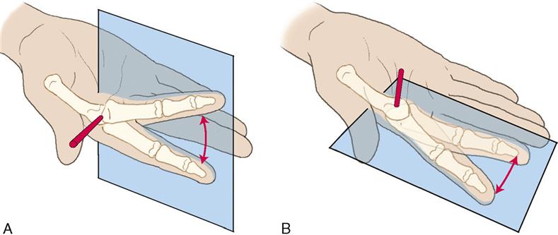
Biaxial Joint: Biaxial joints are like your fingers. They can move in two directions, like bending up and down, and moving side to side. This is why you can wave your finger and move it back and forth.
Multiaxial Joint: Multiaxial joints are like the shoulder and hip joints. They can move in many directions, like a ball rolling in a cup. This means you can raise your arms, spin them around, and move your legs in lots of different ways.

Structural Classification of Synovial Joint
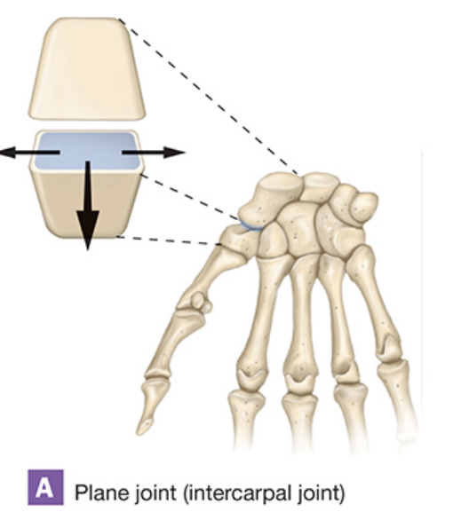
Plane Joint
The ends of the bones that connect at a joint are flat or a little curved. This shape helps the bones slide easily against each other, which is why they're called gliding joints.
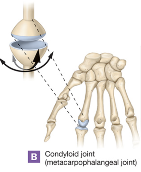
Condylar Joint
A condylar joint is when one bone has a round bump that fits into a round dip on another bone. It's like a ball rolling in a bowl! This shape helps the bones move smoothly together.
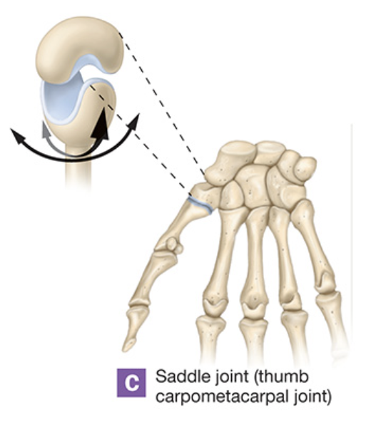
Saddle Joint
Saddle joints are special types of joints in our body. They have two bones that fit together like a saddle on a horse. One side of the saddle is curved in (like the front and back of a saddle) and the other side is curved out (like the sides of a saddle). This helps the bones move in different directions.
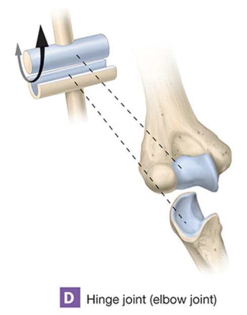
Hinge Joint
A hinge joint is like a door hinge. One part of the bone sticks out a bit, and the other part dips in, just like how a door opens and closes. Good examples of hinge joints are our elbow and knee. They can bend just like how a door can swing open and shut.
Pivot Joint
A pivot joint is a special kind of joint in our body where one bone can spin around another bone. It works like a door that swings open. This type of joint helps with movements like turning your head or your arm. Two examples of pivot joints are the one near your elbow and the one in your neck that helps you shake your head no.

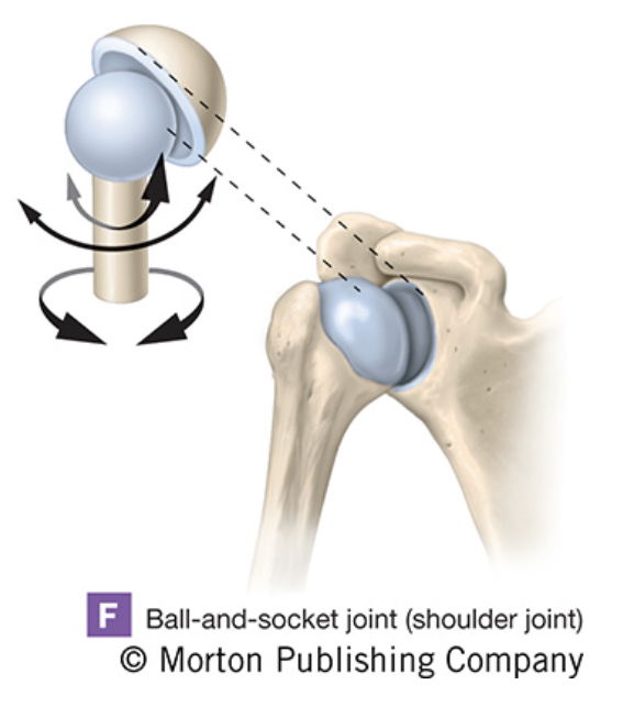
Ball and Socket Joint
A ball and socket joint is like a ball fitting into a cup. This special type of joint is found in your shoulder and hip, letting you move your arms and legs in many directions, just like a ball can roll around in a cup.
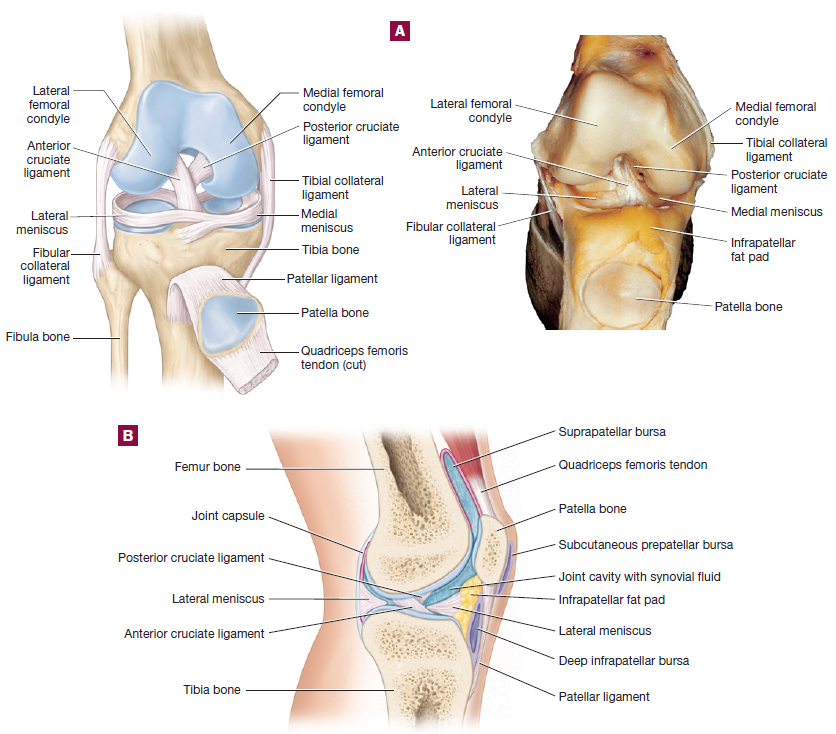
2.7 Knee Joint
The knee joint is a modified hinge joint
Cruciate Ligaments
controls front and back movement of knee joints
Anterior Cruciate Ligament (ACL)
prevents hyperextension of the knee
Posterior Cruciate ligament (PCL)
Stops tibia bone from sliding backwards on the femur bone
Collateral Ligaments
Controls side to side movement of the knee joint
Medial collateral ligament (MCL)
Resists stresses that pull the tibia bone laterally on the femur bone
Lateral collateral ligament (LCL)
Resists stresses that pull the tibia bone medially on the femur bone
3 Bursae in the knee
Suprapatellar bursae
deep infrapatellar burase
subcutaneous bursae
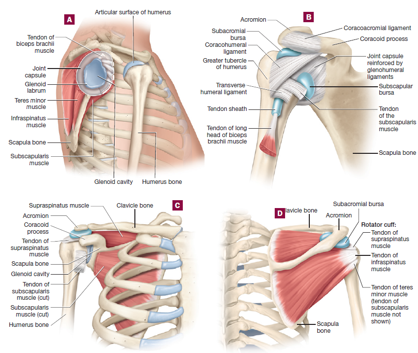
2.8 Shoulder Joint
The shoulder joint is a multiaxial and ball and socket joint
Glenoid Labrum
Fibrocartilaginous ring along the rim of the glenoid cavity
3 Glenohumeral ligaments
reinforces the anterior joint capsule
Coracohumeral ligament
located in the anterior articular capsule between the greater tubercle and coracoid process
Coracoacromial ligament
Located between the coracoid process and acromion
Transverse Humeral Ligament
extends between the greater and lesser tubercles, and forms a tunnel that houses one of the tendons of the biceps brachii muscle
Biceps Brachii Tendon
found in the anterior arm, this tendons passes through the articular capsule of the shoulder joint
Rotator Cuff
group of 4 muscles and tendons - the 3 posterior muscles
supraspinatus (back)
infraspinatus (back)
teres minor muscles (back)
subscapularis muscle (front)
Bursae
2 that help reduce friction in the joint
Subacromial bursa
largest bursae, located below the acromion
Subscapular bursa
3.1 Muscle Tissue Histology
Myology
Study of the muscular system
Actin and Myosin
two proteins that help muscles contract
Three types of muscle tissues
Skeletal (long - voluntary)
Cardiac (small - involuntary)
Smooth (spindle shaped - involuntary)
Striated
overlap of two proteins (actin and myosin)
Non-striated
two proteins not overlapping
Gap Junctions
communication between cells/fibers
Smooth Muscle Tissue: non-striated and involuntary
Smooth muscle tissue
found in sheets or as individual cells
Gap Junction
allows communication between muscle fibers
Cardiac muscle tissue: striated and involuntary
Intercalated disc
constitutes border of each muscle fiber and serves as communication between gap junctions
Skeletal Muscle Tissue: Striated and Voluntary
The entire skeletal muscle tissue is enclosed by a fibrous sheet called the epimysium (outside inside layer)
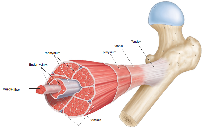
Muscle Fiber
Edomysium - Where muscle fibers are contained
Perimysium - What separates each fasicle
Fasicle - contains groups of muscle fibers
Epimysium - outside inside layer of muscle tissue
Fascia - outside outside layer of muscle tissue
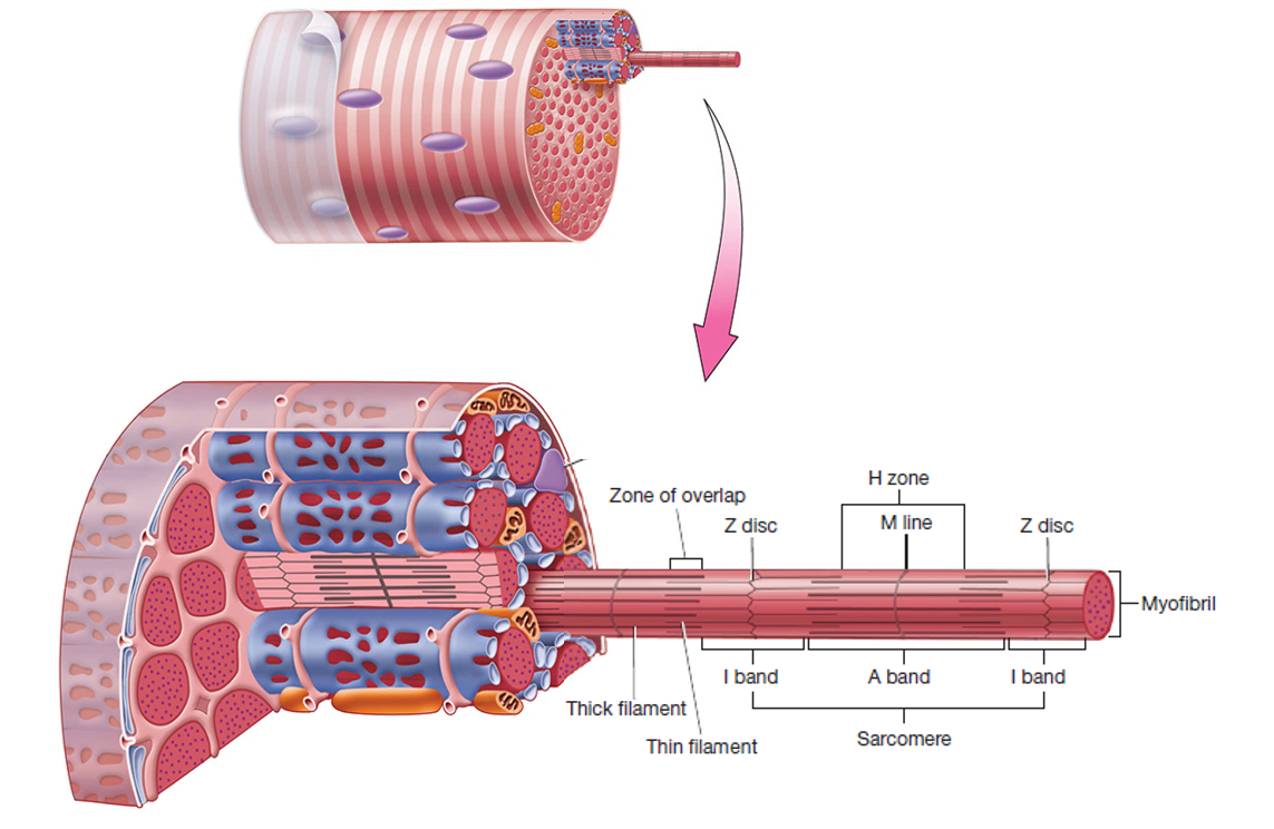
Sarcomere
Smallest functional unit of a skeltal muscle tissue
Mnemonic: ZIMAH
Z disc to z disc forms a sarcomere. One way to remember Z discs are their resemblance of the letter Z.
I bands are the lighter sections on each end of a sarcomere.
M line is the midline in a sarcomere.
A band is the darker section in the center of a sarcomere.
H zone the lighter region in the middle of the A band.
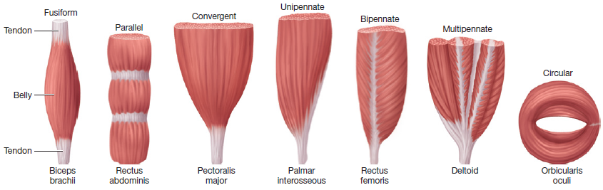
Fusiform
Thick in the middle and tapered at the ends
ex. biceps brachii
Parallel
ex. rectus abdominis
Convergent
Flarred like a wings
ex. pectorallis major
Unipennate
Feather shaped
ex.extensor digitorum longus muscle
Bipennate
Think of a leaf tendon in the middle and fibers flare out
ex. rectus demoris muscle
Multipennate
think leaf with multiple tendons flaring out
ex. deltoid muscle
Circular
arranged in a circle usually around openings (mouth and eye)
ex. oblicularis oris or oculi muscles
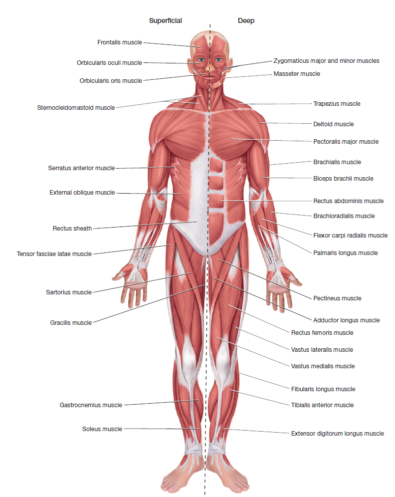
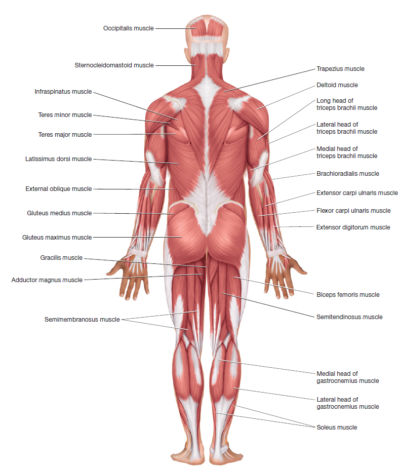
3.3 Axial Skeletal Muscles
A. Scalp Muscles
Galea Aponeurotica: Fibrous connective tissue superficial to the cranium connecting the frontalis & occipitalis muscles
Frontalis Muscle: Superficial to frontal bone
Action: pulls scalp anteriorly, raises the eyebrows & wrinkles the forehead
Occipitalis Muscle: Superficial to occipital bone
Action: pulls scalp posteriorly
B. Orbital & Nasal Region Muscles
Orbicularis Oculi Muscle: Superficial; oculi refers to the area around the eyes
Action: closes the eyelids
Corrugator Supercilii Muscle: Deep; corrugator means to wrinkle & supercilii means eyebrow
Action: frowning muscles by wrinkling the eyebrows
Nasalis Muscle: Superficial to nasal bones
Action: flares the nostrils
C. Oral Region Muscles
Orbicularis Oris Muscle: Superficial; oris refers to the area around mouth; attached to modiolus
Action: closes mouth; puckers lips, & uniquely developed in humans for speech
Zygomaticus Minor Muscle: Superficial; zygomaticus refers to cheekbones; minor means small; located medially to the zygomaticus major muscle, laterally from the levator labii superioris muscle
Action: elevates upper lip for smiling or sneering
Zygomaticus Major Muscle: Superficial; major means big; located superolateral to the orbicularis oris muscle & attached directly to the modiolus
Action: draws angle of mouth up & lateral for laughing
Risorius Muscle: Superficial to masseter muscle; lateral to the lips; attached directly to modiolus
Action: draws angle of mouth lateral for laughing, expressing horror or disdain
Levator Labii Superioris Muscle: Superficial; levator means to elevate, labii superioris refers to upper lip; medially located to the zygomaticus minor muscle & attached superomedial to the orbicularis oris muscle
Action: elevates upper lip
Depressor Labii Inferioris Muscle: Superficial to the mentalis muscle; depressor means downward movement; attached & located inferiolateral to the orbicularis oris muscle
Action: draws lower lip downwards
D. Modiolus
A fibrous tissue located superficial on the lateral side of the lips where facial muscles intersect, like the superficial orbicularis oris, risorius, zygomaticus major muscles, & the deep buccinator muscle
Action: provides stability for mouth movement & facial expression
E. Mental & Buccal Region Muscles
Buccinator Muscle: Deep to masseter muscle
Action: compresses cheeks; aids in food position for chewing or sucking
Mentalis Muscle: Deep; found by the mental region
Action: elevates & wrinkles skin of chin; elevates lower lip as for drinking or pouting
Platysma Muscle: Superficial on the anterolateral side of the neck
Action: creates downward sag of mouth; tenses skin of neck
F. Muscles for Chewing
Temporalis Muscle: Superficial to the temporal bone
Action: closes jaw; elevates & pulls back the mandible
Masseter Muscle: Intermediate; deep to the risorius muscle, superficial to the buccinator muscle
Action: closes jaw; principal muscle for jaw movement