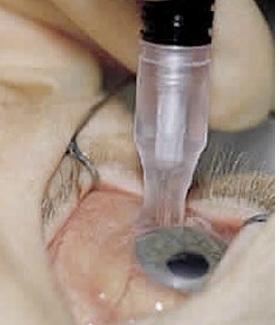
7. Treatment
Treatment Aims
Preserve visual function (acuity and fields)
Preserve the eye (minimise pain)
By reducing intraocular pressure (IOP) to an acceptable level and maintaining it over time
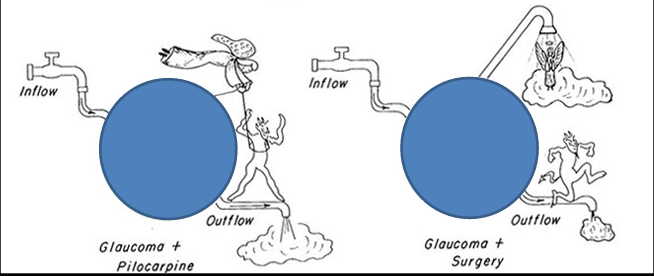
Medical Management Overview
Target IOP depends on optic nerve head (ONH) damage and field loss
On POAG diagnosis, aim to reduce IOP by 1/3 of baseline
Start as monotherapy (fellow eye used as control) and review in 2 weeks (2/52)
If sufficient drop in IOP, commence treatment in fellow eye
If insufficient drop, options include:
Substitute drug
Add another drug (multi-drug therapy or fixed combinations)
Continual monitoring of IOP and visual field (VF)
Failure criteria:
Insufficient lowering of IOP
IOP lowered but VF loss progresses
Non-compliance, side effects, cost, or other factors
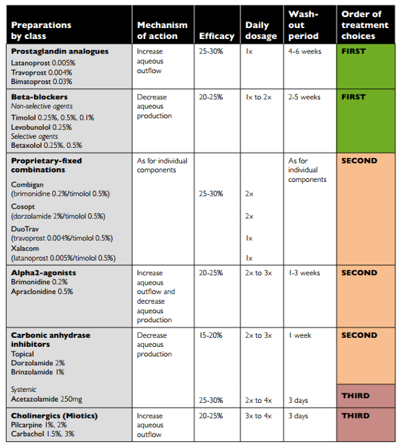
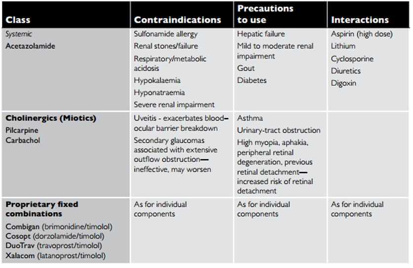
Medications Available in Australia (Table 9.1: Medications in glaucoma management)
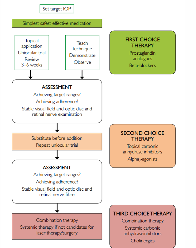
Summary of usage sequence (from table):
FIRST: Prostaglandin analogues; Beta-blockers; alpha2-agonists; fixed combinations may be used
SECOND: Add topical CAIs or alpha2-agonists as needed; consider combinations
THIRD: Add or switch to systemic CAI (acetazolamide) or cholinergics if needed
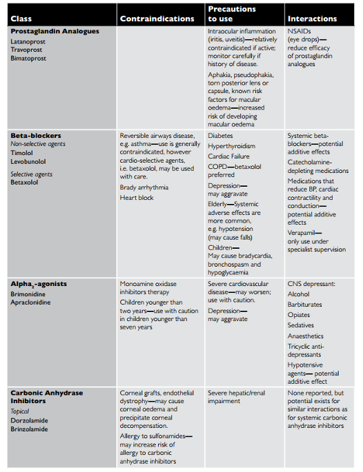
Medications: Contraindications, Precautions, and Interactions (Table 9.3)
Surgical and Laser Management: When Medical Therapy Isn’t Enough
When maximum medical therapy (MMT) fails or is no longer tolerated:
Open the angle
Cataract extraction (Cat & IOL)
Increase/promote outflow through an existing pathway
Peripheral iridotomy/iridectomy (PI)
Argon/Selective Laser Trabeculoplasty (ALT/SLT)
Goniotomy
Create a new outflow pathway
Filtration surgery (Trabeculectomy)
Drainage device (Molteno tube)
Turn off the tap
Cyclodestructive procedures (Trans-scleral laser cyclodiode)
Cataract Surgery and IOL in Glaucoma Care
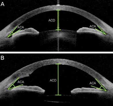
Cataract extraction with IOL can:
Increase anterior chamber depth
Widen the anterior chamber angle
Evidence on cataract removal in glaucoma management is mixed; some controversy remains regarding outcomes in certain glaucoma patients
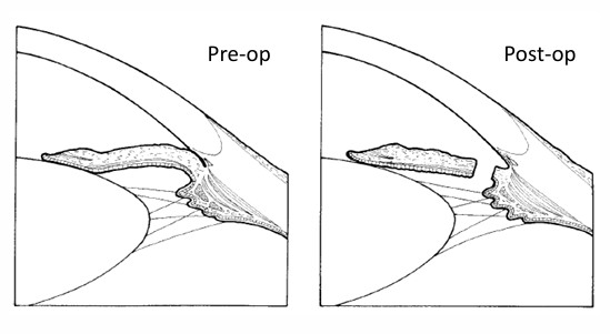
Peripheral Iridotomy vs Peripheral Iridectomy
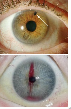
Peripheral Iridotomy (PI) with YAG laser
Indications: Primary angle closure; narrow occludable angles
Technique: Performed under upper eyelid to minimize glare/monocular diplopia; post-op topical steroid for ~1 week
Peripheral Iridectomy
Indications: Primary angle closure with pupil block
Function: Creates a broader pathway for aqueous to enter the anterior chamber
Laser and MIGS Options
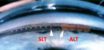
A/SLT (Argon/Selective Laser Trabeculoplasty)
Mechanism: Create discrete burns to trabecular meshwork to enhance outflow
Efficacy: Modest IOP reduction; typically less than 30%;
Often not sufficient as a standalone treatment and may require ongoing medical therapy
useful for non-compliance
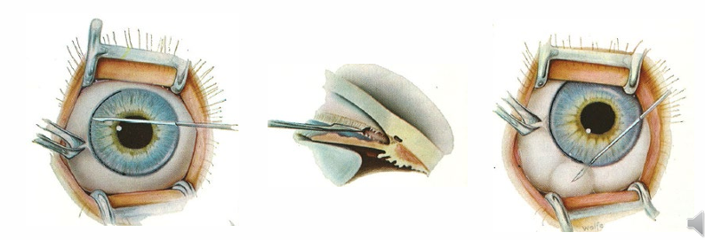
Goniotomy
Indication: Developmental glaucoma with abnormal drainage network
Mechanism: Sweep incision through trabecular meshwork to improve outflow
Potential long-term complications: Corneal decompensation in some cases
Trabeculectomy (trab)
Filtering/drainage surgery
MMT and A/SLT failure
Creates a new pathway for aqueous outflow from AC to sub-tenon’s space
Significant (typically long term) IOP reduction
Antimetabolites to reduce scarring
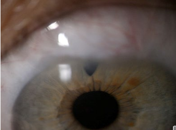
Artificial Drainage shunts
plastic tube which creates a pathway from the AC to sub-Tenon’s space
Failed trabeculectomy or trab not possible
Potential for complications high:
Excessive drainage -> collapsed AC
Tube blockage (blood, iris) -> IOP
Exit bleb seals -> IOP
Tube erosion through conjunctiva
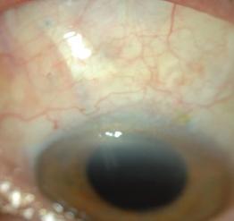
MIGS (Minimally Invasive Glaucoma Surgery)
Stent-based approaches to increase outflow, designed to be less invasive
For patients undergoing cataract surgery
Goals: Reduce medication burden or achieve medication-free control in suitable patients
Not intended for malignant glaucoma or patients on maximum medical therapy (MMT)
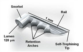
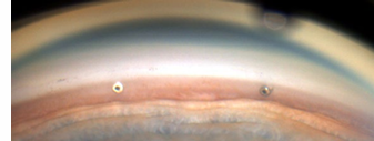
I-Stent (Australia)
Status: The only device currently approved in Australia (as per provided material)
Design: ~1 mm stent implanted in Schlemm’s canal; typically placed into a nearby collector vessel around 9 o’clock position in the eye canal
Cyclodiode:
Lower IOP by reduction of aqueous production (ablation of the ciliary body processes epithelium)
Uncontrollable with MMT or surgery
Often end stage glaucoma
Pain relief, save the eye (even if blind)
Major complications (pthisis/hypotony
last line of defence