Neurology Test 2
PNS Efferents - Autonomic Nervous System
Describe the basic functional and structural organization of the autonomic nervous system.
Efferent division → Autonomic nervous system → Sympathetic & Parasympathetic
Works on the internal environment
Preganglionic neuron: Synapses with the cell body of the postganglionic fiber in a ganglion outside the CNS
Postganglionic neuron: Sends axons that end on the effector organ
Effector organ: Organ affected by the NT (ex: Smooth muscle, cardiac muscle, intestine, etc.)
Somatic Autonomic (Visceral) |
Anatomy (Motor neurons) 1 Motor neuron: Cell body in the spinal cord 2 Motor neurons:
|
What environment does it work on? External environment Internal environment |
Differentiate sympathetic and parasympathetic anatomy, outflow, and neurotransmitters.
Sympathetic Parasympathetic |
Domination Fight or flight Rest and digest |
What does response prepare the body for? Strenuous physical activity Body-maintenance activities |
“Thoracolumbar” “Cranial sacral” |
Role in Dual Innervation Coordinated innervation Direct innervation of target organ Specific control of target neurons |
Length of preganglionic neuron Short Long |
Location of autonomic ganglion In the tissue |
Nerves involved T & L Cranial (III, VII, IX, X) & Sacral |
Parasympathetic:
Most pre/postganglionic neurons pass uninterrupted, directly to the target organ. Specific innervation
Anatomy
Preganglionic neurons involved:
Carry sensory & motor information (both directions)
Oculomotor nerve (III): Ciliary muscles, pupillary sphincter muscle of the eye (dilation/expansion of pupil)
Facial nerve (VII): Submandibular & sublingual salivary, lacrimal (tear duct) & nasal glands
Glossopharyngeal nerve (IX): Parotid glands
Vagus nerve (X): Carries 75% of preganglionic parasympathetic axons. Parasympathetic innervation to thoracic & abdominal region
Sacral spinal cord (S2-S4):
Project via the pelvic nerves to pelvic ganglia located in the bladder, ureters, descending colon, rectum, and reproductive organs
Postganglionic neurons:
In a ganglia near or with the walls of the effector organs
Sympathetic:
Two neuron chain: CNS → autonomic ganglion → effector organ
Dual innervation: Nearly all organs are impacted by both PsNS and SNS
Anatomy
Preganglionic neurons:
Bilateral, in spinal segments T-1 to L-2
Splachchnic nerves to celiac, hypogastric
Ganglia is typically outside of the target
Postganglionic neurons:
Sympathetic chain ganglia:
Innervates smooth muscle of blood vessels and piloerector muscles, sweat glands
Innervate cardiac muscle, smooth muscle of bronchi and iris
8& of nerve fibers within spinal nerves
Prevertebral ganglia:
Celiac ganglion: Ureter, stomach, adrenal, kidney
Hypogastric plexus: Bladder
Adrenal Medulla
Key area for projection of epinephrine and norepinephrine
Allows E and NE to stimulate structures of the body that are not innervated by direct sympathetic fibers
They can reach very distally because they are going through the blood
Chromaffin cells: Synthesis of epinephrine
Understand basic neurotransmission for the ANS and be able to differentiate neurotransmission in the parasympathetic versus sympathetic divisions.
Impact Sympathetic Parasympathetic |
Increase Pupil size (Dilation) Heart rate Contractility (heart) Airway diameter Sweat Salivation Lacrimation Urination Digestion Defecation |
Decreases Salivation GI secretion GI motility Urination Digestion Defecation Heart rate Airway diameter Pupil diameter |
Autonomic neurotransmission
Pregnaglionic fibers: ACh
Parasympathetic - Postganglionic fibers: ACh
Sympathetic - Postganglionic fibers: Most are adrenergic (NE/E), some are ACh
Adrenergic Receptors (GPCR)
Ligand: Norepinephrine or epinephrine
Excitatory or inhibitory depending on the receptor and system
A1 Generally excitatory |
A2 Generally Inhibitory |
B1 Generally excitatory |
B2 Generally inhibitory |
B3 Only found on brown adipose tissue (thermogenesis) |
Activity termination:
Reuptake by the neuron that released it
Enzymatic inactivation
COMMENT
MAO
Define the autonomic nervous system's effects on specific effector organs. Differentiate parasympathetic versus sympathetic effects.
Organ System Action |
Eye Parasympathetic Pupil Constriction Lens accommodation |
Sympathetic Pupil dilation |
Heart Parasympathetic Decreased heart rate |
Sympathetic Increased heart rate |
Lacrimal gland Parasympathetic Increased secretion |
Sweat Glands (mAch receptor) Sympathetic Increased secretion |
Salivary gland Parasympathetic Increased secretion |
Sympathetic Decreased secretion |
Gastrointestinal tract Parasympathetic Secretion Peristalsis Sphincter relaxation |
Sympathetic Inhibits secretion Inhibits motility Sphincter contractionn |
Bladder Parasympathetic Detrusor contraction (causes urination) Internal sphincter relaxation |
Sympathetic Relax detrusor muscle Contract internal sphincter |
Bronchi/bronchioles Parasympathetic Constriction |
Sympathetic Increased diameter (relaxed) |
Understand the idea of an autonomic reflex and what functions utilize a reflex.
Autonomic reflexes allow the ANS to maintain visceral homeostasis
Reflex pathway
Receptor → Sensory neuron → Integrating center → Motor neuron → Effector → Response
Mass discharge (Adrenaline Rush)
The sympathetic nervous system discharges simultaneously as a complete unit
Initiated by: Hypothalamus in response to fear, severe pain, or rage
What are CNS control centers for the ANS?
Cerebrum/limbic system: Thoughts/emotions can influence ANS function via the hypothalamus
Hypothalamus: ANS Integration Center
Nuclei monitor homeostatic things
Directly initiates autonomic responses through direct projections onto preganglionic neurons.
This is done to return to a homeostatic state or go into fight/flight mode
Regulates autonomic responses through projections to autonomic centers in the brain stem
Brain stem: ANS reflex center
Spinal cord: ANS reflex center
Urination, defecation, reproductive behaviors
Parasympathetic vs Sympathetic Summary
Parasympathetic Sympathetic |
Oversimplified function “Rest and digest” “Fight or flight” |
Neurotransmitter ACh ACh, EPI, NE |
Preganglionic neuron Craniosacral Thoracolumbar |
Location of ganglia Close to the organ Close to the cord |
Preganglionic neuron length Long Short |
Postganglionic neuron length Short Long |
Connectivity Specific Ramified |
Regulation of end organs Independent Coordinated |
Essential for life?? Yes No |
PNS Efferents: Motor Control
Define and differentiate the 3 types of movement
Voluntary (Cortex, cerebellum, basal ganglia)
Cerebral cortex: Motor control
Premotor cortex: Mental planning & “staging” of movement
Anterior forms image → posterior translates
Supplemental motor cortex: Integrates movement, “background movements”
Primary motor cortex: Execution of movement
Directing motor neurons to send a contraction message
Primary motor cortex → motor neurons
Basal Ganglia
Nuclei of the basal ganglia:
Striatum: Caudate + putamen
Globus pallidus
Substantia nigra
Subthalamic nucleus
Cerebellum
Red nucleus: Alternative tract to the spinal cord, allows for movement without the primary motor cortex.
Accessory route
Closely associated with cerebellar function
Controls: distal motor groups, upper limbs (things more distal)
Reticular formation
Ponds
Thalamus
Involuntary (spinal cord, reflexes)
These motions happen without a real conscious awareness
Spinal cord
The ventral horn is involved
Rhythmic motor patterns
Know the 4 major CNS regions involved in the integration of motor function
Cerebellum
Cerebral cortex
Basal ganglia
Thalamus
Differentiate the role of CNS regions involved in voluntary movement
Motor cortices: Where is the motor plan formed, translated, or executed?
Planning: Prefrontal cortex
Translation: Premotor/supplemental cortex
Execution: Primary motor cortex → Spinal cord
Understand the basic anatomy and role of the cerebellum in motor control
Cerebellum: Controls timing and coordination of movement (Comparator)
Evaluate what you intend to do & what you do, if these don’t align → cerebellum adjusts
Stimulate the cerebellum? : Nothing happens, but without it, there is a major impact
Differentiate the neuroanatomy and contribution of the Basal Ganglia to motor control.
Direct basal ganglia pathway
Input: Glutamatergic from the cortex
Result: Increase activity (enable movement)
Receptor involved: D1
Indirect basal ganglia pathway
Input: Dopamine from SNc
Result: Decrease activity (inhibit movement)
Receptor involved: D2
Define the corticospinal tract and differentiate from DCML, ALST tracts
Corticospinal tract: Direct pathway to the spinal cord
Controls: Speed and precision of fine motor control
Pathway: Cortex → spinal cord
Most fibers cross the medulla and synapse on interneurons in the spinal cord.
Cells: Betz cell (giant pyramidal cells)
Responsible for direct connections
Red nucleus: Indirect pathway to the spinal cord
Cortex → red nucleus → reticular formation → spinal cord
Be able to identify symptoms of damage to the 3 motor brain regions
Supplemental and premotor areas (hand skills): Motor apraxia
Broca’s area: Base of premotor area
Cortical damage
Positive signs
Spasticity: Too much contraction
Exaggerated spinal reflexes
Babinski response
Negative signs
Hypotonia: Loss of muscle tone
Loss of sensation
Apraxia: Inability to carry out a skilled movement
Aphasia: Inability to produce words
Cerebellum damage
Ataxia: loss of coordination
Past pointing: Moving beyond the point of intention
Intention tremors: AS you're going to move, signals don’t align (ex: both extender and flexor contract)
Hypotonia: Loss of muscle tone
Failures of progression: Coordination error
Understand spinal cord anatomy for motor function
Same as sensory system dermatomes
Myotomes
Cervical (C1-C8): Head neck, shoulders, parts of upper arms, and hands
Thoracic (T1-T12): Arms, hands, and trunk
Lumbar (L1-L5): Waist, thighs, legs, and part of feet
Sacral (S1-S5): Back of legs, buttocks, anus
Sacrococcygeal: Tailbone
White vs Grey matter
White: Tracts
Grey: Unmyelinated neurons
Cell bodies of neurons (interneurons, lower motor neurons)
Types of Cells
Interneurons: Grey matter integration
Motor neurons
Alpha motor neurons (A alpha): Causing contraction
Large
Branches to innervate extrafusal muscle fibers
Alpha motor neuron + extrafusal fibers = motor unit
Gamma motor neurons (A gamma): Helping sensitivity and sensory receptors
Small & fewer than alpha
Innervates intrafusal muscle fibers
Controls muscle tone (for sensitivity)
Interconnections between motor neurons and interneurons are responsible for most of the integrative functions of the spinal cord.
Know muscle essentials for motor control.
Voluntary movement: Information flow
Sensory cortex (detects)
Sensory association cortex (integrate it)
The prefrontal cortex (plan)
Premotor/supplemental cortex (image/translate it)
Primary motor cortex (execution center)
Spinal cord (act)
Skeletal muscle basics
Tendon: Muscle-bone connection
Ligament: Bone-bone connection
Flexion: Contract a flexor muscle → closes joint angle (ex: biceps)
Extension: Contract an extensor muscle → opens a joint angle (ex: tricep)
Extrafusal muscle fibers: Outer muscle layer (what we think of when we think “muscle”)
Intrafusal muscle fibers: Inner muscle layer
Does not contract
Helps form receptors
Controls sensitivity
Differentiate the muscle sensory receptors.
Structure and function (what each one senses)
Muscle spindle
Detects stretch and speed of stretch
Innervated by: Gamma motor neuron
Comes in & winds around the intrafusal muscle fibers
Intrafusal muscle fibers have no actin or myosin → it cannot contract
Attached to extrafusal muscle fiber at each end
Sensory receptor detecting stretch of the muscle & sends it to CNS
Proprioception
Golgi Tendon organ
Tension
Connections to the spinal cord and/or CNS
Muscle spindle
Innervated by gamma motor neuron
Wraps around intrafusal muscle fibers (no contraction here)
Attached to extrafusal muscle fiber
Golgi Tendon organ
Encapsulated at the end of muscle fibers as they attach to tendon
Synpase location: Interneuron
Role in reflexes
Muscle spindle
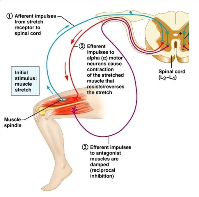
Monosynaptic stretch reflex: One synapse
Sensation: Muscle stretch
Golgi Tendon organ
Lengthening (relaxation) reflex that is protective
Entirely inhibitory
Equalizes contracting force
Some fibers have more tension than others
Have a general knowledge of the 3 reflexes circuits presented, and especially the role of each in movement.
Reflex Arc
Sensory neuron: Detects stimulus (muscle stretch)
Interneurons - (most often) can be excitatory or inhibitory
Sometimes this bypasses
Motor neurons: Muscle contraction
Muscle Stretch Reflex
Components involved
Alpha motor neuron: Directs contraction
Sensory stimulus: Muscle spindle stretch
Synapse location: Directly on an alpha motor neuron
Mechanism
Multiple circuits are involved
Stretch reflex circuit (monosynaptic)
Working on “primary” muscle (ex: Quad)
Splits in the spinal cord
Continues circuit
Synapses at reciprocal inhibition pathway
Reciprocal inhibition
Working on the opposite muscle (ex: Hamstring)
Lateral inhibition circuit: Focuses the signal
Flexor withdrawal reflex: Pain reflex
Mechanism
Excitatory interneuron stimulates contraction
Inhibitory interneuron inhibits opposing muscle pair
Divergent interneuron sends info to the brain
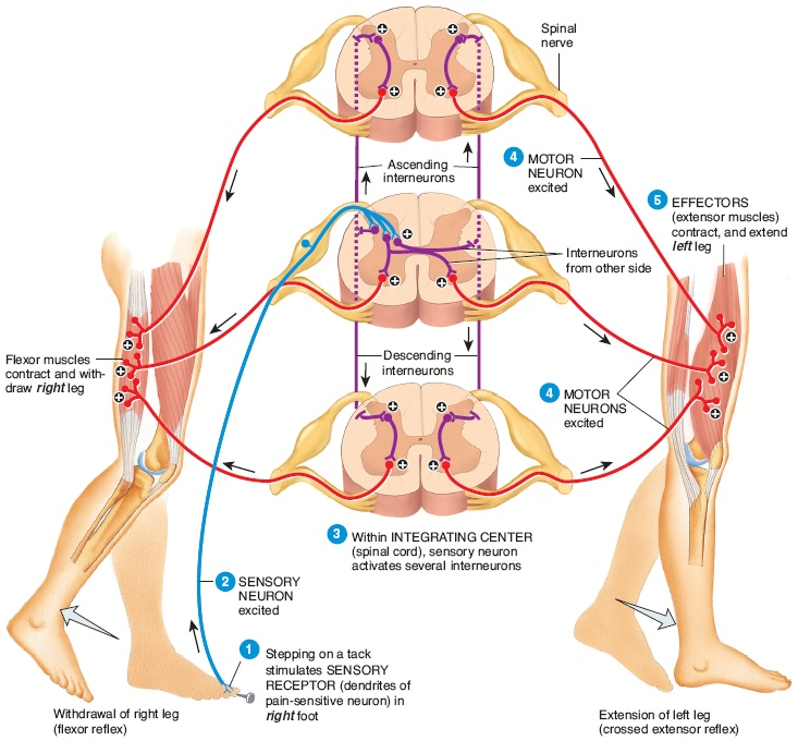
Crosses over & goes to the brain for pain recognition
Pain is synapsing on multiple interneurons
Crossed extensor reflex
Involves the compensation of the opposite limb to the flexor withdrawal reflex
Ex: Step on something → lift foot → take a step back with the other foot
Interneurons cross to the contralateral gray matter activate extensors and inhibit flexors
Delayed effect: Highlights the role of many interneurons
Skeletal Muscle Physiology
Differentiate skeletal muscle from cardiac and smooth
Skeletal muscle
Striation? Striated
Movement: Voluntary
Nucleus: Multiple nuclei in a single cell
Location: Attached to bones
Function: Movement of the body about the external environment
Cardiac muscle
Striation: Striated
Movement: Involuntary
Nucleus: Multiple nuclei in a single cell
Location: Wall of Heart
Function: Pumping blood out of the heart
Smooth muscle
Striation? Unstriated
Movement: Involuntary
Nucleus: Single nuclei per each cell
Location: Walls of hollow organs and tubes
Function: Movement of contents within hollow organs
Understand the key elements of the neuromuscular junction and how they drive muscle contraction
Neuromuscular junction: Where the terminal buttons of the one lower motor neuron synapse to the muscle fiber
Motor unit
Neurotransmitter: ACh
What stops ACh?: Acetylcholinesterase
Formation of the endplate potential
Always excitatory
Initiation of an action potential
Relationship to sarcolemma/transverse (T) tubules
Steps at a neuromuscular junction
AP comes down the axon and is propagated to the terminal
Action potential triggers the opening of voltage-gated Ca2+, Ca2+ flows in
Ca2+ triggers the release of ACh by exocytosis from a portion of the vesicles
ACh diffuses across and binds with nicotinic ACh receptors on the motor end plate of the muscle cell membrane
Binding of ACh to its receptor opens these nonspecific cation channels, leading to a relatively large movement of Na+ into the muscle cell and a small movement of K+ outward
The influx of Na+ results in a depolarization called an end-plate potential. Local current flow occurs between the depolarized end plate and the adjacent membrane
Local current flow opens voltage-gated Na+ channels in adjacent membrane
The resultant Na+ entry drives the potential to threshold, initiating an action potential, which is propagated through the muscle fiber
ACh is subsequently destroyed by acetylcholinesterase, an enzyme located on the motor end-plate membrane (blue dots), acetylcholinesterase terminates the muscle cell’s response
Know the muscle anatomy necessary for muscle contraction
Muscle → muscle fiber → myofibril → Dark A band & light I band
Sarcomere: The basic functional unit of muscle fiber (from Z line to Z line)
Thick and thin filaments are coming together to cause the contraction of the muscle
Z line: thin filaments coming together
A band: thick filaments
I band: thin filaments
Titin filaments: Serves as scaffolding along with M-line proteins to stabilize the thick filaments as they attach to the Z disk
Acts as an elastic spring to augment the muscle’s elasticity & helps the muscle recoil after a stretch
Thick filaments: Made up of myosin filaments
Thin filaments: Made up of actin filaments
Tugs on the thick filaments when excited, which causes the contraction
Differentiate roles of myosin and actin (thick and thin filaments)
Each thick filament is surrounded by 6 thin filaments
Each thin filament is surrounded by 3 thick filaments
Myosin (thick filament)
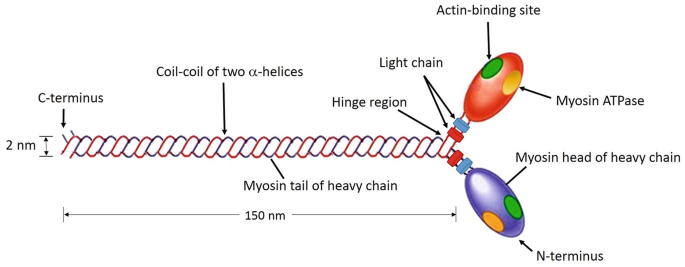
Thick filaments are made up of numerous myosin molecules
Myosin heads:
Binds to active sites on the actin molecules to form cross-bridges
The hinge region can bend and straighten during contraction
ATPase enzymes break down ATP & release energy that is used to bend the hinge region of the myosin molecule during contraction.
Actin (thin filament)
Thin filaments are made up of numerous actin molecules held together by troponin and tropomyosin
Relaxed state
No cross-bridge binding occurs
The active site is blocked
The troponin-tropomyosin complex is blocking the active site
Ca2+ is not present
Excited state
Cross-bridge binding occurs
The active site is no longer blocked
Ca2+ is being released (because the muscle fiber is excited)
Ca2+ binds to the troponin-tropomyosin complex & exposes the active site
Contraction: Myosin (golf club) heads pull on the actin
Troponin
There is one troponin bound to each tropomyosin
In the relaxed state:
Troponin is blocking the active sites of the actin (which would bind to the myosin)
Troponin is inactive
In the excited state:
Troponin moves from blocking the active site of actin, thus myosin can bind
Troponin is activated
Ca2+ binds to troponin, which pulls the complex aside & renders it active.
What is meant by sliding filament mechanism, cross bridge activity, and power stroke?
Sliding filament mechanism
Contraction is accomplished by thin filaments from the opposite sides of each sarcomere sliding closer together between the thick filaments.
This shortens muscle length & causes tension.
Relaxed
H zone: Wide, wide banding power
I band: Wider
A band: Same width
Overall sarcomere: Wide
Contracted
H zone: Shorter
I band: Shorter
A band: Same width
Overall sarcomere: Shorter
Power stroke
Stroking motion pulls (bends) the thin filament toward the center of the sarcomere.
Active site on actin is exposed
Myosin head forms a cross-bridge with actin
Myosin head bends, and ADP and phosphate are released
A new molecule of ATP attaches to the myosin head, causing the cross-bridge to detach
ATP hydrolyzes to ADP + P, which returns the myosin to the “cocked” position
Cross-bridge activity
ATP Necessary
Presence of Ca2+, troponin changes shape
All cross-bridge stroking is directed toward the center of the thick filament
Simultaneous pulling inwards of all 6 thin filaments surrounding a thick filament is occurring
Binding: Myosin cross-bridge bins to the actin molecule
Ca2+ must be present for this
Power stroke: Cross bridge bends, pulling thin myofilament inward
Detachment: Cross bridge detaches at the end of the power stroke and returns to the original conformation
Binding: Cross bridge binds to more distal actin molecule; cycle repeats
Essentially is hoping down the line of actin molecules every cycle repeats
Understand excitation-contraction coupling and the role of Ca2+ and ATP in this process.
Calcium
When present: Pulls troponin-tropomyosin complex away from the active site on the actin (thus allowing it to bind to myosin)
When absent: Troponin-tropomyosin complex is blocking the actin active site (it cannot bind to myosin)
Where does the calcium come from?
Sarcoplasmic reticulum & T tubules
T tubules: Membranous, perpendicular extensions of the surface membrane that gid deep into the muscle fiber at the junctions between the A and I binds of the myofibrils
The action potential travels down here quickly.
Sarcoplasmic reticulum: Fine, membranous network that runs longitudinally and surrounds each myofibril, with separate segments encircling each A band and I band.
Excitation-Contraction Coupling
Ca2+ is necessary for this to occur
AP at NMJ causes release of ACH → AP in the muscle fiber
AP moves across the surface membranes into the interior through T tubules. APs in the T tubules trigger the release of Ca2+ from the sarcoplasmic reticulum
Ca2+ binds to troponin on thin filaments
This allows the actin/myosin to bind
Tropomyosin shifts, revealing myosin cross-bridge sites
Myosin cross-bridges attach
Power stroke (ATP used)
Cross bridge detaches
Still more Ca2+? → Return to step 5 (do another power stroke!)
When APs stop, Ca2+ is taken back up by the sarcoplasmic reticulum. Contraction stops & thin filaments passively reset
Skeletal muscle pathophysiology & movement disorders
Pathophysiology: For each disorder differentiate based on: Definition, basic epidemiology, key symptoms, essential pathophysiology (basic mechanism), and any issues pertinent to pharmacy
Rhabdomyolysis
Idiopathic
Definition: Rare life-threatening muscle degenerative disease
Caused by: overexertion, trauma, toxins, dehydration, medications/drug interactions, prolonged bed rest
Epidemiology: Rare
High risk: Jobs that are outside with excessive heat/exertion, elderly, people taking statins
Statin risk factors: high dose, age, female, renal or hepatic insufficiency, diabetes, taking with other p450 inhibitors
Key symptoms: Myalgia, muscle weakness, dark urine, >11x creatine kinase in blood
Essential pathophysiology: Muscle cell disintegration → myoglobin release increases → kidney failure/death
Increased myoglobin = too much Ca2+ (BAD!)
Pertinent issues: Recent cases
Peripheral neuropathy
Symptomatic or idiopathic
Definition: Umbrella term for over 100 conditions that involve damage to the nerves of the PNS
Epidemiology
Key symptoms: No key symptoms
Symptom severity, progression, and prognosis depend on the cause and type of nerve damage.
Predominantly motor: Muscle weakness/atrophy, cramps, twitching
Sensory: Loss of somatosensation (typically hands/feet)
Sensory-motor
Autonomic (excess sweating, heat intolerance, GI symptoms, BP)
Essential pathophysiology
Pertinent issues
The most common cause is diabetes
Parkinson’s disease
Parkinsonian: Iatrogenic
Can be trauma-induced
Parkinson’s disease: Idiopathic
Definition: Slow progressive degeneration of the nigrostriatal dopamine pathways
Epidemiology: 500,000+ affected
Risk factors:
Age (onset at 60 years)
Gender (male > female)
Key symptoms: Falls become likely
Motor signs: TRAP
Tremor
Rigidity
Akinesia (bradykinesia): slow movement
Posture reflex
Festinating gait: gait with short steps
Hypophonia: quiet speaking
Dysarthria
Dysphagia: Chewing issues
Hypomima
Essential pathophysiology: Slow progressive degeneration of the nigrostriatal dopamine pathway
Disease of the basal ganglia
Substantia nigra: Cell bodies of dopamine producers
Premotor phase
Duration: 5-10 years
Diagnosis? No
Symptoms (precursor):
Hyposmia
Constipation
REM-sleep behavior
Dopamine: progressive degeneration
Motor symptoms: Minimally present (if present)
Non-motor symptoms: Minimally present, but more than motor
Symptomatic PD
Duration: Does not get cured once diagnosis occurs
Diagnosis: Yes
Symptoms (progression):
Cognitive impairment
Postural imbalance
Dysarthria
Dopamine: Progressive degeneration
Diagnosis typically occurs when 80% of the nigral DA levels are lost
Motor symptoms: Rapid increase in presence
Non-motor symptoms: Gradual increase
Pertinent issues
Apply knowledge of dopamine systems to understand the strengths and weaknesses of the DA theory of Parkinson’s disease
Parkinson’s disease likely affects more in the brain than just dopamine. Also, other diseases can affect dopamine receptors
Indirect vs direct paths in the basal ganglia
Direct pathway
Normal conditions:
Glutamatergic input from the cortex to the D1 receptor
Increases activity
Parkinson’s disease:
Glutamatergic input from the cortex to the D1 receptor
Constant activity
Indirect pathway
Normal conditions:
Dopamine input from SNc to D2 receptor
Inhibits activity
Parkinson’s disease:
Loss of dopamine input
Loss of activity inhibition
Normal conditions (summary):
Voluntary movement executed by the primary motor cortex
Glu → spinal cord & brainstem
Nigrostriatal tract
SNpc → striatum
D1 (GABA) → GPi/SNpr
D2 (GABA) → GPe (GABA) → STN (Glu) → GPi/SNpr
BG input to the cortex via the thalamus
GPi/SNpr (GABA) → Thalamus (Glu) → Cortex
Parkinson’s disease (summary):
Voluntary movement executed by the primary motor cortex (to the spinal cord and brain stem)
Nigrostriatal tract (reduced activity!)
No DA to Striatum (Works on D1 and D2)
D1: less active (GABA) → Increased GPi/SNpr activity (GABA can’t work on it to inhibit!)
D2 : Increased activity (GABA) → Decreased GPe (more GABA working on it) → Increaed STN (b/c less GABA working on it) → GPi/SNpr Increased (STN releases Glu)
BG input to the cortex via the thalamus
Parkinson’s: D1 (excitatory) remains the same, but D2 (inhibitory) is nonfunctional, so there is constant excitation → shakiness, stiffness, etc.
Parkinson’s treatment: Differentiate the mechanism of action of the primary treatment approaches by drug classes (DA replacement therapy, DA agonists, muscarinic antagonists)
DA replacement therapy: Increasing dopaminergic neurotransmission
Give precursor → crosses BBB → Converted to DA inside the brain
DA agonists: Increasing dopaminergic neurotransmission
Mimics DA
D2 specific
Muscarinic antagonist: Anticholinergic (treating the tremor)
Appreciate why dopamine is a delicate balance in CNS function
In Parkinsons, the balance between excitation and inhibition in the basal ganglia is lost
Tardive dyskinesia
Iatrogenic
Definition: Involuntary, repetitive excessive movements, especially around the mouth and jaw
Key symptoms: Involuntary, repetitive excessive movements
Tends to be around the mouth & jaw very often
Essential pathophysiology: Prolonged dopamine receptor antagonism
Prolonged DA receptor antagonism → DA receptor function change
Pertinent issues: Causing tardive dyskinesia while treating other psychiatric conditions
Treating psychiatric conditions typically involves antipsychotics (typically block dopamine receptors), some anticholinergics, and SSRIs. If you antagonize the dopamine receptors for an extended period, tardive dyskinesia can result
It can be reversed
Myasthenia gravis
Idiopathic & iatrogenic
Definition: Autoimmune disorder of neuromuscular transmission, that results in skeletal muscle weakness
The small muscles around the face & eyes are affected greatly
Epidemiology: Rare (150-205/1mil)
Risk factors:
Family history of autoimmune disease,
Men > 60; women<40
Genetics
Key symptoms: Fatigue, muscle weakness, ptosis, diplopia, dysphagia
Ptosis: Eyelid drooping
Essential pathophysiology: Degeneration/impairment of neuromuscular junction
Antibodies bind nAChRs → impaired NMJ transmission
Pertinent issues
Treatment: anticholinesterases (increase ACh), immunosuppressants
Multiple sclerosis (MS)
Neither idiopathic nor iatrogenic
Definition: Conduction disorder caused by loss of myelin
Epidemiology: 2.3 million diagnosed worldwide
Diagnosis age: 15-45 years
Risk factors:
Geography
Age
Sex (more women)
Environment (urban): vitamin D deficiency, particularly viral infections, smoking, high sodium intake, circadian disruption
Higher instance the further you are from the equator
Key symptoms
Primary: Direct consequence of conduction disturbances, reflecting where the damage occurred
Paresthesias: numbness/tingling
Ataxia: uncoordinated movement
Weakness
Spasticity
Optic neuritis
Secondary: Complications from the primary symptoms
UTIs
Respiratory infections
Poor nutrition
Depression
Tertiary: Impact of the disease on life
Disease
Inability to work
Social withdrawal
Essential pathophysiology: Inflammatory demyelinating disease of CNS white matter
Loss of myelin
Oligodendrocyte loss
White matter plaques
Axonal loss
Schwann cells (PNS) are protected
Mononuclear cell infiltration
Pertinent issues: Diagnosis of exclusion
Relapse remitting (RRMS, 80% of cases): alternating of recovery and presence of symptoms
Primary progressive (PPMS, 15% of cases): No recovery period, just constant symptoms
ALS
Iatrogenic
Definition: Rapidly progressing and fractal motor neuron disease
Pathophysiology: Degeneration of upper & lower motor neurons
Muscular dystrophy
Idiopathic
Definition: Overarching disorder that includes hereditary, progressive muscle degeneration disorders (100+ muscle degenerative disorders)
Huntington's disease
Iatrogenic
Definition: Hereditary neurodegenerative disease that causes severe chorea and progressive dementia
Psychosis (Schizophrenia): Disorder of thought
Apply knowledge of neurotransmission: Glutamate, DA, 5HT3
Glutamate: Controls dopamine release
Dopamine: Major neurotransmitter that acts on multiple areas of the brain
Mesocortical: Covers all of the cortex & spreads outward
Involves schizophrenia
Mesolimbic: Stays by the base of the brain
Nigrostriatal: Originating at the brain stem and dispersing in the middle of the brain (limbic system?)
Involved with Parkinson’s disease
Serotonin (5HT3):
Understand the pathophysiology of Schizophrenia
Basic epidemiology/stats
Population: 1% (Male = Female)
Development: Neurodevelopmental
Children who later develop schizophrenia have abnormal emotional reactions very early.
Onset: Early adulthood
Environmental risk factors
Prenatal viral infection (2nd trimester flu)
Poor maternal and perinatal nutrition
Perinatal hypoxia and other birth trauma
Illicit drug use
Psychological stress
Advanced paternal age
Symptoms (positive vs negative vs cognitive)
Positive: Addition of a symptom
Delusions (often paranoid): Strong beliefs that misrepresent reality
Thought-related
Hallucinations (often auditory): Perceptual disturbances
Sensory perceptions
Thought disorder
Grandiosity: The patient has a gift or is better than others
Wild trains of thought
Irrational conclusions
Rambling speech
Abnormal, disorganized behavior and/or speech
Catatonia: Added stiffness
Negative: Removal of something
Socially withdrawn
Flattened affect: Lack of emotional reaction or facial expression reactions
Anhedonia: Inability to experience pleasure
Avolition: Lack of motivation to start/complete tasks
Cognitive
Attention: Difficult to focus on the primary task
Memory
Comprehension/Understanding: Lack of logic
Neuropathologies: Likely a heterogeneous collection of several poorly defined/understood diseases
The frontal lobe is thought to be involved
Neurodegeneration?
Enlarged lateral ventricles
Slight reduction in the thickness of cortical gray matter
Hypofrontality: Loss of frontal lobe cortex functionality
Loss of grey matter elsewhere in the brain: insular cortex, thalamus, striatum
Stages of pathology
Premorbid
Cognitive: Present
Prodromal
Cognitive: Present
Negative: Present
Positive: Slight presence
Diagnosis
Cognitive: Present to point of disability
Negative: Present to point of disability
Positive: Spikes, psychotic break occurs
Progressive
Cognitive: Present to point of disability
Negative: Present to point of disability
Positive: Up & down cycles of high and low presence
Residual
Cognitive: Present, begins to decrease
Negative: Present & persisting
Positive: Treatment causes a decrease, but the presence remains
Dopamine vs glutamate vs serotonin hypotheses
Dopamine theory: Hyperactivity of the dopamine system
Follows the mesocortical tract
D2 receptor antagonists were used and relieved positive symptoms
Stimulants (causing excess DA release) produce behaviors similar to a schizophrenic episode in humans
Dopamine receptors
D1 & D5: Gs (stimulatory)
Increase cAMP
Typically postsynaptic
Located in:
Substantia nigra
Striatum
Frontal cortex
Hypothalamus
D2, D3 & D4: Gi (inhibitory)
Decrease cAMP
Presynaptic & postsynaptic
Located in:
Substantial nigra
Striatum
Prefrontal cortex
Limbic system
VTA
Pituitary gland
Limitations
D2 receptor blockade occurs rapidly after antipsychotic dosing, but clinical response typically requires several weeks
D2 antagonist must be continually used to see the effect
Other treatments (for other things) can cause psychotic symptoms
Serotonin theory: Serotonin hypofunction
Agonists of serotonin receptors cause hallucinations
Serotonin antagonists have been successful
Glutamate theory: Glutamate hypofunction
Glutamate controls DA release
Postmortem changes in schizophrenics included:
Decreased glutamate in CSF
Decreased NMDA receptors
NMDA-receptor antagonists produced positive and negative symptoms
Rodent models of reduced NMDAR → decreased social interaction. Antipsychotics (D2 antagonist) reverse this
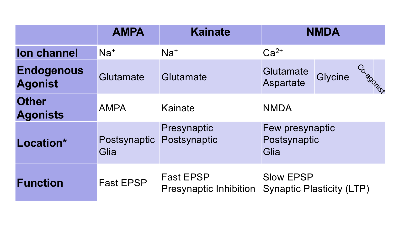
Ionotropic (chart) & metabotropic glutamate receptors
Differentiate the mechanism of action of the primary treatment approaches or drug discovery approaches by drug classes. DA antagonists vs potential Glutamatergic approaches
D2 Antagonist: 1st generation antipsychotics
Block the D2 receptor binding site → Dopamine cannot bind to D2 receptors → DA signaling decreases → Symptoms relieved.
Appreciate why Dopamine is a delicate balance in CNS function
EP movement disorders and tardive dyskinesia
If DA receptor antagonists are used for too long, then DA receptors change & this causes major issues.
Addiction
Understand what addiction is and general related terms
Addiction: Drug addiction is a chronic, relapsing disorder characterized by compulsive drug use, despite having harmful consequences
Repeated drug use changes the brain in a way that impairs the body’s ability to control behavioral processes and interferes with one’s ability to resist drug cravings
Tolerance: Decrease in response after repeated exposure requires more product to reach the same initial feeling
Pushes the curve to the right
Dependence
Psychological: Drug cravings and preoccupation with taking the drug
Physical: Required for conditions, such as cancer patients or terminally ill patients. They are not abusing the drug but may end up addicted.
Withdrawl: Removing the toxin and the body's response shifts the opposite direction to form a hyperresponse.
Reveals that the person is dependent
Relapse: Going back to drugs after being sober for some time
Know the common misused substances and alcohol and their primary mechanism of action
Hallucinogens (Ketamine, LSD, PCP, MDMA “Ecstasy”
The mechanism of action is dependent on the agent
Ketamine: NMDA Receptor antagonist
LSD: D2 agoinst/5HT2A agonist
PCP: NMDA Receptor antagonist
MDMA “Ecstasy”: Serotonergic - agonist, reuptake inhibitor
Marijuana
Mechanism of action: CB1 receptor agonist
Synthetic cannabinoids
Mechanism of action: CB1 receptor agonist
Opiates
Mechanism of action: mu-opioid receptor agonist
Different opioids have different levels of potency at different receptor subtypes.
Sedative-hypnotics
Mechanism of action: APL at GABAA
Stimulants (Cocaine, methamphetamine, amphetamine, methylphenidate, nicotine)
Mechanism of action (amphetamine, cocaine, methamphetamine): Inhibits DAT/NET, VMAT2
Methamphetamine: Also reverses DAT to toxic levels
Mechanism of action (nicotine): nAChR agonist
Alcohol
Appreciate which of the agents discussed today is our biggest problem - greatest harm, most overdoses, greatest use disorder.
SUD or daily use Rates of addiction |
Alcohol 19 million 1-7 to 1-15 |
Marijuana 5.7 million |
Cocaine 2-3 million |
Heroin & Nicotine 1 million 1-3 to 1-5 |
Greatest harm to others: Alcohol
Greatest harm to self: Heroin
Most overdoses: Alcohol
Greatest use disorder: Alcohol
Highest rate of addiction: Heroine, nicotine, tobacco
What is the mesolimbic dopamine system and what differentiates it from the other dopamine systems?
Mesolimbic dopamine system: Activated by addictive drugs
Dopaminergic projection from VTA to NAc (critical site for drug abuse)
Appreciate that all “addictive” drugs increase dopamine but they each have their mesolimbic reward system
Dopamine increase is essential, but not proportional to the “addictive potential” of the drug
Amphetamine & cocaine have higher peaks than heroin, but not as addictive
Seizures
Differentiate seizures vs epilepsy
Seizure: Abnormal and excessive neuronal discharge (hyperactivation of neural activity) with or without a change in the level of consciousness
Epilepsy: Seizure disorder characterized by recurrent, unprovoked seizures (diagnosis = 2 unprovoked seizures > 24 hours apart)
70% idiopathic
30% symptomatic (secondary causes such as injury, tumor, infection, etc.)
Differentiate types of seizures
Focal onset: Aware/Impaired awareness
Psychomotor
May evolve into secondary generalized seizures
Seizure focus → branches to thalamus → spreads throughout the brain
Simple focal (Retains consciousness)
Affect small regions of brain
Symptoms: Motor, somatosensory, autonomic, or psychiatric symptoms
Duration: 15 secs - 3 min
Complex focal (loss of consciousness)
Symptoms: May involve unconscious repetition of simple actions, gestures, or verbal utterances, or simply a blank stare
Unawareness of the seizure, followed by no memory of the seizure
May include an aura
Duration: 15 secs - 3 min
Generalized onset: Impaired awareness
Brain region affected: Both hemispheres
Motor
Tonic-clonic (Grand mal): Typical seizure
Duration: 30-60 secs
Myoclonic
Duration: Seconds
Tonic
Duration: Seconds
Atonic
Duration: Seconds
Non-motor
Absence (Petit mal): Blip/short loss of
Duration: 3-30 seconds
Know the pathophysiology of seizure.
Alterations in excitation or inhibition can lead to seizures
Glutamate: Excitation
GABA: Inhibition
Treatment: Anxiolytic, anti-convulsants, anesthetics, and alcohol enhance GABA receptor activity or block glutamate receptor activity
Basic epidemiology/stats
Prevalence: 4th most common neurological disorder
65 million worldwide with epilepsy
3.4 million in the US with epilepsy
⅓ of cases can’t be treated
Age: Very young & elderly is most common
< 2 has a high occurrence of acute symptomatic seizures
> 65 years has a higher occurrence of unprovoked idiopathic seizures
Symptoms (terms & definitions)
Typically during the postictal phase (recovery period)
Sensory/thought: Blackout, confusion, deafness/sound issues, electric shock feelings, smell, spacing out, out of body experience, vision loss or blurring
Emotion: Fear, panic
Physical: Dizziness, nausea, tonic, clonic, myoclonic, atonic, etc.
Neuropathologies
Understand what EEGs are like in seizure vs normal waves, differentiate absent seizures vs others
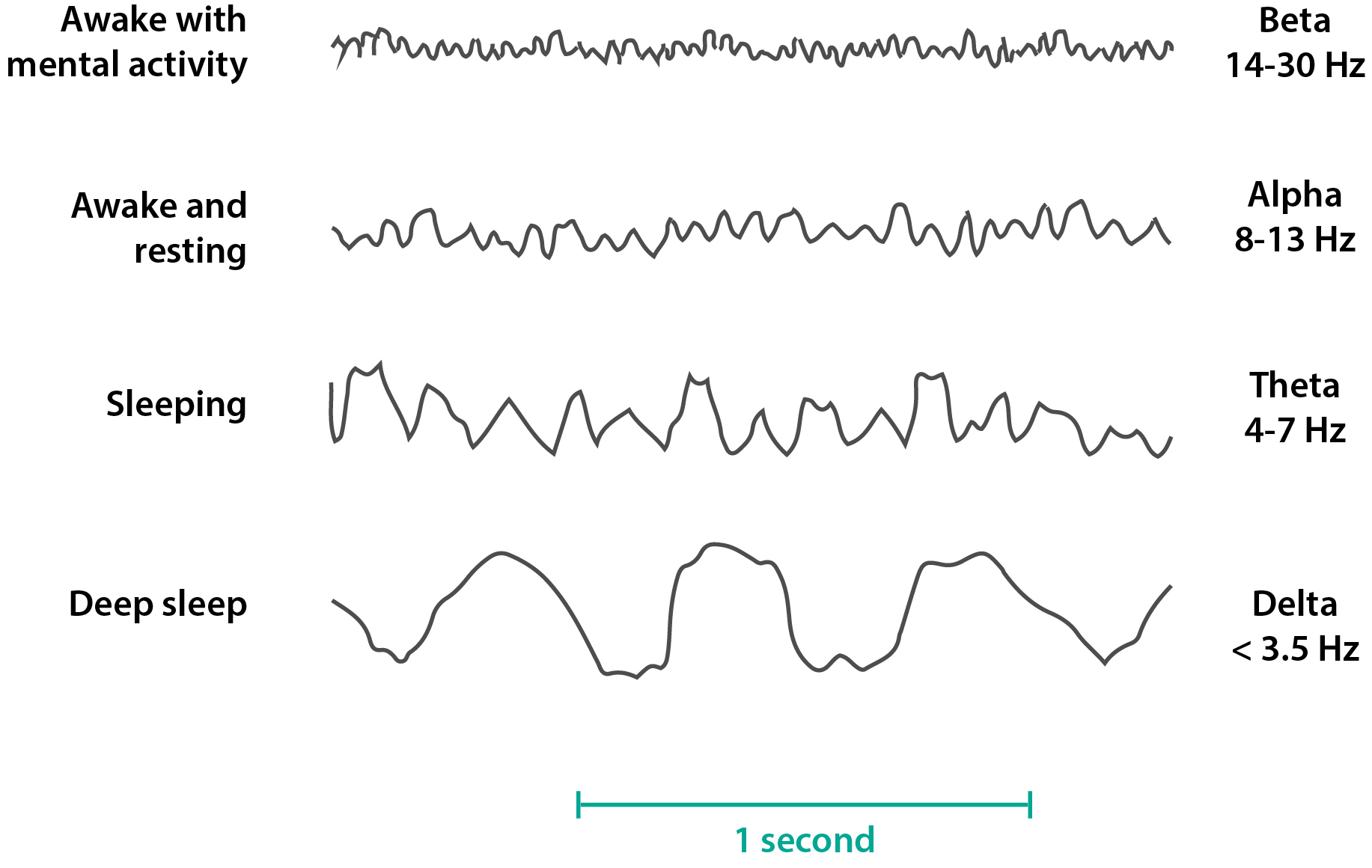
Types of EEG Waves
Alpha
8-13 Hz
Awake, but quiet
Beta
14-80 Hz
Awake & attentive
Asynchronous
Theta
4-7 Hz
Sleep, emotional stress
Some disorders
Delta
<3.5 Hz, higher voltage (large peaks)
Deep sleep, infancy
Severe disorder
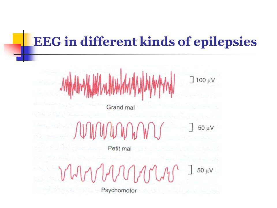
Absent seizure: Spike and dome
Grand mal: Out of control rapid spiking
Understand the 3 main pharmacotherapeutic approaches
Inactive voltage-gated Na+ channels - stop repetitive firing
Goal: Decrease hyperactivity
Enhance GABAergic neurotransmission
GABA is inhibitory
Goal: Decrease hyperactivity
Limit T-type Ca2+ channel (absence seizure only)
Sleep Disorders
Understand brain systems regulating sleep
Sleep is an active process, neurons are still firing
Systems regulating sleep
Arousal system: The reticular activating system in the brainstem regulated by the hypothalamus (hypothalamus and reticular activating system)
Hypothalamus: Promotes wakefulness when stimulated
Slow-wave sleep center: “Sleep on” neurons in the hypothalamus (hypothalamus)
Sleep-promoting neurons activated → GABA released → Inactivates awake neurons
Body awake = sleep neurons inactive
Non REM sleep = sleep neurons very active
Paradoxical sleep center: REM sleep on neurons in the brainstem that switch to paradoxical sleep (REM) (brain stem)
Controls transition from slow wave/deep sleep to REM
Molecules involved in the regulation of the sleep-wake cycle
Awake
Histamine
Anti-histamine causes drowsiness
Orexin (hypocretin): Promotes awakeness, suppressed when sleeping
Noradrenaline: Wake-promoting
Acetylcholine: Wake-promoting
Sleep promoting
GABA: “Sleep on” neurons release
Cytokines: Sleep-promoting
Immune system communicator
Melatonin: Released when dark
Adenosine: Caffeine targets this
Studying sleep: EEG (records brain waves)
Records small electrical fields generated by the synaptic currents in pyramidal cells
Sleep stages
Sleep cycle goes down non-rem (1-4) → REM sleep → up non-rem (4-1)
Non-REM or Slow-wave sleep
Beginning of the sleep cycle, light sleep (5-10 min)
EEG waves: Theta waves
Bursts of rapid, rhythmic brain activity. Body temperature decreases and heart slows.
EEG waves: Mixed activity (sleep spindles)
Deep, slow brain waves emerge. The transition between light and very deep sleep
EEG waves: Progress into delta waves
Slow wave or delta sleep (deep sleep)
EEG waves: Delta waves, slow waves
REM sleep (paradoxical sleep)
At the end of each sleep cycle (10-15 min)
Dreaming occurs here
EEG Waves: Similar to the awake state
Differentiate between types of sleep disorders.
Insomnia: A dissatisfaction with sleep quantity or quality with complaints of difficulty initiating sleep, maintaining sleep, or early morning awakening
Prevalence: 6-10% of US adults
Older women
Risk factors: Temperamental, environmental, genetic, and physiological
Hypersomnolence disorder: Excessive quantity of sleep, deteriorated quality of wakefulness, and sleep inertia
Risk factors: Stress, alcohol use, viral infection, genetics
Narcolepsy: Irrepressible need to sleep, lapsing into sleep, or napping occurring within the same day (Autoimmune disease)
Prevalence: 0.02-0.04%
Possible slight male preponderance
Onset: 15-25 years & 30-35 years; lifetime condition
Risk factors: Other familial sleep disorders; viral infections; head trauma; genetic; T cell genes (because it is autoimmune)
Breathing-related sleep disorders
Obstructive sleep apnea: Repeated episodes of upper airway obstruction during sleep that results in apneas or hypopneas
Loss of O2 when sleeping due to the tongue blocking the airway
Prevalence:
J distribution (Increases with age)
1-2% in kids
>20% in older adults
2:1 - 4:1 - male:female
Higher frequency in men
Risk factors: Obesity, sleep position, substance use, adenotonsillar hypertrophy (kids), genetic syndromes that reduce upper airway, genetics
Central sleep apnea: Repeated episodes of apneas or hypopneas during sleep
The area of the brain that controls your breathing when sleeping (pons) does not function properly
Sleep-related hypoventilation: A rare condition of decreased respiration during sleep which results in insomnia and sleepiness
Circadian rhythm sleep-wake disorder: A persistent or recurrent pattern of sleep disruption that is primarily due to an alteration of the circadian system
Risk factors: mutation in the “clock genes”, melatonin mutations, blindness
Delayed sleep phase types: Individual who is delayed in falling asleep at night and waking up >2 hours (sleeps late)
Advanced sleep phase type: Individual who is premature in falling asleep at night and waking up > 2 hours (sleeps early)
Irregular sleep-wake type: Lack of discernible sleep-wake circadian rhythm (3 periods of shorter sleep through the day)
Non-24-hour sleep-wake type: Sleep-wake cycle is not aligned with the 24-hour day
Shift work type: Excessive sleepiness at work and impaired sleep at home
Parasomnias: Abnormal behavioral, experiential, or physiological events occurring in association with sleep, specific sleep stages, or sleep-wake transitions
Non-REM Sleep Arousal disorders: Incomplete awakening; “sleepwalking”
Nightmare disorders: Repeated occurrence of extended, extremely dysphoric, and well-remembered dreams
Restless leg syndrome: An urge to move the legs along with an uncomfortable sensation
Explain the clinical manifestations of sleep disorders
Insomnia: Sleep issues > 3 nights/week for > 3 months
Hypersomnolence disorder: Excessive quantity of sleep, deteriorated quality of wakefulness, and sleep inertia
Narcolepsy: Irrepressible need to sleep, lapsing into sleep, or napping occurring within the same day
Criteria:
Recurrent periods of an irrepressible need to sleep, lapsing into sleep, or napping occurring within the same day (3x/week for > 3 months)
Presence of >1 of the following:
Episodes of cataplexy (loss of muscle tone)
Orxein deficiency in cerebrospinal fluid
Nocturnal REM sleep latencies <15 min
Pathophysiology: T cells may kill orexin neurons (wakeful neurons) → narcolepsy occurs
Circadian rhythm sleep-wake disorder: Alteration of the suprachiasmatic nucleus (which controls the circadian rhythm)
Identify the causes and consequences of select sleep disorders
Narcolepsy: Deficiency/destruction of orexin neurons
Orexin neurons: Maintain wakefulness
Obstructive sleep apnea: Physical changes in the body, having a floppy airway
Central sleep apnea: Changes in the brain
Sleep-related hypoventilation: PHOX2B gene & obesity
Circadian rhythm sleep-wake disorder: Alteration in the circadian system
Alzheimer’s disease and other dementias
Describe and differentiate types of neurocognitive disorders
Delirium: Acute condition that is characterized by disturbance of attention or awareness accompanied by a change in baseline cognition
DSM-V:
Disturbance in attention and awareness
Disturbance develops rapidly and tends to fluctuate throughout the day
Additional cognitive disturbances (e.g. disorientation, language, perception)
Exclusion criteria
A and C are not due to preexisting neurocognitive disorder
Evidence that disturbance is a direct physiological consequence of another condition (e.g. substance use, medication withdrawal, surgery, and infection)
Prevalence: 1-2% of the overall population, but much higher in the elderly (14% of 85+)
Onset: Rapid
Risk factors: Surgery, infection, hospitalization
Things that activate the immune system
Duration: One week
Resolution: Best outcomes if treated rapidly; can progress to stupor, coma, death. Increases risk for long-term neurocognitive disorders in the future
Mild neurocognitive disorders: An acquired decline in one or more cognitive domains (noted by self, peer, clinical, or diagnostic test assessment)
DSM-V:
Evidence of modest cognitive decline in one or more cognitive domains
Cognitive deficits do not interfere with the capacity for independence
Exclusion criteria
Cognitive deficits do not interfere in the context of delirium
Cognitive deficits are not better explained by another mental disorder (e.g. schizophrenia, MDD)
Specify: With or without behavioral disturbances (e.g. psychotic symptoms, apathy, agitation; due to another condition)
Major neurocognitive disorders: An acquired decline in one or more cognitive domains (noted by self, peer, clinical, or diagnostic test assessment)
DSM-V:
Evidence of significant cognitive decline in one or more cognitive domain
Cognitive deficits interfere with the capacity for independence
Exclusion criteria
Cognitive deficits do not interfere in the context of delirium
Cognitive deficits are not better explained by another mental disorder (e.g. schizophrenia, MDD)
Specify: With or without behavioral disturbances (e.g. psychotic symptoms, apathy, agitation; due to another condition)
Dementia: Overarching condition that affects cognitive domains, mainly affecting learning and memory
Alzheimer’s Disease: 70% of dementia cases
Involves trouble creating new memories
Risk factors: Increased age, vascualr disease, genetics, head injury, lower education, sex, substance use, depression, sedentary lifestyle
Protective factors: Exercise, mediterranean diet, social engagement, mental activity, higher education
Vascular dementia: 17% of dementia cases
Involves trouble retrieving memories
Cause: Damage to subcortical fibers
Reduced blood flow to the brain
Risk factors: Hypertension, Hypercholesterolemia, Insulin-resistant diabetes, tobacco use
Dementia with Lewy bodies (#3)
Parkinson’s disease (#4)
Causes of Dementia
Degenerative disease: Alzheimer's, Lewy body Disease
Endocrine: Thyroid, cortisol
Metabolic: Electrolyte imbalance
Exogenous agents: toxins, drugs
Neoplasia
Trauma: Brain injury
Infection
Affective psychiatric disorders: MDD, schizophrenia
Stroke
Identify changes in distinct neurocognitive domains and underlying neural circuitry
Learning and memory
Region:
Frontal lobe
Hippocampus
Thalamus: Medial diencephalon
Hypothalamus: New memory retrieval
The strongest deficits occur here, it is very susceptible to disease
Most impacted: Hippocampus, episodic memory!!
Short & long-term memory
Executive function
Region: Mainly frontal lobe
Functions: Planning, decision-making, working memory, self-behavior control, flexibility, multitasking
Complex attention: Focusing on a primary task
Regions:
Frontal lobe
Parietal lobe: Selective attention
Reticular activating system: Arousal
Social cognition: Recognizing own & others emotions
Regions:
Orbitofrontal cortex
Temporal lobe: Empathy control
Perceptual motor function
Language
Explain the clinical manifestations of Alzheimer’s disease
Development
Early onset: Genetic
Causative genes
B-Amyloid precursor protein (APP)
Presenilin 1
Presenilin 2
Late onset: Idiopathic/sporadic
Susceptibility genes
Apolipoprotein E
Symptoms
Neurocognitive domains affected: (Learning memory → executive function → complex attention → social cognition)
Declarative memory is affected (not non-declarative!)
Apraxia: Impaired movements
Co-morbidities
Agitation (50-70%)
Anxiety (30-50%)
Depression (25-50%)
Disinhibition (20-35%)
Delusions (15-50%)
Hallucinations (10-25%)
Aggression (25%)
Sexual disinhibition (5-10%)
Discuss brain changes and genetic risks of Alzheimer’s disease: defining the role of APP, Aβ peptide, presenilins, and apolipoprotein E
Cholinergic hypothesis: Cholinergic neurons are negatively affected (in the nucleus basalis Meynert)
Cholinergic neurons signal to cortex → attention & arousal
Less cholinergic neurons = less attention/arousal = visible deficits
Excitotoxicity hypothesis (glutamate): Too much glutamate
Intraneuronal neurofibrillary tangles: Tangled clusters of tau proteins → no stabilization of microtubules → disintegration of microtubules
Accounts for early symptoms of memory loss & issues with higher cortical function
Regions affected: Forebrain, hippocampus, entorhinal
Cause: Clumping of tau proteins
Result: Disintegrating microtubules
Extracellular neurotic plaques: Clusters of APP in the brain (it is sticky)
Amyloid material (APP): Dense core, the primary material in these plaques
Sticky material that builds up and develops into a toxic oligomer
Typically cleaved by a-secretase
When b-secretase & gamma-secretase are involved, this can be bad
Dystrophic neurites: Surround the amyloid core
Reactive astrocytes: Recruited when the plaques are detected
Microglia: Immune response in the brain
Microglia & reactive astrocytes contribute to the problem because they increase inflammation.
Presenilin protein - gamma secretase
Presenilin proteins regulate the secretase
Presence of Presenilin 1/2 causes Alzheimer’s (raises risk greatly)
Presenilin 1 (Chromosome 14)
Presenilin 2 (Chromose 1)
Apolipoprotein E (Gene for late-onset Alzheimers): Regulates synthesis and secretion of microglia & astrocytes, lipid accumulation
Apo E2: Decreased risk for Alzheimer's
Apo E3: The most common gene possessed by people with Alzheimer's, but doesn’t necessarily increase risk.
Apo E4: Increases risk for Alzheimer's
Anxiety, Depression, and Bipolar Disorders
Describe and differentiate between types of anxiety, mood, and bipolar disorders.
Anxiety: Share features of excessive fear and anxiety and related behavioral disturbances
Fear of fear
Separation anxiety (1-2%, 5% kids)
Selective mutism (<1%): Failure to speak in certain situations, such as school or in crowds
Selective phobia (3-16%, culture/age): Fear of specific things, such as spiders
Social anxiety disorder (7% in the US, 1% in the world): Anxiety about interactions with others
Panic disorder (2-3%):
Agoraphobia (0.5-1%)
Generalized anxiety disorder (GAD): The most common
DSM-V Criteria
Excessive anxiety and worry, occurring more days than not for at least 6 months, about several events or activities.
The individual finds it difficult to control the worry
The anxiety and worry are associated with three (or more) symptoms, *only one required in children*
Restlessness or feeling on edge
Easily fatigued
Difficulty concentrating or mind going blank
Irritability
Muscle tension
Sleep disturbances
Anxiety, worry, or physical symptoms cause clinically significant distress or impairments in function
Exclusion criteria
Disturbance is not caused by substance use or another medical condition
Disturbance is not better explained by another mental disorder
Incidence and prevalence
Lifetime risk: 9% +
Median age of onset: 30 years
Comorbidity: Up to 50% with some conditions
Men: High comorbidity with depression and substance use disorder
Women: High comorbidity with depression
Depression
Major depressive disorder (MDD)
DSM-V Criteria
Must have at least one of these occurring daily for 2 or more weeks
Depressed mood
Loss of interest and pleasure (anhedonia)
Must have at least five of these occurring daily for 2 or more weeks
Depressed mood
Loss of interest and pleasure (anhedonia)
Increase or decrease:
Appetite/weight
Sleep
Activity/psychomotor
Decrease:
Energy
Concentration
Increase:
Guilt
Morbid death thoughts
c. Exclusion criteria: Depression is not due to psychosis, manic episodes, illness, medication/drug, or bereavement
Incidence and prevalence
Population: 5-7% of population
350 million people worldwide
Age: All ages
Most suicides are in youth (15-29)
Genetics: Familial
21% rate across 1st degree family members
30-50% rate across identical twins
Candidate genes:
Serotonin-linked polymorphic region
Serotonin 1A receptor
Methylenetetrahydrofolate reductase
Dopamine D4 receptor
DNA Hypermethylation:
Brain-derived neurotrophic factor
Serotonin transporter
Alternative treatments
Electroconvulsive therapy
Ketamine: Involves glutamate
Transcranial magnetic stimulation: Stimulates neurons
Deep brain stimulation
Typical (melancholic)
Decrease:
Appetite/weight
Sleep
Increase:
Activity/psychomotor
Atypical (most common form)
Decrease
Activity/psychomotor
Increase
Appetite/weight
Sleep
Psychotic: Depression characterized by psychotic symptoms
Anxious depression
Postpartum depression
Seasonal affective disorder
Bipolar
A disorder characterized by periods of mania alternating with periods of depression
DSM-V Criteria
Must have for at least 1 week, or hospitalization may occur before the one-week mark
Abnormally and persistent elevated or irritable mood with increased energy/activity
Must have 3 or more symptoms (4 or more if “a” is irritable mood)
Increase
Grandiosity
Talkative
Racing thoughts
Distractibility
Goal-directed activity
Negative consequences
Decrease
Sleep
c. Cause marked impairment in social or occupational function or hospitalization occurs
d. Exclusion criteria: Mood shifts are not due to medications/drugs or another medical condition
Incidence & Prevalence
Population: 1-2% of population
25% of the population with anxiety & depression
Genetic: 85% familial
12% rate across 1st degree family members
Candidate genes:
Glutamate neurotransmission
Neurogenesis/brain development
Serotonin transporter
Catechol-methyl-transferase
Monoamine oxidase
Tryptophan hydroxylase
Treatment
Lithium, antipsychotics, and certain anticonvulsants can treat manie
Explain the clinical manifestations of the distinct disorders
Anxiety
Symptoms
Inability to be still and calm
A feeling of panic, fear, uneasiness
Vigilance: Hyperarousal and awareness of surroundings
Difficulty sleeping
Feelings of panic
Tension/muscle tension
Sweating
Dry mouth
Shortness of breath
Dizziness
Palpitations
Depression
Symptoms
A: Must have at least one of these occurring daily for 2 or more weeks
Depressed mood
Loss of interest and pleasure (anhedonia)
B: Must have at least five of these occurring daily for 2 or more weeks
Depressed mood
Loss of interest and pleasure (anhedonia)
Increase or decrease:
Appetite/weight
Sleep
Activity/psychomotor
Decrease:
Energy
Concentration
Increase:
Guilt
Morbid death thoughts
Relationships with other illnesses
Secondary to other diseases
Hypothyroidism
Neurodegenerative diseases
Inflammatory conditions (trauma, cancer, diabetes)
Complicates treatment of medical conditions
Severe trauma
Cancer
Diabetes
Cardiovascular disease
Typical (melancholic)
Decrease:
Appetite/weight
Sleep
Increase:
Activity/psychomotor
Atypical (most common form)
Decrease
Activity/psychomotor
Increase
Appetite/weight
Sleep
Psychotic: Psychotic symptoms
Bipolar
Type 1
Manic episode: Required for diagnosis
1+ week duration
Impairs functioning
Severe mania
Depressive episode: Not required for diagnosis
Longer duration
Severe depressive episode
Type 2
Manic episode: Required for diagnosis
4+ consecutive days
Hypomania (not as severe)
Depressive episode: Required for diagnosis
Longer duration
Severe depressive episode
Name key brain structures involved in fear conditioning, anxiety disorders, and mood disorders.
Fear conditioning: Useful for studying fear and anxiety
Amygdala: Fear center in the brain
Increase in activity
Hippocampus: Learn fearful association
No change in activity
Insular cortex: Similar to the amygdala, provides emotional context
Increase in activity
Processes emotions
Anxiety
Amygdala: Fear center in the brain
Increase in activity
Hippocampus: Learn fearful association
No change in activity
Less prevalent once anxiety is established, but the hippocampus forms the fearful association.
Insular cortex: Similar to the amygdala, provides emotional context
Increase in activity
Processes emotions
Depression
Hippocampus: Decrease in size
Amygdala: Increase or decrease in reactivity
Frontal cortex: Decrease in connectivity to the amygdala
Typical (melancholic)
Increase in the HPA Axis (Negative feedback loop that controls cortisol release): There is a lack of cortisol rhythm in the body.
Atypical (most common)
Increase in hypersensitivity
Sensitive to criticism and feedback
Amine hypothesis
Raphe nuclei: Source of serotonin
Locus coeruleus: Source of norepinephrine
Long-term effects of antidepressants
Increased adrenergic or serotonergic receptor density or sensitivity
Increased G protein coupling and cyclic nucleotide signaling
Induction of neurotrophic factors
Increased neurogenesis in the hippocampus
Bipolar
Hippocampus: Increase in activity
Frontal cortex: Decrease in activity
Amygalada: Increase or decrease in reactivity
Explain changes in brain function that are associated with anxiety, mood, and bipolar disorders.
Anxiety
Serotonin decrease
Treatment: Increase serotonin through SSRIs and other antidepressants
Common treatment
GABAA: Key in brain excitability
Treatment: Increase GABAA activation through the use of benzodiazepines
Less common treatment because benzos are addictive
Very short-term
Depression
Depression may be caused by a multitude of factors in different patients
STAR*D Trial: Some patients responded to certain things, while others didn’t
Amine hypothesis: Depression is related to a deficiency of monoamines in the brain
Amine neurotransmitters (such as serotonin, dopamine, etc.) are related to improved mood
Patients were treated with iproniazid (MAO inhibitor) and they displayed inappropriate levels of happiness, and when they were treated with reserpine (depletes catecholamines) they displayed symptoms similar to that of depression
Dopamine, serotonin, and norepinephrine are primary contributors to MDD
Cytokine hypothesis: Increased cytokines are a key factor in the development of depression
Cytokines: Proteins released when there is inflammation in the body, common during illness and injury
Symptoms of depression are very similar to symptoms the body experiences when sick
Bipolar
Hippocampus: Increase in activity
Frontal cortex: Decrease in activity
Amygalada: Increase or decrease in reactivity