Structure and function of synapse bio
Structure and function of synapse
A synapse is the point where one neurone communicates with another or with an effector. They are important in linking different neurones together and therefore coordinating activities.
Structure of a synapse
 Synapses transmits information, but not impulses, from one neurone to another by means of chemicals known as neurotransmitters.
Neurones are separated by a small gap, called the synaptic cleft, which is 20-30nm wide. The neurone that releases the neurotransmitter is called the presynaptic neurone.
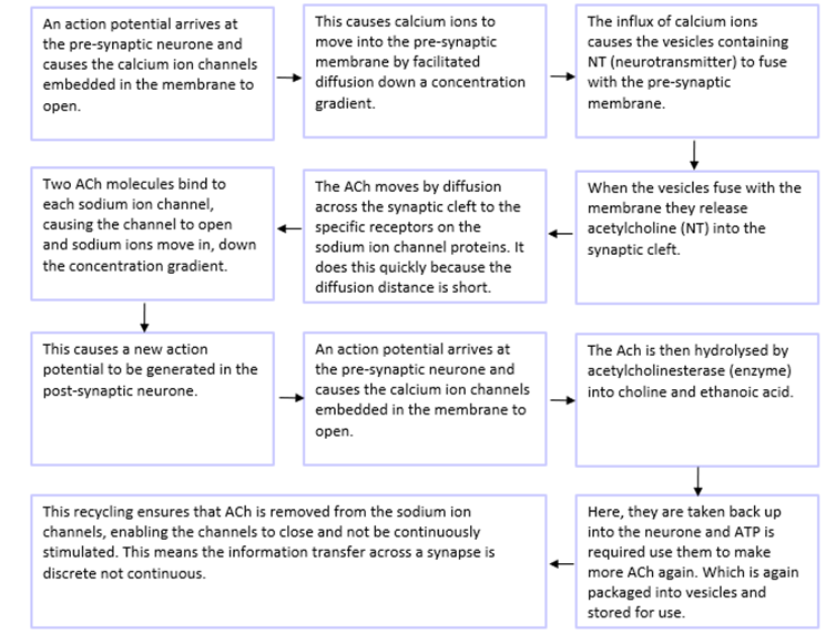
The axon of the neurone ends in a swollen portion known as the synaptic knob.
Signals are transferred from on neurone to another across a synapse in the form of a chemical message.
A synapse or synaptic cleft is the gap between 2 neurones.
The neurone that comes before the synapse is called the pre-synaptic neurone.
The neurone that come after the synapse is called the post-synaptic neurone.
The end of the pre-synaptic neurone is bulbus and enlarged compared to the rest of the axon, and it is called the pre-synaptic knob.
Inside the pre-synaptic neurone there are lots of mitochondria and endoplasmic reticulum. This is because the processes occurring in the knob rely on a good supply of ATP and proteins need to be produced in large quantities.
The protein produced is called acetylcholine (Ach) and this is a neurotransmitter.
this neurotransmitter is packaged and stored in vesicles in the synaptic knob.
When the neurotransmitter is released to the synaptic cleft the vesicle fuses with the pre-synaptic membrane and the Ach will diffuse across the cleft to the specific receptors on the post-synaptic membrane
On the post-synaptic membrane, the specific receptors have 5 subunits that undergo a conformational change when the Ach binds. These receptors are sodium channels, that open and allow positively charged sodium ions to move into the post-synaptic neurone once open.
Key terms:
An excitatory synapse is one that cause the generation of a new action potential.
A cholinergic synapse is one that has Ach as the neurotransmitter. Cholinergic synapses occur in vertebrates in the CNS and neuromuscular junctions (between the nerve and muscle).
 Features of the synapse
Unidirectionality
synapses can only pass information in one direction-from the presynaptic neurone to the postsynaptic neurone. In this way, synapses act like valves.
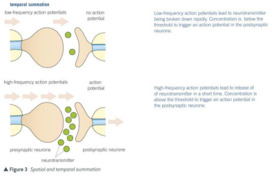
Summation
Low-frequency action potential often leads to the release of insufficient concentrations of neurotransmitter to trigger a new action potential in postsynaptic neurone. They can, however, do so in process called summation. This entails a rapid build-up of neurotransmitter in the synapse by one of two methods:
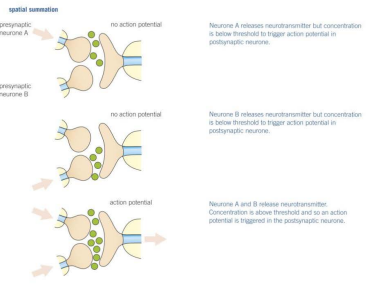 Spatial summation- in which a number of different presynaptic neurones together release enough neurotransmitter to exceed the threshold value of the postsynaptic neurone. Together they therefore trigger a new action potential.
Temporal summation-in which a single presynaptic neurone releases neurotransmitter many times over a very short period of time. If the concentration of neurotransmitter exceeds the threshold value of the postsynaptic neurone, then a new action potential is triggered.
Inhibition
Some synapses make it less likely that a new action potential will be created on the postsynaptic neurone. These are known as inhibitory synapses. They operate as follows:
The presynaptic membrane releases a type of neurotransmitter that binds to chloride ion protein channels on the postsynaptic membrane.
The neurotransmitter causes the chloride ion protein channels to open.
Chloride ions move into the postsynaptic neurone by facilitated diffusion.
The binding of the neurotransmitter causes the opening of nearby potassium protein channels.
Potassium ions move out of the postsynaptic neurone into the synapse.
The combined effect of the negatively charged chloride ions moving out is to make the inside of the postsynaptic membrane more negative and the outside more positive.
The membrane potential increases to as much as -80mV compared with the usual -65mV at resting potential, meaning EVEN MORE sodium ions are required to move into the membrane to get action potential generated.
This is called hyperpolarisation and makes it less likely that a new action potential will be created because a larger influx of sodium ions needed to produce one.
Function of synapses
Synapses transmit information from one neurone to another. In doing so, they act as junctions allowing:
A single impulse along one neurone to initiate new impulses in a number of different neurones at a synapse. This allows a single stimulus to create a number of simultaneous responses.
A number of impulses to be combined at a synapse. This allows nerve impulses from receptors reacting to different stimuli to contribute to a single response.
A chemical (the neurotransmitter) is made only in the presynaptic neurone and not in the postsynaptic neurone.
The neurotransmitter is stored in synaptic vesicles. When an action potential reaches the synaptic knob in the membranes of the vesicles fuse with the presynaptic membrane to release the neurotransmitter.
When release, the neurotransmitter diffuses across the synaptic cleft to bind to specific receptor proteins which are found only on the postsynaptic neurone.
The neurotransmitter binds with the receptor proteins, and this leads to a new action potential I the postsynaptic neurone. Synapses that produce new action potentials in this way are called excitatory synapses.