Hemoglobin
hemoglobin is protein that carries oxygen in blood to transport it
oxygen is NP and insoluble in aqueous solution
hemoglobin is in red blood cells when blood is oxygenated, pressure of O2 high in lungs hemoglobin grabs hold of the blood and blood travels to capillaries and hemoglobin drops of O2
process repeats
hemoglobin is heterotetramer with quaternary structure
meaning 4 subunits that are not identical
2 alpha subunits
7 helices A-H but missing D
2 beta subunits
8 helices A-H
each subunit has a heme (prostatic group non protein that binds O2)
has 4 hemes so bind O2 in 4 places
suitable for O2 transport
myoglobin: 8 helices A-H
1 heme and only tertiary structure
suitable for O2 storage
stored in muscles and has a strong affinity for O2
useful when concentration of oxygen drops in blood like during anaerobic respiration myoglobin releases it then it diffuses through muscle cells and mitochondria pick it up for ATP sythensis
higher concentration of myoglobin in deep see mammals, the longer the animal can go without O2
binding affintiy between Protein & Ligand
enzyme and substrate type situtation but with protein and ligand
Ligand is a small molecule that binds to binding site of protein, is suited for its protein
needs to have charge, shape, size, hydrophobicity must complement protein
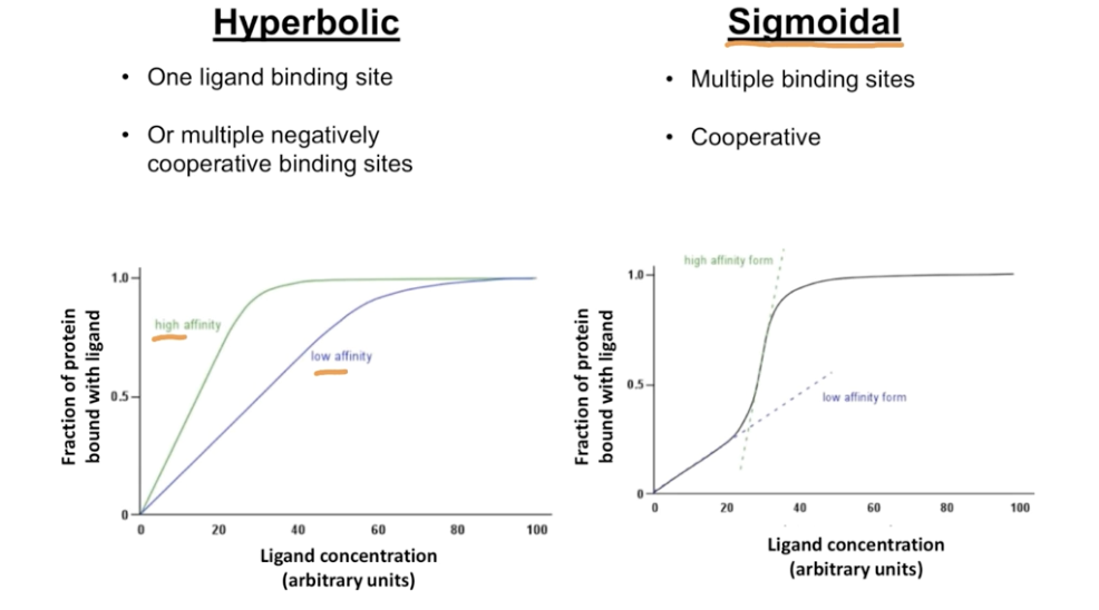
by plotting fraction of protein bound to ligand and ligand concetration increases can see how well a protein binds to ligand
as start adding ligand the staured the solution becomes giving more chance to bind to protein
at start this is rapid but curve starts to slow as binding sites decreases
when concentration is high enough 100% of protein will be bound to ligand
P50: point at which ½ of protein is bound to ligand
x-axis is in terms of pressure for O2
for hemoglobin it is 28 torr
for myoglobin it is 3 torr
hyperbolic curve: one ligand binding site or multiple negatively cooperative binding sites: myoglobin
binding sites work indenpendtly of each other
sigmoidal curve: multiple binding sites and cooperative: hemoglobin
steep slope means high affinity for ligand
why is hemoglobins sigmoidal curve important
has to regulary bind and drop of O2, needs the high and low affinity to do this
cooperativity: binding of 1 ligand affects affinity of remaining sites, initially difficult to bind ligand but once one is bound then more and more can bind
2 conformations of hemoglobin
Tense (T-state): deoxyhemoglobin, low O2 binding affinity
favored during low Pressure of O2
in tissues
Relaxed (R-state): oxyhemoglobin, high O2 binding affinity
favored during high Pressure of O2
in lungs
multiple conformations means its tertiary/quaternary structure have multiple ways of being arranged in 3-D form
for hemoglobin binding of one O2 at one subunit increases affinity of O2 for the other subunits
hemoglobin is most stable in T or R state
heme: oxygen binding site that is prosthetic group, non-proteinaceous molecule, found in each monomer added to protein during or after translation, uses iron to bind O2
iron is found in the middle of the porphyrin ring structure
has coordinate covalent bonds, this bond is a type of covalent bond where 1 atom donates both electrons, occur between metal ions and ligands
Fe and 4 nitrogens
iron 2+ state binds oxygen reversible and iron 3+ can’t
Fe2+ in heme participates in 6 different bonds when oxygenated
has octahedral geometry
4 bonds with Nitrogen
1 with histidine
1 with O2 at an angle places it next to another histidine residue called distal His, His E7 or His 64
iron has coodiranate bond with side of histidine residue called proximal histidine or His 93 or His F8
His93 is the proximal histidine in myoglobin chain
for alpha chain it is His 87
for beta chain it is His 92
proximal and distal naming for histidine in relationship to heme
nitrogen in distal His acts as a H bond donor for the oxygen which is the H bond acceptor
diatomic oxygen can’t be a H bond acceptor but is in this case because of the polar iron and oxygen bond
quaternary change happens when O2 binds to heme or is released from it
heme is oxygenated has planar conformation with iron in middle of it
heme is deoxygenated iron is repelled from porphyrin ring in direction towards proximal His, domed shape results
relays a change to position of F alpha helix
alpha 1 and beta 1 & alpha 2 and and beta 2 subunits are assoicated tightly due to hydrophobic effect forming dimers, held together by more than 30 residues
dimer is chemical structure that is formed by linking of 2 similar sub units
alpha 1 and beta 2 & alpha 2 and beta 1 are held together by 19 residues
interactions keeping dimers together are stronger
if hemoglobin was treated with urea (denaturing agent) it would separate into its dimers
upon oxygenation the distance between the beta subunits grows more narrow
factors that stabilize R and T states
low O2 T state favored; domed shape of heme
stabilized by larges # of ion pairs relative to R state at alpha 1& beta 2 and alpha 2&beta 1 interfaces
hemoglobin likes to arrange polypeptide in a particular way when no ligand bound
high O2 R state favored; planar shape of heme
ion pairs that stabilize T state are broken
proximal histidine is key for cooperatively as it shifts F-helix upon O2 binding
replacing proximal histidine with glycine the position of F helix was unaffected, no cooperativity and increase in O2 affinity (experiment done by Barrick and team)
different IMF take place in R and T state
T state has greater # or ion pairs
The proximal histidine forms a covalent bond with heme iron, while the distal histidine forms a hydrogen bond with O2
negative allosteric effectors of hemoglobin, H+, CO2, and BPG
there is something in blood that decreases hemoglobin’s affinity for O2
pure hemoglobin has higher affinity for O2
allostery: binding of ligand at one site affects binding of ligand at another
positive stabilizes ligand-binding conformation
O2 for hemoglobin as it stabilizes the ligand binding conformation and is homotrophic same as effector (ligand)
negative destabilizes ligand binding conformation
destabilize R-state
CO2, H+, BPG found in blood
negative heterotrophic effectors as they are different then ligands
CO2 one of the byproducts of cellular resipration, 3 ways CO2 transported back (get rid off
7-10% dissolves in plasma of blood
70% dissolves in blood as HCO3-
generates negative allosteric effectors protons
20% carried away by hemoglobin, called carbaminohemoglobin (second negative allosteric effector)
protons
red blood cells have carbonate anhydrase which catalyses
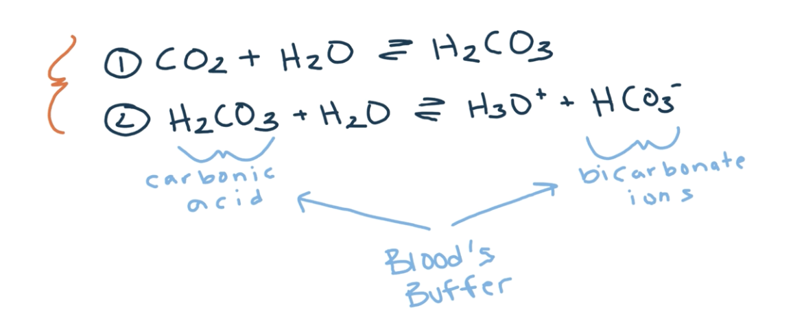
the protons generated from this reaction, is transported by hemoglobin about 20%
when pressure of O2 high in lungs hemoglobin favors R state, and get O2
equilibrium favors R state or oxygenated form of hemoglobin
p50 of hemoglobin is lower
when pressure of O2 low in tissues T state favored for hemoglobin
when the hydration of CO2 happens in tissues, the pressure of CO2 increases, increasing protons, decreasing pH, causes pronated form of hemoglobin to increase, making it give up O2
protons don’t bond in the same place as O2 but in different residues
p50 of hemoglobin is higher
increase in protons decreases hemoglobins affinity for O2, so hemoglobins p50 rises
Bohr effect: effects of pH on O2 binding curve
as pH decreases p50 increases
salt bridge between R of histidine 146 and R group of aspartic acid 94, stabilizes T state
happens only when protonated
happens when pH decreases like in T state in tissues
myoglobin doesn’t display Bohr effect, isn’t effected by pH, while hemoglobin is
the histidine side chain pKa increases upon O2 release
its R group pKa is 6, but it can change based on environment changes called pKa perturbation
holds on to proton more when closer to aspartic acid
carbaminohemoglobin
deoxygentated form of hemoglobin
CO2 reacts with N terminin of four globin subunits to form a carbamate end which carries a neagtive charge
carbamate ends can participate in salt bridges that stabilize T-state
release of protons also contribute to Bohr effect
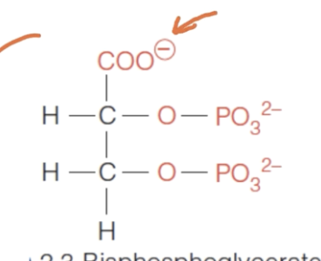
2,3 BPG
binds to central cavity of hemoglobin (this changes with T and R state, larger in T state)
has a negative 5 charge so interacts with positive charges, so binds to conjugate acid forms of basic side chains and N terminal amino groups
lysine, histidine, and N terminus in central cavity
8 positively charge groups in B subunits positioned in central cavity that stablize BPG
allows significant release of O2 (case for all the negative allostrotic effectors
fetal hemoglobin and BPG
still has alpha 1 and 2 but instead of beta it has gamma 1 and 2
when fetus inside mom the p50 is lower then adult hemoglobin
needs the stronger binding affinity to O2 to be able to steal it from the adult hemoglobin
not very good at dropping but doesn’t need to be because fetus is smaller then adult
very good at picking up O2
has serine at 143 residue instead of histidine in beta subunit
and 6 positive charges to stabilize it so doesn’t bind as tightly