Psych 1010: Unit 3
Every psychological phenomena is rooted from biological functions
Biological psychology: Studies the relationship between the nervous system and behaviour
Researchers are biological psychologists and/or neuroscientists
Brain mapping phrenology: Skull shape thought to reflect brain size and cognitive function (Discredited and has dark history such as eugenics, and criminal tendencies)
Brain damage: understand how the brain works by seeing how it doesn't function
Computerized Tomography: shows brain tumors
Positron Emission Tomography (PET) Scan: Shows brain activity
Magnetic Resonance Imaging: shows brain tissue
FMRI (Functional) MRI: shows changes in metabolic activity overtime (which parts of the brain are more activity than others)
Electroencephalography (EEG): Measures and records electrical activity of brain
Magnetoencephalography (meg): measures magnetic fields to determine which areas from the brain are generating seizures
Deep brain stimulation (DBS): modify brain function through implanted electrodes (e.g. parkisons)
Transcranial magnetic stimulations (TMS): applies magnetic fields to the surface of the skull to either enhance or interrupt brain function
Interpreting brain imaging. They are not photos of the brain in action. How brain activity during an activity of interest increases, decreases or differs relative to a control. Experimental – control. Brain area activity on brain scan could mean neurons are inhibiting rather than exciting. Scientifically, extremely persuasive (like expert testimony) – problematic when introduced into courtroom (e.g., to prove diminished culpability)
Neurons: are building blocks
Action potentials: nerve impulse or emotional signals that travel down an axon
Glial cells: support, nourish and protect neurons (body guards)
Neurons meet at synapses
Neurons communicate through neurotransmission
Neurons: they are nerve cells, specialise in communication. They transmit information in the form of electrical signals
Below is the order of processes
Dendrites: Receives information
Cell body: builds up neuron
Axon: tails that transmit information
End of axon containing synaptic vesicles filled with neurotransmitters (what are these; they transmit information
Synapse: space between the neurons through which neurotransmitters travel to connect to another neurons
Glial cells (means glue): they make myelin which builds signalling in the brain by insulating (improves signal transmission)
Myelin sheath: relates to the insulation from glial cells to the axon; poor insulation=poor signalling
How does a neuron fire: action potential
All-or-none law: A limit must be reached for a neuron to focus (cannot be less or more)
Step 1: resting potential; neuron is polarised (negative inside, positive outside). Before neuron fires
Selectively permeable - gates don't allow sodium ions (Na+) to pass through
Step 2: action potential: brief electrical charge that travels down neurons (When stimulated, neuron depolarized; gates open, Na+ rushes in)
Step 3: repolarization: Potassium flows out the repolarizing the axon
Step 4: return to resting potential
Step 5: refractory period (neuron wont fire, needs resting period, will not fire even if there is simulation)
Electrochemical communication
Electrochemical communication: when an electrical signal reaches the end an axon, it triggers release of neurotransmitters into the synapse (chemical)
These chemicals are neurotransmitters; bind to the neurons’ dendrites (which receive signals) resulting in the transmission of a signal
Excitatory: message that make it more likely a neuron will fire
Inhibitory: message that make it less likely that a neuron will fire
Between neurons: Chemical
Within neurons: Electrical
Neurotransmitters: Chemical messengers that help neurons communicate with each other
Influence emotions and mood (serotonin and dopamine)
Control movement
Regulate sleep
Learning and memory
Implicated in mental illness (e.g. giving more serotonin to treat depression)
Neurotransmission Processes
Release: Action potential (from neurons): trigger neurotransmitters release from vesicles into the Synaptic cleft. Then the neurotransmitters bind to receptors to the postsynaptic neuron (lock and key).
Reuptake: Excess neurotransmitters are moved or reabsorbed; broken down, taken back into the presynaptic neuron.
Some drugs block reuptake, prolonging NT effect, agonist)
Neurotransmitters helpers and blockers
Agonist: mimic or enhance the effect of an neurotransmitter (Cocaine enhances dopamine by having
Antagonist: block or impede the normal activity of a neurotransmitters
How to understand agonist/antagonist: Any thing that enhances an neurotransmitter like cocaine is an agonist (it blocks the absorption of dopamine but it makes an influx of it in between the synapses, hence why it is an agonist
E.g. opioids are agonist, naloxone are antagonist (don't allow binding to the neurotransmitter)
Schizophrenia associated with excess dopamine-> dopamine antagonist to help with schizophrenia
Parkinson associated with low dopamine -> prescribed dopamine agonist (enhance the effect of dopamine neurotransmitters)
Neurotransmitters
Glutamate: most common; excitatory (Plays function in learning and memory)
GABA: inhibitory, dampening neural activity (slows it down)
Glutamate and GABA: work together to manage the levels (Nicotine is a stimulate: glutamate increases, gaba decreases)
Coffee: glutamate increase , gaba decreases
Acetylcholine: Attention, memory, sleep, arousal, (Alzheimer's-> neurons containing acetylcholine are destroyed, leads to memory loss; aricept-> boosts acetylcholine levels, a treatment methods
Benadryl; slows down acetylcholine by blocking it
Dopamine: pleasure and reward, voluntary movement, Attention
Parkinson->deficit of dopamine; needs dopamine agonist (aids dopamine receptors)
schizophrenia + symptoms -> excess dopamine; needs dopamine antagonist (stops dopamine neurotransmitters)
Synapse cannot be carried out due to cocaine, lots of dopamine in the system (is cocaine a agonist or inhibitory)
Serotonin: mood, sleeping and pain
Increase serotonin by running, light exposure, foods which help reproduce serotonin
Drugs help with serotonin to treat depression
MDMA massively releases serotonin
SSRI (Selective serotonin reuptake inhibitor; more synapse (can't get reabsorbed=more serotonin= agonist)
The brain
Neural Plasticity: Brain is adaptable and can change
Myelination: makes neurons faster, brain regions more efficient
Pruning: reorganisation of brain to be more connection (e.g. pruning an apple removing connections)
Plasticity decreases in adulthood
Intergenerational trauma: PTSD associated with changes in brain structure, function and chemistry which may be passed down makes brain more vulnerable to trauma
Major regions of the brain
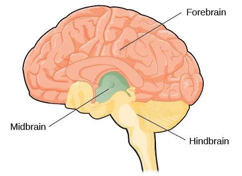
Hindbrain; oldest part of brain, primitive, controls function like eating and sleeping. Has four parts
Medula: vital functions like heartbeat, breathing, blood pressure
Pons: sleep and arousal
Cerebellum: motor coordination (e.g. timing of leg and arms movements, stroke affects this)
Reticular activating system: key in arousal, directing attention,(dysregulated in this system results in ADHD brains)
Midbrain and forebrain
Midbrain: Responsible for movement, transmit information that enables seeing and hearing (between brain and the eyes and the ears, sensory)
Forebrain: has the most complex cognitive activities, sensory and associative function and voluntary motor activities. Has cerebral cortex and limbic system: has the Thalamus, Hypothalamus, amygdala and hippocampus
Cerebral cortex: For higher mental processes: Has 4 regions (lobes).
What are the four lobes: Frontal: planning, decision making. Parietal: sensation (somatosensory). Temporal: auditory. Occipital: vision
Lateralization: function which relies on one side of the brain compared to another side; Left: speech, actions like facial expression and motion detection. Right hemisphere: coarse language skills, simple speech, writing, tone of voice,
Frontal lobe: planning, execute functions, motor most sophisticated information processing. Broca area: language formation. Motor cortex: responsible more body movement. Prefrontal Cortex: thinking, planning, and language, the “ceo”
Parietal lobe: somatosensory cortex; sensory for pressure and temperature: Communicates info to the motor cortex every time we reach grasp or move our eyes
Temporal lobe: responsible for understand language , storing autobiographical memories, auditory core (has the Wernicke’s area which is for language comprehension)
Occipital lobe: specialised for vision processing and higher order visual function (complex shapes, naming objects) Located back of the brain
Limbic system
Emotional center- also a role in smell, motivation and memory
Hypothalamus: regulates and controls internal bodily states (homeostasis); control pituitary glands (Body temp, hunger, sexual behaviour)
Thalamus: relays information from the sense organs to primary sensory cortex
Amygdala: Pays role in fear, aggression, excitement arousal, damage to this region makes it difficult to feel fear
Hippocampus: spatial memory, damage to this region causes inability to form new memories (memories are not stored here)
Nervous System
Peripheral Nervous system:
Somatic nervous system conveys information from Central Nervous System to muscles
Autonomic nervous system: control all involuntary movement of the body
Sympathetic fight or flight
Parasympathetic rest and digest
When one is active, the other is inactive
The polygraph: uses physiological measurements linked to ANS to detect “depiction”
You can fool a polygraph by activating the sympathetic nervous system by breathing, pinching yourself, anything that can trigger the baseline.
Endocrine system: glands which produce hormones to regulate bodily functions, regulates emotions
The hypothalamus: links the nervous system and endocrine system via the pituitary gland
Pineal gland secretes melatonin can calcify with age or alzheimer’s
Pituitary gland: releases hormones the influence growth
Oxytocin: responsible for reproductive functions implicated in trust and love