Week 7: Tissues Working Together 1: Mucosa and Serosa
Mucosa (Mucous Membrane):
Lines the digestive, respiratory, urinary and reproductive tracts. Basically lining any tube which is connected to the outside of the body.
Coated with secretions from the mucous glands.
The definition of the mucosa is based on the tissue type within the mucosa and the location of the mucosa.
Most of the time, a mucosa is made up of a:
Innermost surface epithelium
Underlying Loose CT Layers called laminate propia (location specific name)
However, for example, in the small intestine (part of the GIT), there is a third tissue included in the mucosa of the GIT.
So it has a:
Innermost surface epithelium
Underlying Loose CT Layer
Plus a thin band of smooth muscle called the muscularis mucosae (muscle of mucosa)
Mucosa in the small intestine:
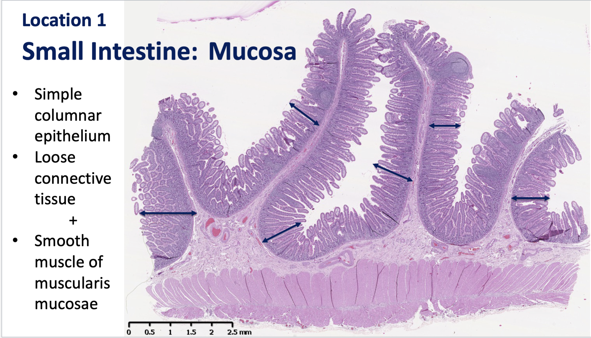
This is a LM of a section through the wall of the small intestine.
In this, we see the outer part of the wall of the small intestine at the bottom.
The arrows show the actual mucosa (purple appearance). Purple colour shows it is highly cellular as there is a nuclei with numerous cells present.
The simple columnar epithelium we also see on the surface of the mucosa. And directly under is the CT.
At higher magnification, there is a pink line that shows the smooth muscle (muscularis mucosae) specific to the small intestine.
Also arm-like protrusions present (really small), but this shows that villi is present.
Function of Mucosa In Small Intestine:
It acts as a site of exchange between the epithelium and capillaries. Contents of the lumen which need to cross the epithelium and get into blood capillaries.
Many epithelial and CT components contribute to this function.
CT characteristics of the Mucosa in Small Intestine
Abundant AGS present, allows for diffusion/transport of absorbed materials from epithelium into blood capillaries and lacteals.
AGS is viscuous as a gel nature > slows movement of large materials like bacteria, and allows free movement of small molecules
Numerous defense cells > neutralise and remove invading micro-organisms that have passed the epithelial layer.
Also has fine fibres instead of bundles, which gives structural support without blocking diffusion pathways.
Epithelial characteristics:
Simple Epithelium > Single layer of cells for ease of access
Columnar > Cells have cytoplasmic space for organelles to modify/process materials crossing epithelium
We can see that there are 2 cell types in epithelium, indicating 2 functions.
Cell Characteristics:
Have enterocytes with microvilli on the apical surface, which increases the surface area for absorption.
Goblet cells, which secrete mucus to protect the barrier against environmental damage.
Tight junctions to limit intercellular movement across epithelium to protect against seepage. Increases selectivity of absorption to limit bacterial invasion.
Mucosa in the Urinary Bladder:
The mucosa in here has:
A transitional epithelium instead.
Loose CT
Also has lamina propia.
Mucosa here acts as an impermeable barrier to prevent urine from getting into the blood circulation and body tissues. The epithelium predominantly contributes to this function.
Connective Tissue Characteristics:
Has abundant AGS like in the intestine, but the purpose is different. AGS here diffuses/transports oxygen, nutrients and removes waste between the capillaries and epithelium to maintain the epithelium in an optimal condition.
Few defense cells. There are fewer cells because the epithelium already provides a multiple layered cell (somewhat impermeable) prevent the invasion of organisms, so less defense cells are needed.
Also has fine fibres like intestine, which provides structural support but does not block pathways for diffusion.
Epithelial Characteristics:
Multiple layers of cell > acts as a physical barrier to prevent entry across epithelium. If a cell wants to cross from the lumen, it has to go around all these layers of cell.
Surface cells also have the ability to change shape, hence why its called a transitional epithelium,. This means it can cope with changes in volume of the bladder lumen. (So if the lumen is empty, the cells are rounded up to reduce space. Then when urine fills up, the cells can stretch to accomodate the change in fluid).
Cell Characteristics:
Tight Junctions > Seal of intercellular space so materials cannot go across the epithelium through the cells.
Surface cells have specialised membrane plaques, which makes the membrane impermeable to small molecules. Materials cannot cross the epithelium via the transcellular pathway.
Comparison of Lamina Propria in Intestine and Bladder:
Basically, the density of cells reflects function.
In the intestine laminate propia, there is a lot of absorption happening, and lots of defense cells are present to remove harmful products and materials.
Whereas in the laina propia in the bladder, there is no absorption occurring. In fact, there are few defense cells needed as the epithelium is doing most of the work as a barrier preventing those harmful products/materials.
Serous Membrane (Serosa)
Lines the body cavities closed to the exterior of the body. I.E. Peritoneal (Abdominal), Pleural (Lungs) + Pericardial (Heart) Cavities:
The definition of the serous is also based on the tissue type and location of it.
Epithelium in every serosa is the same type. Its called a covering mesothelium (simple squamous epithelium).
Also has underlying thin band of CT like mucosa. Its generally loose CT but can be different
Function: Lines enclosed body cavities and covers outer surfaces of organs in body cavities.
Has location specific names like:
Peritoneal = Abdominal
Pleural = Lungs
Pericardial = Heart
Peritoneum = Organ covering + lining of Abdominal Cavity
Mesentry = Double-layer serosa that covers organs in abdominal cavity
Functions of the epithelium + CT:
Mesothelium provides a smooth surface, gives a gliding movement of organ to reduce workload
Has a slippery fluid coating dervied from blood in vessels in serosal CT.
CT provides flexible support to mesothelium; fibres provide strength + robusticity to serosa
CT also stores lipids in adipocytes.
Serosa in the Epicardium of the heart (outermost layer of the heart)
Has blood vessels
Adipose Tissue - Protects vessels and heart works less harder cause of less resistance
Mesothelium provides that smooth slippery surface, allows heart to beat without much friction.
Review the characteristics of epithelium & connective tissue & the concept of tissue membranes.
Link specific structural components of epithelium & connective tissue with their functions.
Apply structure/function linkages to explain the general & location-specific roles of a mucosa & a serosa.
Appreciate that the various roles undertaken by a mucosa or serosa rely on cooperation between individual tissues (& cells) & reflect efficiency of location specific function.