Lesson 4: Vector
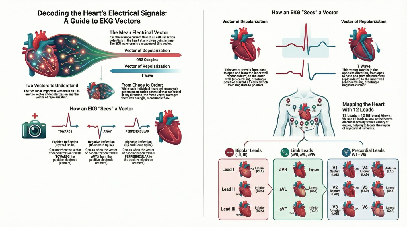
Lesson 4: Mean Electrical Vector
Represents average current flow of action potentials at a specific time.
EKG waveform measures the mean electrical vector.
EKG Deflections
Positive deflection: Vector of depolarization towards positive electrode.
Negative deflection: Vector of depolarization away from positive electrode.
Biphasic deflection: Vector travels perpendicular to positive electrode.
EKG Leads
Categories:
Bipolar leads: I, II, III
Limb leads: aVR, aVL, aVF
Precordial leads: V1-V6
Mapping leads to coronary arteries for diagnosing myocardial ischemia:
Right coronary artery: Inferior heart (II, III, aVF)
Circumflex artery: Left lateral heart (I, aVL, V5, V6)
Left coronary artery: Anterior heart (V1-V4)
Vectors in Action Potentials
Two key vectors:
Vector of depolarization
Vector of repolarization
Each myocyte generates an action potential moving in various directions.
Deflections and Current Direction
Positive deflection -> vector towards positive electrode.
Negative deflection -> vector away from positive electrode.
Biphasic deflection -> vector perpendicular to positive electrode.
Depolarization and Repolarization
Depolarization (QRS Complex):
Direction: Base to apex, endocardium to epicardium.
Polarity: From internally (-) to internally (+).
Produces a positive current.
Repolarization (T Wave):
Direction: Apex to base, epicardium to endocardium.
Polarity: From internally (+) to internally (-).
Produces a negative current.
Repolarization vector often points in the same direction as depolarization due to opposite direction and negative current relationship.