VF Revision - Alex Black
Visual Field Defects
Topographical Organization:
Fibers within the visual pathways correspond closely to specific areas within the visual field.
Visual pathways can be divided into four territories.
Variable terminology exists, which may refer to either visual field space or retinal location.
Visual Field Definitions:
Right Visual Field (VF): Composed of:
Right nasal fibres
Left temporal fibres
Superior Visual Field (VF): Composed of:
Right and left inferior fibres.
Visual Field Plots:
Right Eye (RE) is typically presented on the right side of diagrams.
blind spot on right.
Left Eye (LE) is represented on the left side of diagrams.
blind spot on left.
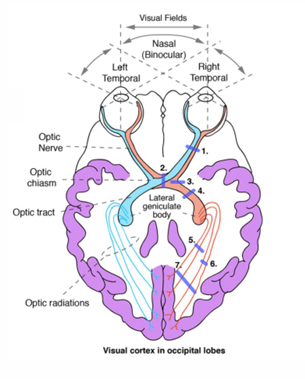
VF Territory 1 Overview
Anatomical Regions Represented:
Lids, brows, nose, rim of the trial frame.
Cornea, Lens, Vitreous body.
Outer retina conditions:
Macular Degeneration (MD)
Diabetes
Retinitis Pigmentosa (RP)
Choroidal conditions affecting outer retina.
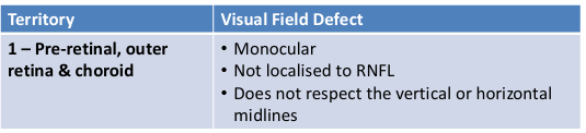
Visual Field defect 1:
Involves pre-retinal, outer retinal, and choroidal defects.
Characteristics:
Monocular (affecting one eye).
Not localized to the Retinal Nerve Fiber Layer (RNFL).
Does not respect the vertical or horizontal midlines.
VF Territory 2 Overview
Optic Nerve Head: Conditions like:
Glaucoma
Anterior Ischemic Optic Neuropathy (AOIN)
Papilledema.
Optic Nerve Damage: Caused by:
Optic Neuritis
Toxic damage
Hereditary factors
Orbital compression.
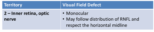
Visual Field Defect 2:
Affects inner retina and optic nerve.
Characteristics:
Monocular.
May follow distribution of RNFL.
Respects the horizontal midline.
VF Territory 3 Overview
Common Patterns of Defects:
Bitemporal Hemianopias: Usually beginning superiorly.
Anterior Junctional Scotoma.
Homonymous Hemianopias: Incongruent forms.
Characterised by:
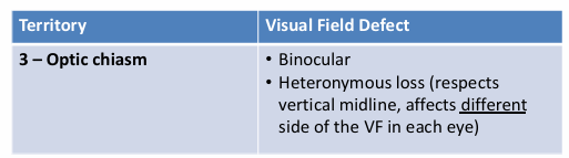
Heteronymous loss (respects vertical midline, different effects on each eye).
Usually manifests as bitemporal hemianopias, typically beginning inferiorly.
Visual Field Defect 3:
Involves the optic chiasm.
Binocular (affecting both eyes).
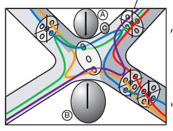
VF Territory 4 Overview
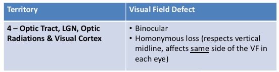
Defects Related to Specific Brain Structures:
Incongruous Homonymous Hemianopia: Seen in the occipital tract.
Incongruous Homonymous Sectoranopias: Related to the Lateral Geniculate Nucleus (LGN).
Superior Congruous Quadrantanopia: Involvement of temporal lobe or optic radiations.
Inferior Congruous Quadrantanopia: Associated with the parietal lobe or optic radiations.
Congruous homonymous hemianopias
macular sparing or splitting
sparing or loss of temporal crescent
homonymous paracentral scotoma
Visual field defect 4:
Affects optic tract, LGN, optic radiations, and visual cortex.
Characteristics:
Binocular.
Homonymous loss (respects vertical midline, affects the same side of the VF in both eyes).
Describing visual field defects
Accurate Description Requirements:
Essential for clinical records and referrals regardless of the underlying cause.
Four key description criteria:
Monocular or binocular defect.
Depth of defect:
Shallow (mild depression, small total deviation values) to dense / absolute (severe depression, large total deviation value).
expected central dB value: 30 ish
25 ~ shallow
1 ~ moderate
if > 0 dB = absolute defect → px cannot see the brightest light
if = 0 dB → px can first see target at the brightest light.
Location/Pattern of defect:
Various presentations including diffuse, cluster, superior quadrant, nasal hemifield.
Use relevant visual field terminology, such as:
Homonymous, Heteronymous, Altitudinal, Nasal step, Arcuate defect, Congruous, Incongruous, Macular sparing/splitting.
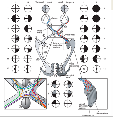
Examples of Defects and Their Locations
Example 1:
Defect Description: Nasal absolute scotomas not respecting horizontal midline.
Likely Location: Rim defect.
Example 2:
Defect Description: Far peripheral ring absolute scotomas not respecting horizontal or vertical midline.
Likely Location: Rim defect.
Example 3:
Defect Description: Mid peripheral ring absolute scotomas not respecting horizontal or vertical midline.
Likely Location: Rim defect.
Example 4:
Defect Description: Clover leaf pattern.
Likely Location: Initial attention followed by extensive inattention.
Example 5:
Defect Description: RE diffuse mild generalized depression.
Likely Cause: Incorrect correcting lens.
Central 24-2 Threshold Test
Test Details:
Fixation Monitor: Gaze/Blind Spot.
Fixation Target: Central.
Fixation Losses: 1/15, 2/16 (various examples).
False Positive Errors: Ranging from 0% to 6%.
False Negative Errors: 0% to 3%.
Test Duration: Varies, e.g., 06:14, 06:00.
Fovea: Generally reported as OFF.
Stimulus: III, White.
Background: 31.5 ASB.
Strategy: SITA-Standard.
Cases of Visual Field Defects
Comprehensive lists of reported defects include:
Congruous homonymous hemianopsia: Macular sparing or splitting (temporal crescent sparing).
Incongruous homonymous hemianopias: Observed in various lesion sites affecting the optic tract, LGN, etc.
Cases of significant conditions such as glaucoma affecting optic nerve head and visual pathways.
Defining lesions causing various patterns of visual loss, e.g., lesions in the occipital lobe leading to sectoranopia or quadrantanopia.
Visual Field Defect Case Analysis
Comprehensive examples include:
Right eye (RE) superior dense defect with left eye (LE) normal.
Left eye arcuate scotoma linked to glaucoma.
Dense peripheral defects caused by retinitis pigmentosa affecting photoreceptors.