Human Reproductive System
Core Syllabus Requirements & Research Objectives
Puberty and Hormonal Regulation
Definition and Onset:
Puberty is the developmental stage during which a child's body physically matures into an adult body capable of sexual reproduction.
It marks a growth period where sex organs mature and produce sex cells (gametes).
Onset timing varies individually: in boys, puberty typically begins at ; in girls, it begins at .
Changes encompass physical (visible external structural shifts), physiological (internal functional modifications), emotional, and hormonal alterations.
Endocrine Initiation Pathway:
The hypothalamus in the brain initiates puberty by releasing Gonadotropin-Releasing Hormone ().
acts on the anterior pituitary gland, stimulating the secretion of Follicle Stimulating Hormone () and Luteinizing Hormone ().
In females, stimulates ovarian follicle development, initiating the menstrual cycle, while triggers ovulation and subsequent progesterone production.
In males, stimulates spermatogenesis, while drives testosterone secretion from Leydig cells in the testes.
Hormones Active During Puberty:
Growth Hormone (): Increases growth rate and causes somatic growth spurts.
Luteinizing Hormone (): Induces ovulation in females and stimulates the production of oestrogen, progesterone, and testosterone.
Follicle Stimulating Hormone (): Stimulates maturation of germinal cells into ova or sperm.
Progesterone: Maintains the endometrial lining of the uterus to support blastocyst implantation.
Oestrogen & Testosterone: Steroid hormones responsible for developing secondary sexual characteristics.
Comparative Physical Changes in Females and Males:
Female Physical Changes (Oestrogen-driven) | Male Physical Changes (Testosterone-driven) |
|---|---|
Rapid growth in height (up to ) | Rapid growth in height (up to ) |
Axillary (armpit) hair growth | Axillary (armpit) hair growth |
Pubic hair growth (pubarche) | Pubic hair growth (pubarche) |
Skin becomes oilier; acne/pimples may develop | Skin becomes oilier; acne/pimples may develop |
Breast development (thelarche; begins at , fully mature at ) | Facial hair growth (beard/moustache at ); voice deepens (, squeaky during breaking) |
Widening of hips and pelvic frame | Broadening of shoulders and chest |
Ovaries develop and initiate ovum production; vagina grows; menstruation (menarche) starts ~ post-thelarche | Penis and testes enlarge; testes initiate sperm production (from ) |
Emotional and Social Dimensions:
Puberty induces mood swings, changes in social dynamics with peers and family, and the emergence of personal identity.
Male Reproductive System Anatomy and Physiology
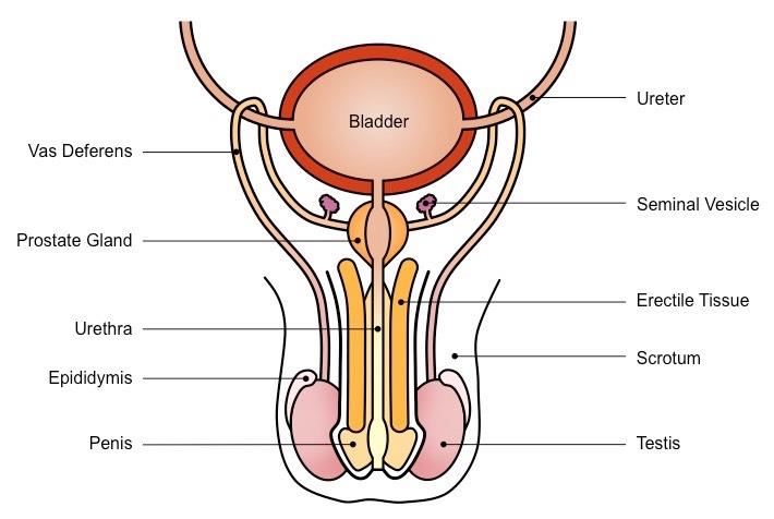
Penis:
Male copulatory organ consisting of a shaft and a terminal conical head called the glans penis.
The glans penis is partially covered by loose skin called the foreskin (prepuce), which is removed during circumcision.
The internal shaft contains three longitudinal columns of spongy erectile tissue:
Corpora Cavernosa: Two paired dorsal/upper columns of erectile tissue.
Corpus Spongiosum (Corpus Cavernosum Urethrae): One ventral/lower column surrounding the urethra.
During sexual arousal, increased blood flow fills the vascular spaces (blood sinuses) of the spongy erectile tissue, causing the penis to swell, stiffen, and become erect.
Engorgement of spongy tissue maintains urethral patency during erection, allowing fluid transport.
Function: Transfers semen into the female vagina during copulation.

Urethra:
Urinogenital duct passing through the penis that carries urine from the urinary bladder and semen from reproductive glands to the exterior, though never simultaneously.
Urinary Bladder:
Muscular sac that stores urine before excretion.
Sperm Duct (Vas Deferens):
Muscular tube that transports sperm via peristaltic contractions from the epididymis to the urethra.
Prostate Gland:
Exocrine gland positioned surrounding the base of the bladder.
Secretes a milky, alkaline fluid that provides semen with its characteristic odour.
Prostatic fluid promotes maximum sperm motility and neutralizes acidic residues in the male urethra and female vagina.
Seminal Vesicles:
Glandular structures posterior to the bladder.
Secrete a thick, yellowish, sticky fluid rich in fructose, serving as the primary energetic substrate (food source) for sperm locomotion.
Cowper's Glands (Bulbourethral Glands):
Two small glands situated on either side of the urethra inferior to the prostate.
Produce alkaline secretions that clear urethral urine residue, enhance sperm motility, and lubricate the glans penis prior to intercourse.
Epididymis:
A tightly coiled, convoluted duct approximately in length situated along the posterior border of each testis.
Formed by the convergence of seminiferous tubules.
Functions as a storage reservoir where immature sperm mature and acquire motility over time.
Lined by a mucous membrane that secretes fluid to assist sperm movement.
Scrotum:
An external pouch of skin enclosing the testes outside the abdominal cavity.
Maintains testicular temperature at below core body temperature (), an environment necessary for viable spermatogenesis.
Testes (Singular: Testis):
Primary male sex organs (gonads) suspended within the scrotum.
Internally partitioned into lobules, each packed with convoluted seminiferous tubules.
Combined length of seminiferous tubules in a single testis can reach up to , maximizing surface area for high-volume sperm generation.
Functions: Production of male gametes (spermatozoa) and secretion of testosterone.
Testicular Microanatomy and Spermatogenesis
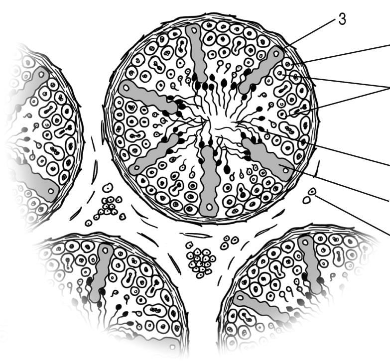
Histological Cell Types of the Testis:
Germinal Epithelial Cells: Diploid cells lining the inner surface of seminiferous tubules that undergo meiotic division to produce haploid spermatocytes and mature spermatozoa.
Sertoli Cells: Large, elongated helper cells located near the basolateral membrane of seminiferous tubules. They provide metabolic support, protection, and nutrients to developing germ cells during spermatogenesis. Sertoli cells are activated by to initiate sperm maturation. Absence of functional Sertoli cells causes male infertility.
Leydig Cells (Interstitial Cells): Clusters of endocrine cells situated in the connective tissue spaces outside the seminiferous tubules. They produce and secrete testosterone when activated by .
Spermatozoa (Sperm Cells): Microscopic, motile haploid male gametes produced within the germinal epithelium via meiosis.
Spermatogenesis and Semen:
Spermatogenesis is the complete process of sperm cell production via meiotic division within the germinal epithelium of the seminiferous tubules.
Semen is the fluid ejaculated from the male urethra, comprising spermatozoa suspended within seminal plasma (secretions from seminal vesicles, prostate, and Cowper's glands).
Unreleased sperm inside the male reproductive tract are phagocytosed and broken down by the body.
Mammalian sperm cells cannot survive prolonged exposure to air, necessitating internal fertilisation.
Physiological Roles of Testosterone:
Primary Sexual Development: Drives embryonic testicular descent into the scrotum, initiates spermatogenesis, causes penis and testicular enlargement, and heightens libido.
Secondary Sexual Characteristics: Regulates male secondary sex traits including facial/body hair distribution, vocal cord enlargement (voice deepening), and anabolic growth.
Anabolic Effects: Stimulates skeletal muscle hypertrophy by accelerating protein synthesis, and promotes bone elongation at epiphyseal growth plates early in puberty, followed by epiphyseal fusion later in adolescence.
Structure of the Sperm Cell and Sexual Intercourse

Morphology of Spermatozoa:
Head Region: Contains a dense haploid nucleus storing paternal chromosomes. The anterior tip is covered by the acrosome, a modified lysosome containing hydrolytic enzymes (such as hyaluronidase) capable of digesting the protective outer layers (corona radiata and zona pellucida) of the ovum during fertilisation.
Middlepiece (Midpiece): Contains spirally arranged mitochondria tightly wrapped around the axial filament. These mitochondria generate adenosine triphosphate () via cellular respiration to power flagellar beating.
Tail (Flagellum): Long, thin whip-like structure that executes undulatory wave movements, propelling the sperm cell through cervical mucus, uterine cavity, and oviducts.
Mechanism of Sexual Intercourse:
Prior to copulation, parasympathetic signals cause blood sinuses within the corpora cavernosa and corpus spongiosum of the penis to fill with blood, producing an erection.
Vaginal walls secrete lubricating fluids to facilitate penile insertion.
At sexual climax, sympathetic stimulation triggers ejaculation, propelling semen (containing hundreds of millions of sperm cells) through the vas deferens and urethra into the upper vagina near the external cervical os.
Female Reproductive System Anatomy and Physiology

Uterus (Womb):
A hollow, thick-walled, inverted pear-shaped muscular organ located in the female pelvis between the bladder and rectum.
Functions as the site of embryonic implantation and foetal development throughout pregnancy.
Uterine Wall Composition:
Serous Layer (Perimetrium): Outer thin membranous visceral layer.
Muscular Layer (Myometrium): Thick middle coat of involuntary smooth muscle fibers capable of forceful contractions during labour.
Endometrium: Inner mucosal lining rich in blood vessels and glands. Undergoes cyclic changes under ovarian steroid influence. If fertilisation does not occur, its functional layer sloughs off during menstruation.
Fallopian Tube (Oviduct):
Paired muscular tubes extending laterally from near each ovary into the superior portion of the uterus.
The open ovarian end terminates in a expanded funnel (infundibulum) with finger-like projections (fimbriae).
Lined with ciliated epithelium and smooth muscle cells that move the ovum toward the uterus.
Functions as the primary site of fertilisation.
Ovary:
Paired primary female sex organs (gonads) measuring approximately to in length, attached to the uterus via ovarian ligaments.
Function dual roles as primary gametogenic organs (producing ova) and endocrine glands (secreting oestrogen and progesterone).
Ovaries alternate each month to mature and release a single secondary oocyte.
Cervix:
The narrow inferior neck of the uterus projecting into the upper vagina.
Contains a central cervical canal connecting the uterine lumen to the vagina.
Directs sperm deposited in the vagina into the uterine cavity, allows clearance of menstrual discharge, and dilates up to during childbirth.
Vagina:
A fibromuscular, elastic canal extending from the cervix to the body exterior.
Contains rugae (transverse folds) that allow distension during copulation and childbirth. Serves as the copulatory receptacle and birth canal.
Clitoris:
A small, highly sensitive erectile structure located at the anterior junction of the labia minora.
Homologous/analogous to the male penis, containing rich nerve endings and spongy erectile tissue.
Ovarian Histology, Folliculogenesis, and Development
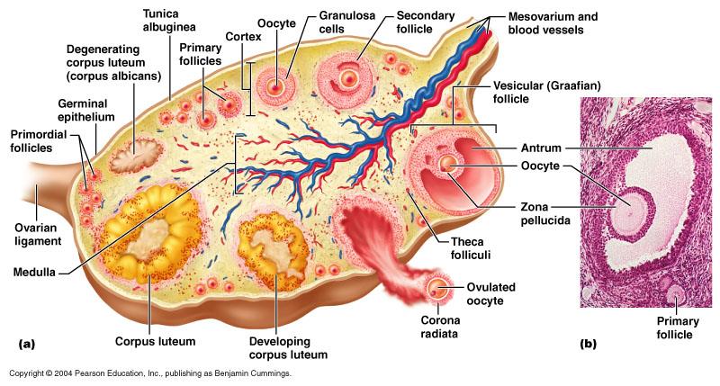
Ovarian Microanatomy:
Outer surface bounded by a simple cuboidal germinal epithelium overlying the tunica albuginea.
Divided into an outer cortex (containing developing follicles and oocytes) and an inner medulla (containing vascular, lymphatic, and neural networks).
Folliculogenesis:
Female infants are born with approximately primary follicles within their ovarian cortices.
Under periodic stimulation by during reproductive life, batches of primary follicles begin maturation into secondary follicles, though typically only one reaches full maturity each cycle.

The Graafian Follicle:
A fully developed mature fluid-filled ovarian follicle.
Contains a central primary/secondary oocyte surrounded by the zona pellucida (glycoprotein coat) and corona radiata (granulosa cell layer).
Features a large fluid-filled cavity called the antrum, filled with liquor folliculi, encased by internal (theca interna) and external (theca externa) vascular layers.
Granulosa cells of the Graafian follicle secrete high quantities of oestrogen.
Ovulation and Corpus Luteum Dynamics:
A mid-cycle surge of causes the Graafian follicle to rupture at the ovarian surface, releasing the ovum into the peritoneal cavity near the oviduct funnel.
Following ovulation, transforms the collapsed follicle wall into a temporary yellow endocrine gland: the corpus luteum.
The corpus luteum secretes large amounts of progesterone along with oestrogen to prepare and maintain the vascularized endometrium during the luteal phase.
If fertilization does not occur, the corpus luteum survives for before degenerating into a fibrous scar (corpus albicans).
Steroid levels drop following corpus luteum breakdown, removing hormonal support from the endometrium and triggering menstruation.
If fertilization occurs, the developing embryo secretes Human Chorionic Gonadotropin (), maintaining the corpus luteum until the placenta takes over progesterone production around of gestation.
Endocrine Regulation of the Reproductive Systems
Comparative Roles of Pituitary Gonadotropins ( and ):
Hormone | Action in Females | Action in Males |
|---|---|---|
Stimulates ovarian follicle growth and maturation; drives follicle cells to convert precursor steroids into oestradiol (oestrogen). | Stimulates Sertoli cells within seminiferous tubules to synthesize Androgen-Binding Protein () and inhibin, promoting spermatogenesis. | |
Triggers final oocyte maturation, rupture of Graafian follicle (ovulation at Day 14), and luteinization of follicular remnants into the corpus luteum. | Binds to Leydig (interstitial) cells, stimulating the synthesis and secretion of testosterone. |

Male Negative Feedback Control:
Elevated serum testosterone levels feed back to the hypothalamus and anterior pituitary, inhibiting , , and secretion.
Inhibition of decreases Leydig cell testosterone production, restoring physiological hormone balance.
Excessive testosterone or exogenous synthetic anabolic steroid abuse triggers severe negative feedback, suppressing pituitary release. Lacking stimulation, Sertoli cell function declines and spermatogenesis ceases, leading to testicular atrophy and infertility.
Elevated sperm counts stimulate Sertoli cells to release inhibin, which selectively acts on the anterior pituitary to lower output, slowing down sperm production rates.
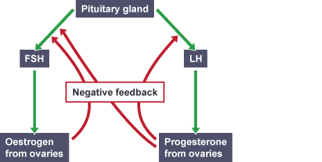
Female Negative Feedback Control:
Low systemic oestrogen and progesterone levels at the end of a cycle stimulate pituitary release of .
Rising oestrogen secreted by maturing follicles exerts moderate negative feedback on , preventing over-development of excess follicles.
High concentrations of oestrogen produced by a dominant Graafian follicle switch to positive feedback, triggering a massive surge of that induces ovulation.
Post-ovulatory progesterone secreted by the corpus luteum exerts strong negative feedback on both the hypothalamus and pituitary gland, shutting down and release to prevent new follicle maturation while the uterus prepares for implantation.
The Menstrual Cycle and Ovarian Cycle

Chronological Breakdown of the Menstrual Cycle ( Average):
Days 1–5 (Menstrual Phase / Menstruation):
Circulating levels of oestrogen and progesterone are at baseline low values.
Deprived of hormonal maintenance, the functional layer of the endometrium breaks down, sloughs off, and is discharged through the vagina along with blood and mucus.
levels begin a slight rise near Day 5 to stimulate a new cohort of ovarian follicles.
Days 5–13 (Repair / Proliferative / Follicular Phase):
Rising promotes ovarian follicle enlargement.
Developing follicular granulosa cells secrete increasing amounts of oestrogen.
Oestrogen acts on the uterine wall, repairing and thickens the endometrium, stimulating arterial growth and glandular proliferation.
Day 14 (Ovulation Phase):
Peak concentration of oestrogen induces a sharp surge in pituitary output.
The Graafian follicle ruptures, releasing the secondary oocyte into the pelvic cavity for pickup by the oviduct fimbriae.
The endometrial lining continues thickening.
Days 15–28 (Secretory / Luteal Phase):
The ruptured follicle converts into the corpus luteum, which secretes high amounts of progesterone alongside oestrogen.
Progesterone converts the thickened endometrium into a receptive, highly vascularized, and nutrient-glandular secretory tissue.
If fertilisation does not occur: the unfertilized ovum disintegrates within , the corpus luteum degenerates around Days 24–26, progesterone and oestrogen output drops, and endometrial sloughing initiates Day 1 of the subsequent cycle.
If fertilisation occurs: embryonic preserves the corpus luteum, sustaining elevated progesterone levels and preventing menstruation.
Cellular Lifespans and Reproductive Milestones:
An ovulated egg remains viable for fertilisation for approximately .
Injected spermatozoa can survive and remain fertilizing-capable within the female reproductive tract for .
Menopause: Permanent cessation of ovarian follicular activity and menstrual cycles, typically occurring around of age.
Fertilisation, Early Development, and Implantation
Fertilisation (Conception):
Process wherein a single haploid sperm cell successfully penetrates and fuses its nucleus with that of a haploid secondary oocyte.
Occurs within the upper region of the oviduct (fallopian tube).
The ovum is moved along the oviduct length toward the uterus by coordinated beating of ciliated epithelial cells and peristaltic muscular contractions.
Prevention of Polyspermy:
Upon entry of the first sperm cell head, cortical granules release contents that alter the vitelline membrane/zona pellucida, forming a impenetrable fertilization barrier that blocks entry of additional sperm.
Zygote to Blastocyst Transformation:
Sperm-egg nuclear fusion restores diploidy ( chromosomes), forming a zygote.
The zygote travels down the oviduct, undergoing rapid mitotic cleavage divisions without increasing overall size, transforming into a solid ball of cells (morula) and then a hollow blastocyst.
Implantation:
Approximately 6–7 days post-fertilisation, the blastocyst embeds into the thickened endometrial lining of the uterus.
Trophoblastic cells of the blastocyst absorb nutrients from glandular secretions until functional placental connection establishes.
Gestation Stages:
Total human pregnancy duration is approximately ().
Embryo: Term applied to the developing human from fertilisation through the first of gestation.
Foetus: Term applied from (9 weeks) onward, when all primary organ systems have formed.
Gestation and Pregnancy Hormones
Key Hormones Governing Pregnancy:
Hormone | Primary Source | Major Gestational Function |
|---|---|---|
Luteinizing Hormone () | Anterior Pituitary | Induces Graafian follicle rupture (ovulation) and converts remnants to corpus luteum. |
Follicle Stimulating Hormone () | Anterior Pituitary | Stimulates primary ovarian follicle growth and oocyte maturation. |
Progesterone | Corpus Luteum (early); Placenta (after ) | Maintains vascularized endometrial lining, suppresses uterine smooth muscle contractions, and prevents menstruation during pregnancy. |
Oestrogen | Ovarian Follicle, Corpus Luteum, Placenta | Promotes uterine hypertrophy, enhances blood flow, maintains uterine lining, and triggers foetal organ development. |
Oxytocin | Posterior Pituitary | Induces rhythmic uterine smooth muscle contractions during labour; stimulates myoepithelial contraction for milk ejection (let-down) during lactation. |
Prolactin | Anterior Pituitary | Stimulates alveolar epithelial growth and milk synthesis within mammary glands. |
Extra-Embryonic Membranes, Placenta, and Umbilical Cord
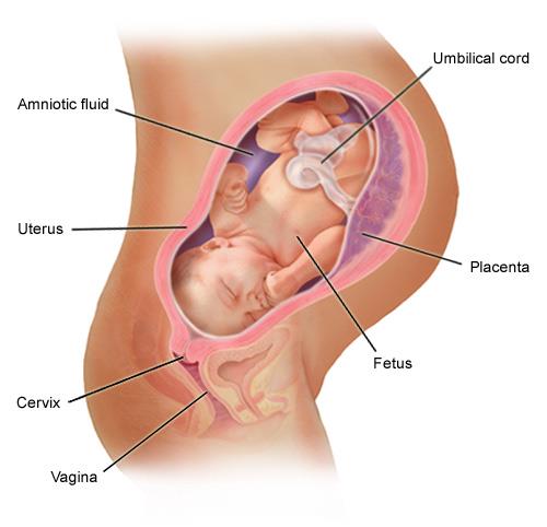
Placenta:
A temporary discoid organ formed jointly from foetal chorionic tissue and maternal endometrial tissue (decidua basalis).
Typically attached to the superior, lateral, anterior, or posterior uterine wall. Implantation near or over the internal cervical os is termed placenta previa (low-lying placenta).
Term Dimensions: Mass of approximately , diameter of , thickness of , and functional exchange surface area of nearly 15\text{ m}^2$.\n * Structural unit: **Chorionic Villi** — finger-like vascular projections of foetal tissue bathed in maternal blood pools within intervillous spaces.\n * **Maternal and foetal blood systems NEVER mix directly.** Exchange occurs across thin placental membranes via diffusion and active transport.\n\n\n\n* **Functions of the Placenta:**\n 1. **Nutrition:** Transfers dissolved nutrients (glucose, amino acids, fatty acids, vitamins, ions) from maternal circulation to foetal blood.\n 2. **Excretion:** Transports metabolic waste products (urea, uric acid, creatinine) from foetal blood to maternal blood for disposal.\n 3. **Gas Exchange:** Delivers maternal oxygen ( ext{O}_2 ext{CO}_2).\n 4. **Microfiltration & Passive Immunity:** Selective barrier preventing entry of most bacteria and macromolecules. Allows maternal Immunoglobulin G ( ext{IgG}) antibodies to cross, providing passive immunity. Certain pathogens (e.g., Rubella virus / German measles, Human Immunodeficiency Virus / HIV) and toxins can cross.\n 5. **Endocrine Secretion:** Takes over synthesis of progesterone and oestrogen from the corpus luteum around the 12th week of pregnancy, maintaining gestational stability.\n\n* **Chorionic Villus Sampling (CVS):**\n * A prenatal diagnostic procedure involving the extraction of a sample of chorionic villi cells from the placenta.\n * Used early in pregnancy to screen for genetic diseases, chromosomal abnormalities (e.g., Down syndrome), and metabolic disorders.\n\n* **Amnion and Amniotic Fluid:**\n * The **amnion** is a membrane enclosing the fluid-filled amniotic cavity around the embryo.\n * **Amniotic Fluid Composition:** Initially isotonic saline water; by weeks 12-14, contains dissolved proteins, carbohydrates, lipids, phospholipids, electrolytes, and urea.\n * **Functions of Amniotic Fluid:**\n * Serves as a fluid cushion and shock absorber, protecting the foetus against external abdominal impacts.\n * Prevents adhesion of foetal skin to surrounding amnion membranes.\n * Provides temperature regulation and buoyancy, allowing unhindered movement required for muscular and skeletal development.\n * Foetal swallowing of fluid promotes gastrointestinal tract maturation; foetal inhalation/exhalation of fluid drives normal lung and alveolar expansion.\n\n* **Amniocentesis:**\n * A diagnostic procedure where a needle inserted through the maternal abdominal and uterine walls under ultrasound guidance aspirates a sample of amniotic fluid.\n * Sloughed foetal skin cells within the fluid are cultured to detect chromosomal defects, metabolic disorders, neural tube defects, or determine sex.\n\n\n\n* **Umbilical Cord Anatomy and Physiology:**\n * A flexible conduit connecting the foetal umbilicus to the placenta.\n * Enased by amniotic epithelium and filled with a gelatinous connective tissue called **Wharton's jelly**.\n * Contains three primary blood vessels:\n * **Two Umbilical Arteries:** Carry deoxygenated, nutrient-depleted blood laden with metabolic waste products ( ext{CO}_2, urea) from the foetal internal iliac arteries to the placenta.\n * **One Umbilical Vein:** Carries oxygenated, nutrient-rich blood from the placenta to the foetal heart.\n\n* **Blood Composition Comparisons:**\n\n| Umbilical Arteries (2) | Umbilical Vein (1) |\n| :--- | :--- |\n| Deoxygenated blood | Oxygenated blood |\n| Foetus ightarrow ightarrow Foetus direction |\n| High waste products ( ext{CO}_2 ext{O}_2); low waste |\n\n| Maternal Arteries | Umbilical Arteries |\n| :--- | :--- |\n| High ext{O}_2 ext{O}_2 concentration |\n| Low ext{CO}_2 ext{CO}_2 concentration |\n| High nutrient concentration | Low nutrient concentration |\n| Low metabolic waste concentration | High metabolic waste concentration |\n\n# Birth, Lactation, and Mammary Gland Physiology\n\n\n\n* **Parturition (Birth Process):**\n * Initiated by hormonal signals: maternal progesterone levels fall, while the posterior pituitary releases high amounts of **oxytocin**.\n * Oxytocin stimulates strong, rhythmic contractions of the myometrium.\n * The cervix softens, effaces, and dilates to a maximum of 10 ext{ cm}.\n * Uterine contractions push the foetus downward, pressing against the cervix, driving delivery through the dilated cervix and vagina.\n * **Caesarean Section (C-Section):** Surgical delivery of a baby through abdominal wall and uterine incisions, performed when vaginal delivery poses clinical risks to mother or child.\n\n* **Mammary Glands and Lactation Mechanics:**\n * **Anatomy:** Structural lobules of glandular alveoli embedded in adipose tissue, supported by suspensory ligaments (Cooper's ligaments), draining into lactiferous ducts, lactiferous sinuses, and exiting via the nipple.\n * **Gestation Stage:** High progesterone and prolactin levels circulate. High progesterone directly blocks prolactin receptors at the mammary alveolar level, preventing active lactation prior to delivery. Small amounts of pre-milk fluid (**Colostrum**) are produced.\n * **Colostrum:** A thick, yellowish fluid produced during late pregnancy and early postpartum days. Highly concentrated in maternal secretory antibodies ( ext{IgA}), proteins, and minerals, providing passive protection and nutrition to the neonate.\n * **Postpartum Stage:** Loss of the placenta causes circulating progesterone to drop precipitously while prolactin remains elevated. This removes receptor inhibition, signaling alveolar cells to synthesize milk.\n * **Suckling Reflex:** Neonatal suckling sends afferent neural impulses to the hypothalamus, stimulating anterior pituitary prolactin release (sustaining milk synthesis) and posterior pituitary oxytocin release (inducing myoepithelial contraction in lactiferous sinuses, driving milk ejection / let-down reflex).\n\n# Contraceptive Methods and Reproductive Health\n\n* **Classification of Contraception:**\n 1. **Natural / Fertility Awareness Methods:** Tracking menstrual cycle timing and body temperature to identify ovulation, practicing periodic abstinence during fertile windows (e.g., Rhythm method).\n 2. **Barrier Methods:** Physical obstacles preventing sperm from reaching the cervical canal.\n * *Male Condom:* Thin latex/polyurethane sheath rolled over the erect penis.\n * *Female Condom (Femdom):* Internal polyurethane sheath lining the vagina.\n * *Diaphragm / Cervical Cap:* Shallow dome-shaped silicone caps inserted into the vagina covering the external cervical os.\n 3. **Intrauterine Devices (IUD):** T-shaped devices inserted into the uterine cavity by a practitioner.\n * *Hormonal IUD:* Releases progestin locally, thickening cervical mucus and keeping the endometrium thin.\n * *Copper IUD:* Releases copper ions that act as a spermicide and induce a localized endometrial reaction.\n\n \n\n 4. **Chemical / Hormonal Methods:** Synthetic steroid administration preventing ovulation.\n * *Oral Contraceptive Pills:* Combined oestrogen and progestin pills taken daily that inhibit ext{FSH} ext{LH} release, blocking ovulation.\n * *Injectable Contraceptives:* Long-acting progestin injections (e.g., Depo-Provera) administered every 3 months.\n * *Transdermal Patches, Vaginal Rings, and Subdermal Implants.*\n 5. **Surgical / Permanent Sterilisation:**\n * *Vasectomy:* Surgical cutting and ligation/cauterization of both vas deferens in males, preventing sperm passage into ejaculated semen.\n * *Tubal Ligation:* Surgical cutting, clamping, or tying of both fallopian tubes in females, preventing gamete transport.\n 6. **Emergency Contraception:** High-dose progestin pills ("morning-after pill") taken post-unprotected intercourse to delay or prevent ovulation.\n\n* **Comparative Analysis of Contraceptive Methods:**\n\n| Contraceptive Type | Method & Mechanism | Advantages | Disadvantages |\n| :--- | :--- | :--- | :--- |\n| **Condom** (Barrier) | Latex sheath covers penis; traps semen, preventing sperm entry into vagina. | Highly effective if used correctly; **protects against STDs/STIs**. | Can tear/break if misused; latex allergy; shelf-life expiration risks. |\n| **Diaphragm** (Barrier) | Flexible silicone cup inserted over cervix prior to intercourse. | No hormonal side effects; reusable, inexpensive. | Lower efficacy than hormonal options; **does not protect against STDs/STIs**. |\n| **Contraceptive Pill** (Hormonal) | Daily oral intake of oestrogen + progesterone; suppresses ext{FSH/LH} to stop ovulation. | Highly effective; regulates menstrual cycles and dysmenorrhea. | Requires strict daily compliance at same hour; potential systemic side effects. |\n| **Hormone Injection** (Hormonal) | Deep intramuscular progestin injection given every 3 months. | Highly effective; long-lasting; eliminates daily pill compliance. | Potential weight gain, mood shifts, or delayed fertility return; no STD protection. |\n| **IUD** (Intrauterine Device) | T-frame inserted into uterus; copper ions kill sperm or progestin thins lining. | Near 100 ext{\%}99 ext{\%}3-12 ext{ years}; rapidly reversible. | Insertion pain/cramping; risk of heavier initial bleeding; **no STD protection**. |\n| **Vasectomy** (Surgical) | Bilateral transection and ligation of vas deferens. | Permanent; near 100 ext{\%} effective; does not alter sexual performance. | Surgical risks; non-protecting against STDs; difficult/expensive to reverse. |\n| **Tubal Ligation** (Surgical) | Surgical transection or clipping of fallopian tubes. | Permanent; extremely effective; no ongoing maintenance. | Invasive surgical procedure; irreversible; non-protecting against STDs. |\n| **Rhythm Method** (Natural) | Tracking menstrual cycle dates; abstaining during predicted fertile window. | Zero financial cost; no medical side effects; culturally acceptable. | **Least effective method** due to cycle variability; no STD protection. |\n\n# Infertility, Surrogacy, and Male Circumcision\n\n* **Infertility Treatments:**\n * Defined as the inability to conceive after 12 months of regular unprotected intercourse.\n * Treatments include ovulation-inducing medications (e.g., clomiphene citrate), surgical restoration of tubal/duct patency, and Assisted Reproductive Technology (ART).\n * *Intrauterine Insemination (IUI):* Direct catheter insertion of washed sperm into the uterine cavity around ovulation.\n * *In Vitro Fertilisation (IVF):* Surgical retrieval of oocytes, fertilisation with sperm in a laboratory dish, and transfer of resulting embryos into the uterus.\n\n* **Surrogacy:**\n * An arrangement wherein a woman (surrogate) carries a child to term for intended parents.\n * Regulated under South African legal frameworks: formal court confirmation of a surrogate motherhood agreement is required prior to fertilisation. The surrogate relinquishes all parental rights upon birth, vesting full legal parenthood in the intended parents.\n\n* **Medical Male Circumcision (MMC):**\n * Surgical excision of the penile foreskin (prepuce), exposing the glans penis.\n * Performed for clinical/health benefits (reduces risk of acquiring HIV, Human Papillomavirus, and urinary tract infections), religious, or cultural reasons.\n * MMC does not reduce male fertility or sperm output.\n\n# Teratogens and Health Factors During Pregnancy\n\n* **Effects of Maternal Smoking:**\n * Inhaled nicotine (vasoconstrictor), carbon monoxide (binds hemoglobin to form carboxyhemoglobin), and heavy metals cross the placenta.\n * Restricts uterine artery blood flow, lowering oxygen and nutrient delivery to the foetus.\n * Increases foetal resting heart rate.\n * Elevates risks of spontaneous abortion (miscarriage), stillbirth, premature rupture of membranes, low birth weight (< 2500 ext{ g}), placental abruption, and neonatal respiratory distress.\n\n* **Effects of Maternal Alcohol Consumption & Foetal Alcohol Syndrome (FAS):**\n * Ethanol freely crosses the placenta. Foetal liver enzymes (alcohol dehydrogenase) are immature, causing high and prolonged foetal blood alcohol concentrations.\n * Alcohol acts as a potent teratogen, inducing cell death and disturbing cell migration in the developing central nervous system.\n\n\n\n* **Foetal Alcohol Syndrome (FAS) Clinical Profile:**\n * *Somatic Growth:* Pre- and post-natal growth retardation.\n * *Craniofacial Anomalies:* Microcephaly (abnormally small head and brain), short palpebral fissures, smooth philtrum, and thin upper lip border.\n * *Central Nervous System Damage:* Structural brain defects, microencephaly, intellectual disability, impaired motor skills, attention deficit, hyperactivity, and speech/language delays.\n * *Organ Defects:* Congenital heart defects, spinal abnormalities, and joint contractures.\n\n* **Effects of Illicit and Prescription Drugs:**\n * Cross the placenta, increasing rates of spontaneous abortion, placental abruption, intrauterine growth restriction (IUGR), premature labor, birth defects, neonatal withdrawal syndromes, and maternal death.\n\n# Review Exercises and Tutorial Practice\n\n* **Penis Microanatomy & Sinuses (Tutorial Focus):**\n * *Significance of Blood Sinuses:* The presence of large vascular sinuses within the corpora cavernosa and corpus spongiosum allows rapid accumulation of blood under pressure during sexual arousal. Compression of outflow veins maintains penis rigidity (erection), enabling vaginal penetration and keeping the urethra open for semen passage.\n\n* **Seminal Fluid Components & Functions:**\n 1. *Fructose (Seminal Vesicles):* Provides metabolic ATP energy for sperm flagellar motility.\n 2. *Alkaline Prostatic Fluid (Prostate Gland):* Neutralizes acidic environments of the male urethra and female vagina, ensuring sperm survival.\n 3. *Mucus / Pre-ejaculary Lubricant (Cowper's Glands):* Clears urethral urine residues and lubricates the glans penis.\n\n* **Steroid Abuse & Fertility Calculation Exercise:**\n * Exogenous anabolic steroid intake increases circulating synthetic testosterone ightarrow ightarrow ext{FSH} ext{LH} ightarrow ext{FSH} ightarrow spermatogenesis halts, causing testicular atrophy and azoospermia/infertility.\n\n* **Follicle Diameter Measurement Data Analysis:**\n\n| Day of Menstrual Cycle | Average Follicle Diameter ( ext{mm}) |\n| :--- | :--- |\n| Day 5 | 6.9 ext{ mm} |\n| Day 10 | 13.3 ext{ mm} |\n| Day 13 | 18.8 ext{ mm} |\n| Day 14 | 22.0 ext{ mm} |\n| Day 20 | 3.4 ext{ mm} |\n| Day 25 | 2.6 ext{ mm} |\n\n* *Hormone driving growth from Day 5 to 14:* ext{FSH}.\n* *Explanation of drop from Day 14 (22.0 ext{ mm}3.4 ext{ mm}):* Day 14 marks ovulation; the mature Graafian follicle ruptures, releasing the secondary oocyte and follicular fluid, causing the collapsed follicular structure to shrink as it transforms into the corpus luteum.\n* *Magnification Formula:* ext{Magnification} = rac{ ext{Measured Image Size}}{ ext{Actual Size}}$$. Expressed rounded to 1 decimal place with appropriate units.