H Biology Unit 5 Cell Division Study Guide
The Cell Cycle and Mitosis
Why do cells need to divide?
DNA Overload
A large cell would not have enough DNA to address the needs of a growing cell
Large Volume to SA ratio
A large ratio causes problems for the cell.
The Cell Cycle
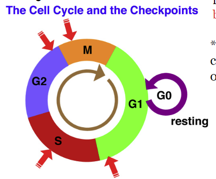
The life of the cell is divided into three main parts: Interphase, mitosis, and cytokinesis
Interphase: Consists of three parts: Growth 1 (G1), Synthesis (S), and Growth 2 (G2). The main events of interphase are cell growth and DNA replication. DNA is in the form of chromatin.
During G1, the cell is doing its job. This also contains cell growth.
G0, called the resting stage, is when the cell does not divide but still performs normal functions (eg heart cells/neurons enter G0, and remain there for the rest of their life.
During the S phase (synthesis), the cell makes a complete copy of its DNA
G2, (growth 2) the cell prepares for mitosis by making more organelles, and growing the cytoplasm.
Mitosis AKA karyokinesis (etymology: nucleus movement) does not include interphase and cytokinesis: Use the acronym PMAT: Prophase, Metaphase, Anaphase, and Telophase. The goal of mitosis is to produce a clone of the cell.
Prophase (pro- first or before) - Chromatin coils into chromosomes, nuclear membrane disappears, centrioles begin to form spindle fibers. DNA is in the form of sister chromatids or duplicated chromosomes.
Metaphase (meta sounds like middle) - Spindle fibers attach to the centromeres of chromosomes and pull them to line up in the middle
Anaphase (ana soudns like away)- Sister chromatids separate and move to opposite poles of the cell
Telophase (telo means distant/end)- Spindle fibers disappear, chromatids begin to unwind into chromatin, cytokinesis begins, nuclear membrane reappears. Cleavage furrow appears.
Cytokinesis: Division of the cell (cytoplasm and organelles), completes cell division. DNA is in the form of single chromatids.
Interphase usually takes about 18-20 hours. (5-6 hours for S phase, G2 2-4 hours). Mitosis takes about 2 hours. The total cell cycle takes about 22-24 hours.
Checkpoints happen during G1, S, and M. If everything does not pass, the cell combusts itself (apoptosis)


Bacteria reproduces in a similar process called binary fission, which uses budding. It is not technically mitosis because they lack a nuclear envelope.
Vocabulary
Sister Chromatid - When the DNA is copied during S phase, the duplicated chromosome will contain two identical DNA strands
Centromere- The central region that connects two sister chromatids
Chromatid - When sister chromatids are separated during mitosis, they become independent chromosomes, each made out of one DNA strand
Chromosome- Supercoiled DNA wrapped around proteins called histones
DNA - Made out of nitrogen base
Centrioles- Organelles that produce spindle fibers
Spindle fibers - Microtubules (proteins) that help move chromosomes to opposite sides of the cell (poles)
Regeneration - Regrow lost parts yayyy
Autosomes - Chromosomes 1-22
Cancer
Cancer is caused by damaged DNA. Environmental factors such as chemicals, radiation, or viruses can damage DNA. Habits such as sunbathing or smoking greatly increase the chances of DNA damage. Mutations can also be inherited genetically.
Most irregular cells are caught at checkpoints and killed through apoptosis. However, with a faulty checkpoint, the irregular will continue to divide forever, causing a tumor.
Benign Tumors
Tumors that do not spread to other parts of the body. They usually remain in a capsule (thin layer of skin) and can be safely removed.
They could possibly turn malignant but are usually removed before.
Malignant Tumors
Tumors which can metastasize, spread through the body and start new tumors
Karyotypes
Karyotypes are pictures of people’s chromosomes. A woman can take a amniocentesis to detect genetic disorders in her baby.
A human karyotype has 46 chromosomes arranged in pairs
The first 22 pairs are called autosomes and code for the same traits in both males and females. Autosomes are placed in order based on banding patterns and centromere position from largest to smallest.
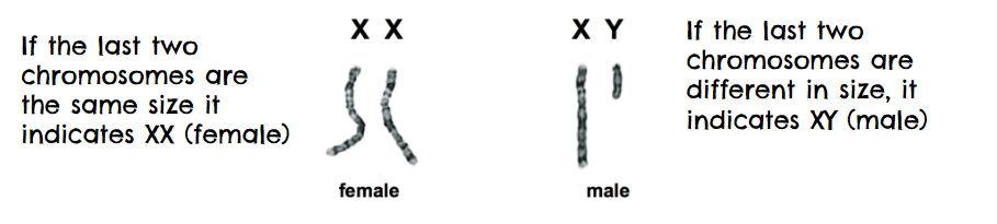
The individual’s gender is determined by looking at the 23rd pair of chromosomes, the sex chromosomes. If the sex chromosomes are XX, the individual is female. If the sex chromosomes are XY, the individual is a male.
Most chromosome abnormalities are caused by nondisjunction (failure of homologous chromosomes to separate). There are two main types. They mainly happen with faulty meiosis, when chromosomes fail to separate properly.
Trisomy: When there is more chromosomes than needed (47)
Monosomy: When there is less chromosomes than needed (45)
Some genetic diseases when a part of a chromosome is missing (deletion), region of chromosome is repeated (duplication), or moved to another chromosome (translocation)
Genetic Diseases
Turners Syndrome - One missing sex chromosome (45 instead of 46 chromosomes)
Edwards SYndrome - Extra 18th Syndrome
Klinefelter: Extra X Chromosome
Down Syndrome - Extra 21st Chromosome
Meiosis
Two kinds of cells: Somatic, and gametes
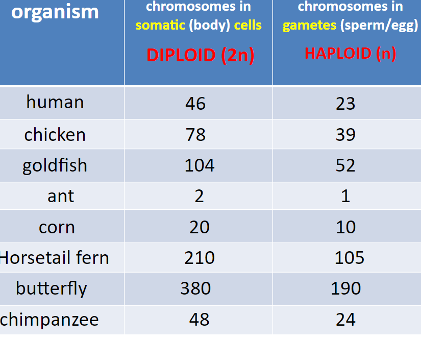
Somatic cells are called diploid. They are body cells and the symbol is 2n. Human somatic cells have 46 chromosomes. They are made by mitosis.
Gametes are called haploid. They are sex cells and the symbol is n. Human gametes have 23 chromosomes. They are made by meiosis in the gonads (ovaries/testes)
 The fusion of a sperm and egg make a zygote. A zygote is a diploid.
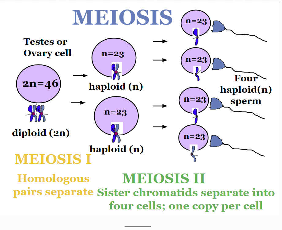
During spermatogenesis, each somatic testes cell makes 4 sperm. 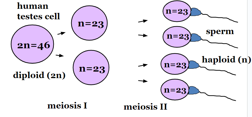
During oogenesis, each somatic ovary cell makes one egg and 3 polar bodies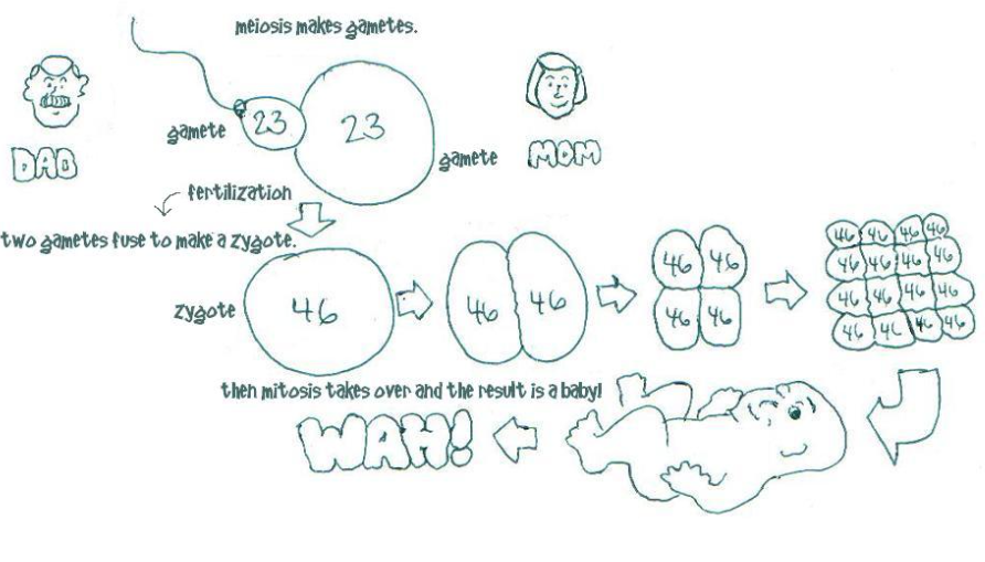
Phases of Meiosis - # 1
Meiosis 1 is cell division that reduces the number of chromosomes by half.
Interphase 1, is similar to mitosis. The chromosomes replicate during S phase. Each duplicated chromosome consist of two identical sister chromatids attached at their centromeres. Centriole pairs also replicate during G2. Nucleus and nucleolus visible.
PMAT
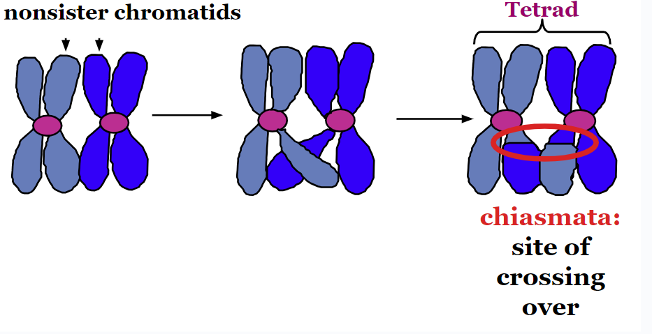
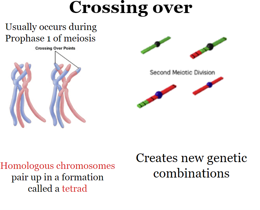
Prophase (90%) of meiosis. Synapsis (fusion of chromosomes) occurs: homologous chromosomes come together to form a tetrad. Tetrad is two homologues with four chromatids.
HOMOLOGOUS CHROMOSOMES -Pair of chromosomes (maternal and paternal) that are similar in shape and size.
One chromosome in the pair is called a homologue. Homologous pairs are called tetrads.
Each locus (position of a gene) is in the same position on each homologue.
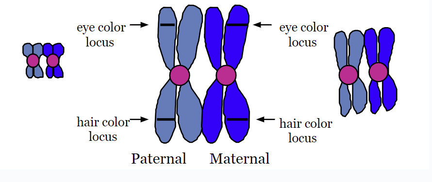
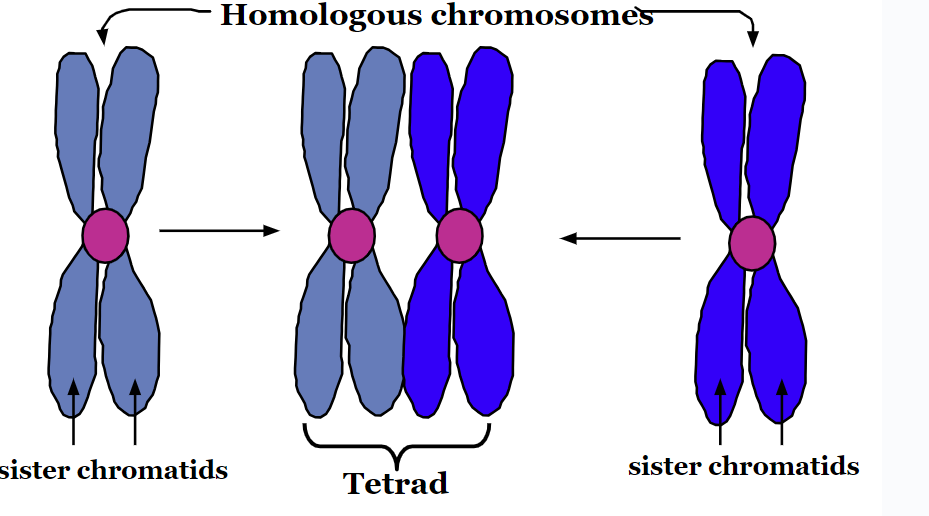
Humans have 23 pairs of homologous chromosomes.
Crossing Over
Segments of non-sister chromatids break apart and reattach to the other chromatid.
Chiasmata: The site of crossing over
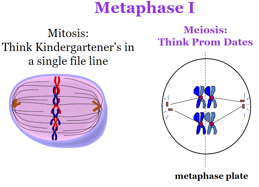
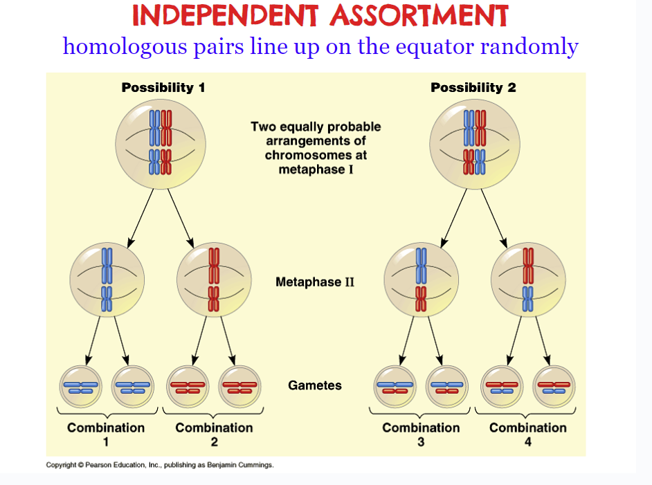
Metaphase 1 - Shortest phase. The tetrads align on the metaphase plate. Here is when independent assortment occurs. This causes variation, because they line up on the equator randomly. The amount of variation can be calculated using 2^n.
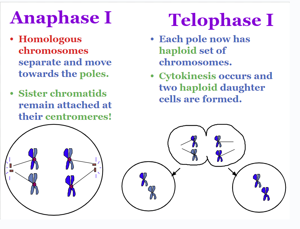
Anaphase 1 - Homologous chromosomes separate and move towards the poles (using spindle fibers). Sister chromatids remain attached to the centromere, unlike in mitosis.
Telophase 1 - Each pole now has a haploid set of chromosomes. Cytokinesis occurs, and two daughter cells are formed.
Phases of Meiosis - #2 (Remember there are two cells at this point)
Meiosis 2 does not have interphase! There is no DNA replication during this phase. Meiosis ll is very similar to mitosis.
Prophase - Centrioles begin to form spindle fibers
Metaphase - Chromosomes are pulled into a single file line on the metaphase plate.
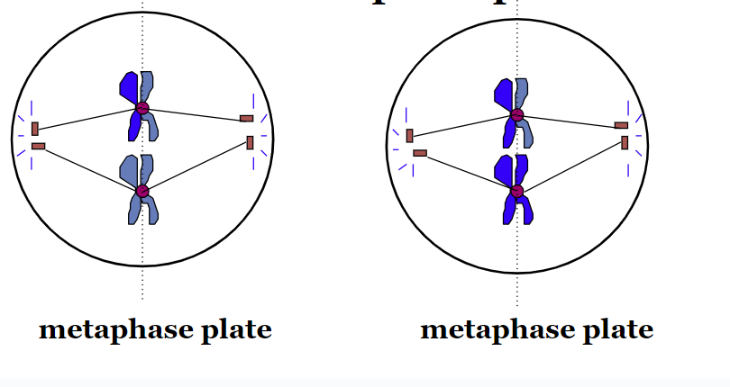
Anaphase - Sister chromatids separate
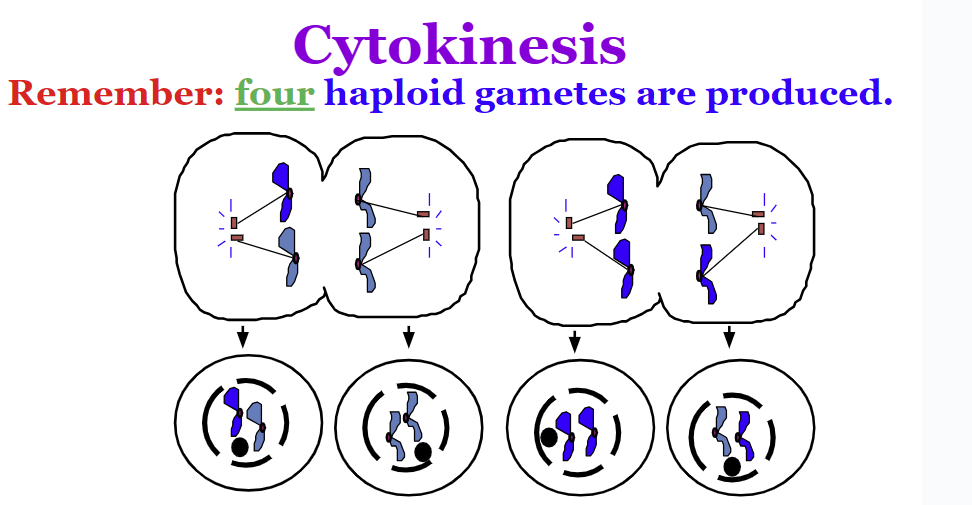
Telophase - Nuclei form and cytokinesis begins
Cytokinesis- Four haploid gametes are produced.
Where does genetic variation come from in meiosis?
Crossing Over
Independent assortment
Random fertilization
A cell containing 20 chromosomes (diploid) at the beginning of meiosis would, at its completion, produce cells containing how many chromosomes?
10 (2n→n to make haploid cells)
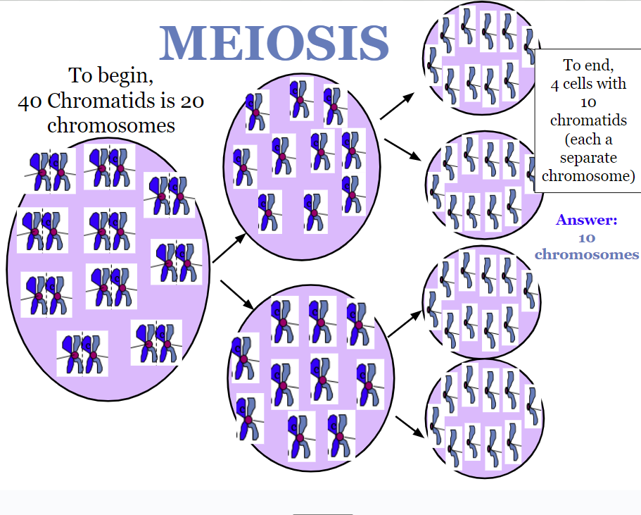
A cell containing 40 chromatids at the beginning of meiosis would, at its completion, produce cells containing how many chromosomes?
40 chromatids = 20 chromosomes. To end, four cells with 10 chromatids to make 10 chromosomes