✨⭐BIO 2 EXAM 3 FREEFORM NOTES⭐✨
Date: 3/19/25
Animal Nutrition
EXTRA CREDIT DUE MONDAY
•Every meal reminds us that we are heterotrophs, dependent on a regular supply of food
•In general, animals fall into three categories:
–Herbivores eat mainly autotrophs (plants and algae)
–Carnivores eat other animals
–Omnivores regularly consume animals as well as plants or algal matter
•An adequate diet must satisfy three needs:
–Fuel for all cellular work
–Organic raw materials for biosynthesis
–Essential nutrients, substances that the animal cannot make for itself
•Main feeding mechanisms: suspension feeding, substrate feeding, fluid feeding, bulk feeding
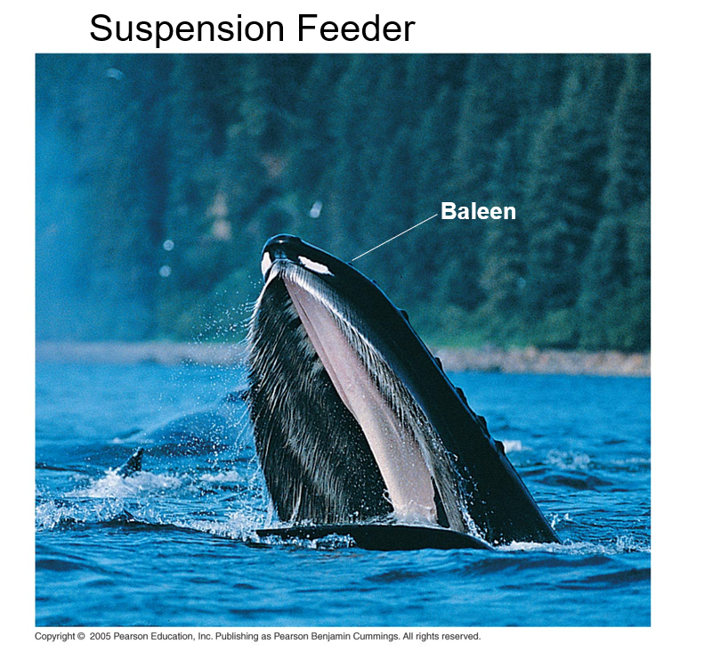
they create a suction in the water pulling in the nutrients
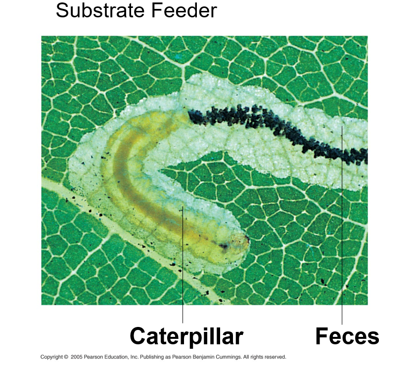
They live on or in the thing they’re eating. Maggots are also this as they crawl into the thing and go YUM YUM to the carcus
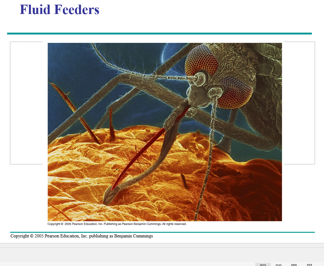
Mosquitoes, ticks, butterfly’s, leeches, hummingbirds. They only consume liquids —- things that were never solid
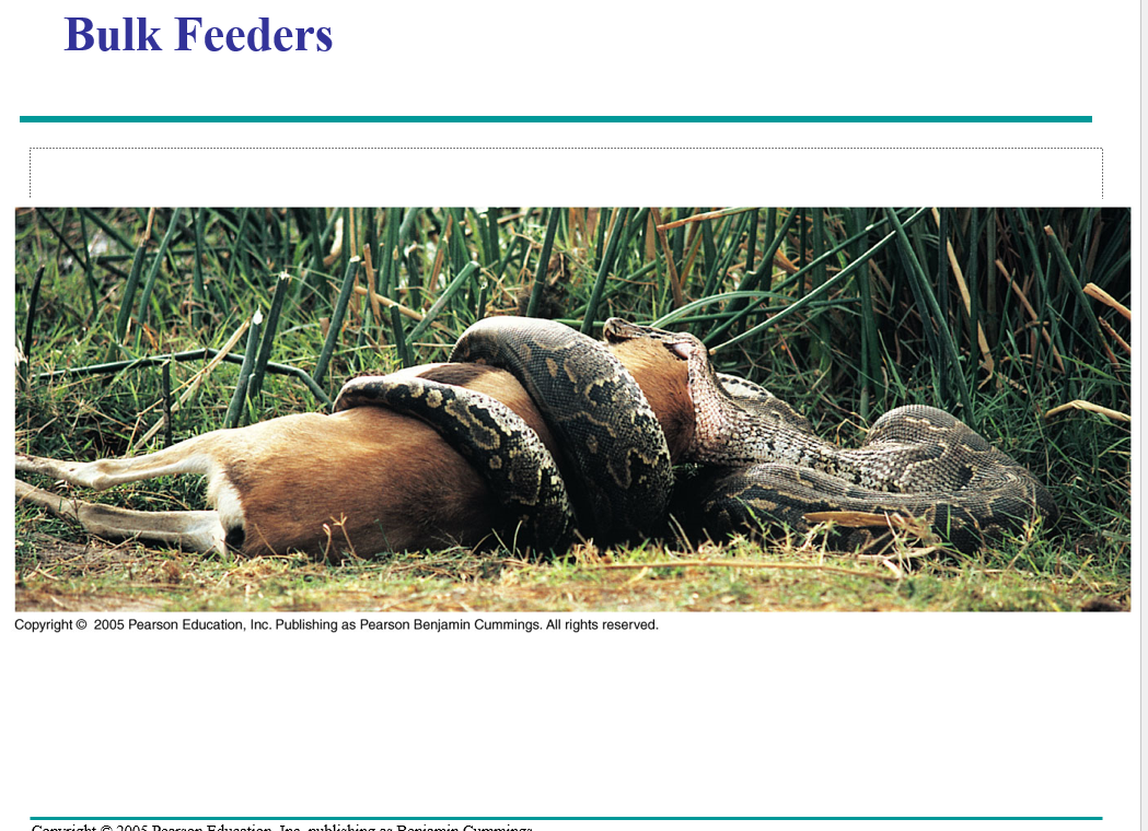
They eat things in chunks — this would be us/humans
Nearly all of an animal’s ATP generation is based on oxidation of E rich molecules: carbohydrates, proteins, and fats
Animals store excess calories as glycogen in the liver and muscles and as fat
glugose is a major fule for cells
hormones regulate glucose metabolism
When fewer calories are taken in than are expended, fuel is taken from storage and oxidized
Pancreas is Dr. Daft’s favorite organ. Small intestate is his second favorite.
Undernourishment occurs in animals when their diets are chronically deficient in calories — regardless of food intake. this just depends on the calories
–Glycogen and fat are used up – begin breaking down proteins for fuel
–Muscles decrease in size and brain becomes protein-deficient
–Occurs in drought, war, or anorexia
Overnurishment, or obesity, results from excess caloric intake stored as fat
the WHO recognizes obesity of obesity as a major global health problem
•Obesity contributes to a number of health problems, including diabetes, cardiovascular disease, and colon and breast cancer
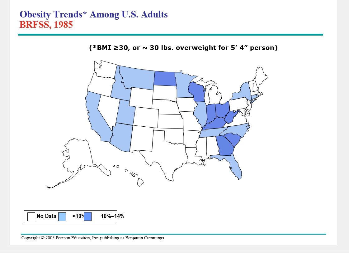
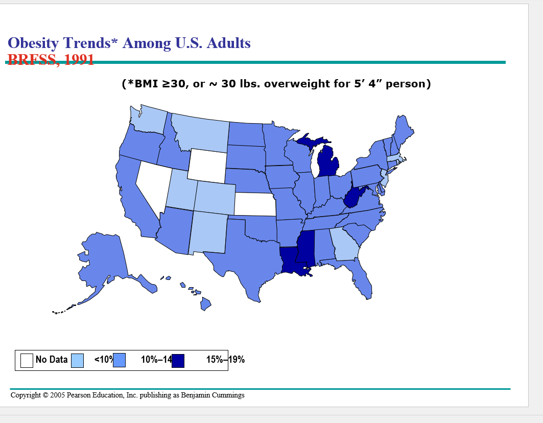
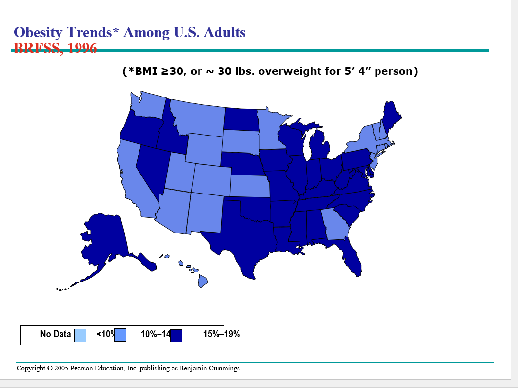
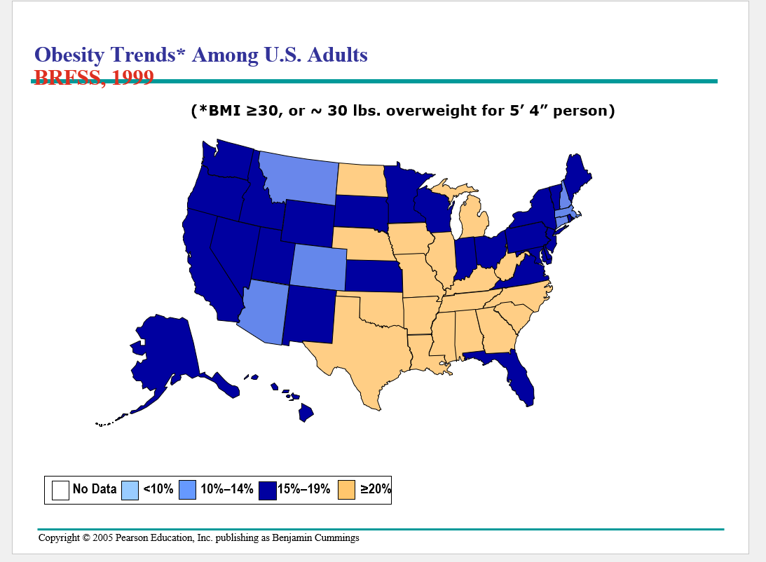

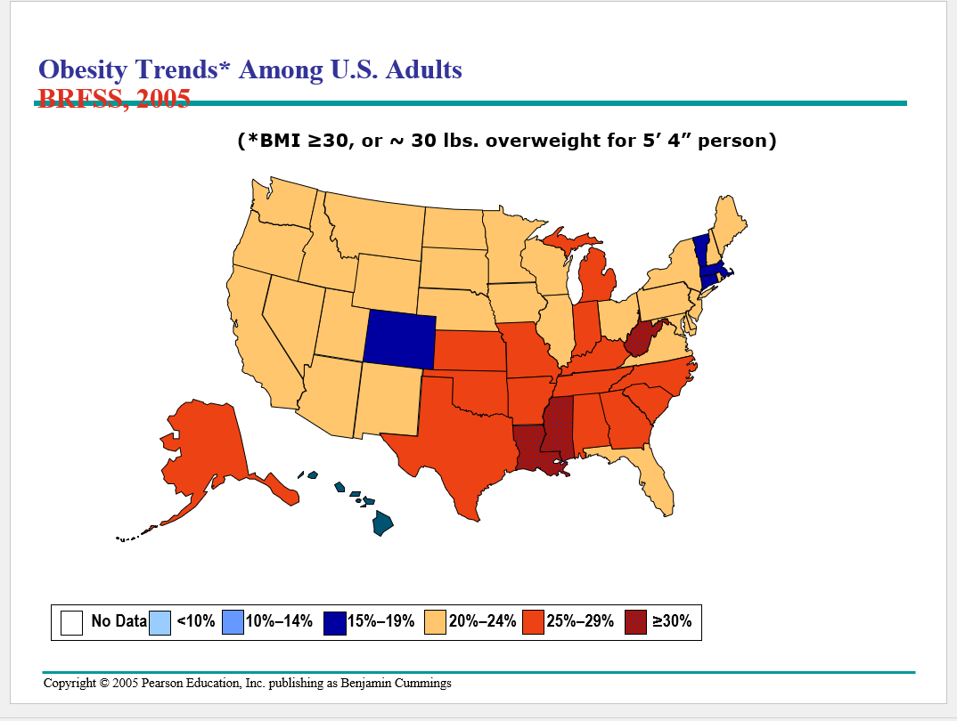
It just keeps going up in the southeast
Researchers have discovered several of the mechanisms that help regulate body weight
over the long term, homeostatic mechanisms are feedback circuits that control the body’s storage and metabolism of fat
Hormones regulate long-term and short-term appetite by affecting a “satiety center” in the brain
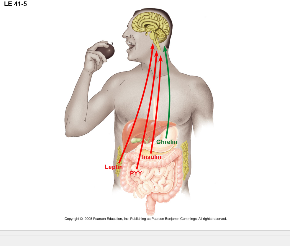
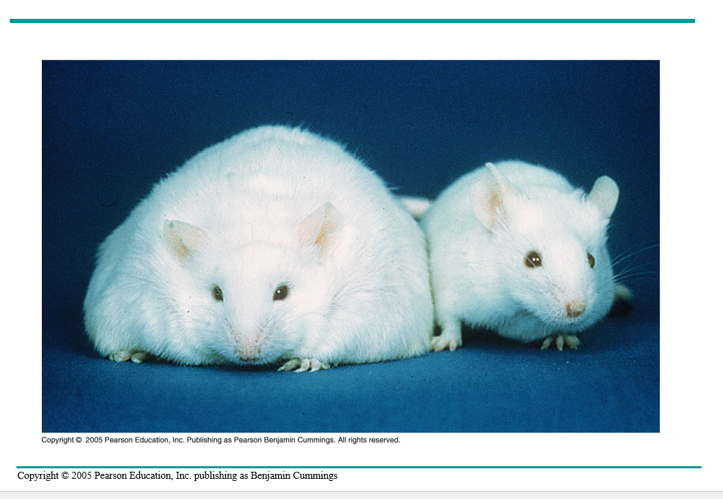
The complexity of weight control in humans is evident from studies of the hormone leptin
Mice that inherit a defect in the gene for leptin become very obese
Can decrease weight by injection of leptin
Can occur in humans
Fecal transplants baby! — ground up, strained poop, that’s shot up either the poop shoot or down the food shoot
The problem of maintaining weight partly stems from our evolutionary past, when fat hoarding was a means of survival
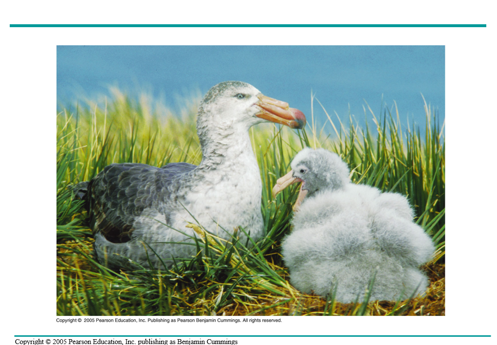
A species of birds called petrels become obese as chicks due to the need to consume more calories than they burn
An animal must obtain carbon skeletons from its food to build complex molecules
•Besides fuel and carbon skeletons, a diet must supply essential nutrients in preassembled form
•A malnourished animal is missing one or more essential nutrients in its diet
Can a person be obese and still be malnourished? — ON EXAM
Yes, because malnutrition is when you don’t have the essential nutrients, but obesity happens with too much caloric intake
•Herbivores may suffer mineral deficiencies if they graze on plants in soil lacking key minerals (phosphorous)
•Animals require 20 amino acids and can synthesize about half from molecules in their diet
•The remaining amino acids, the essential amino acids, must be obtained from food in preassembled form
•Eight amino acids (9 in infants) are essential
•A diet that provides insufficient essential amino acids causes malnutrition called protein deficiency
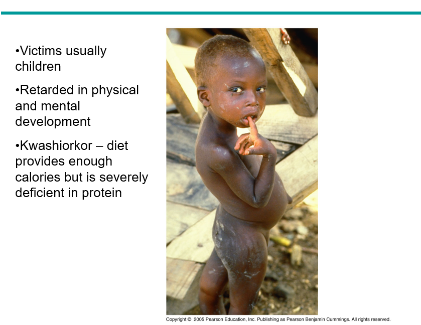
they’ll have a fluid excess due to the lack of protien, leeding to the bulid up on water due to osmolality suff
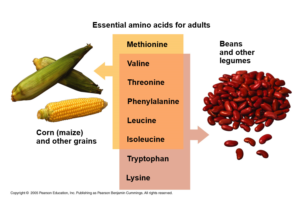
Rice is really similar to corn-- this is why rice and beans are basically in every culture.
Theretically, one could sustain on nothing but this.
Date:3/21/25
Animals can synthesize most of the fatty acids they need
the essential fatty acids are certain insaturated fatty acids
Human—linoleic acid must be present in the diet
Deficiencies in fatty acids are rare
Vitamins are organic mol. req. in the diet in small amts.
13 vitamins essential to humans have been ID’d
vitamins are grouped in two catagories: fat, and water soluble
•Water-soluble – overdoses excreted in urine
•Fat-soluble – overconsumption can be toxic
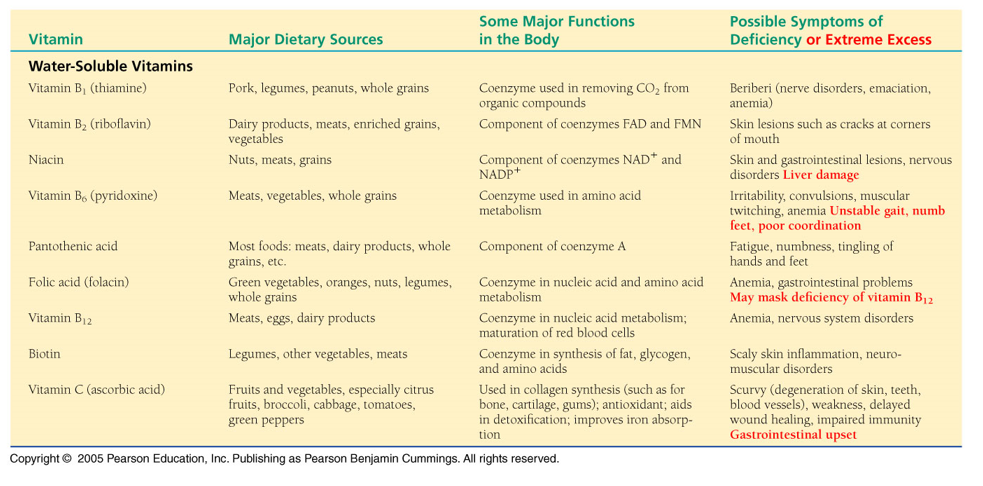
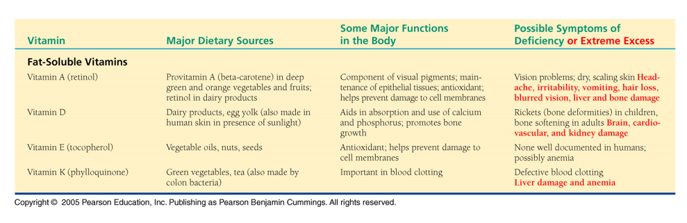
We get a lot of Vit.K from the bacteria in our guts
Minerals are simple inorganic nutrients, usually required in small amounts
•Most people ingest more salt than needed (20X required amount of sodium)
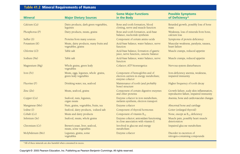
in your body you have bulk and trace elements
just BC trace, does not meen not important.
Iron is trace, but it causes big probs w/out it (anemia and such)
Neural tube defects — tissue fails to enclose the developing brain and spinal cord
1970-s — more frequently in low socioeconomic communities
Thought malnutrition may be responsible
Vit supplements (folic acid) reduces risk
•All 20 amino acids are needed to make animal proteins. Why aren’t they all considered essential amino acids? — WILL BE ON TEST
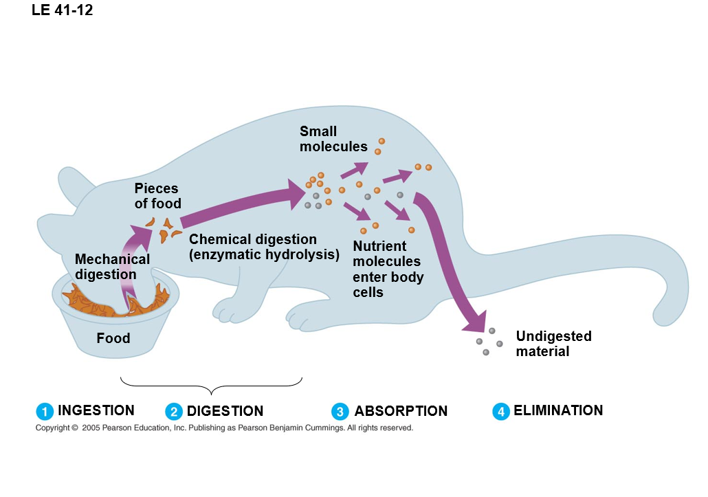
•Ingestion is the act of eating
•Digestion is the process of breaking food down into molecules small enough to absorb
–Enzymatic hydrolysis
•Absorption is uptake of nutrients by body cells
•Elimination is the passage of undigested material out of the digestive compartment
Cellulose = fancy word for fiber
if colin absorbs too much water =constipation
if colin absorbs not enough H2O diahhreah
How do animals apply their digestive processes to food w/out digesting their own cells and tissue
•Most animals process food in specialized compartments
•These compartments reduce risk of self-digestion
In intracellular digestion, food partials are engulfed by endocytosis and digested within food vacules
Newly formed food vacuoles fuse with lysosomes — contain hydrolytic enzymes
•Mixes food with the enzymes and allows digestion to occur safely within a compartment
•Sponges digest their food entirely by the intracellular mechanism
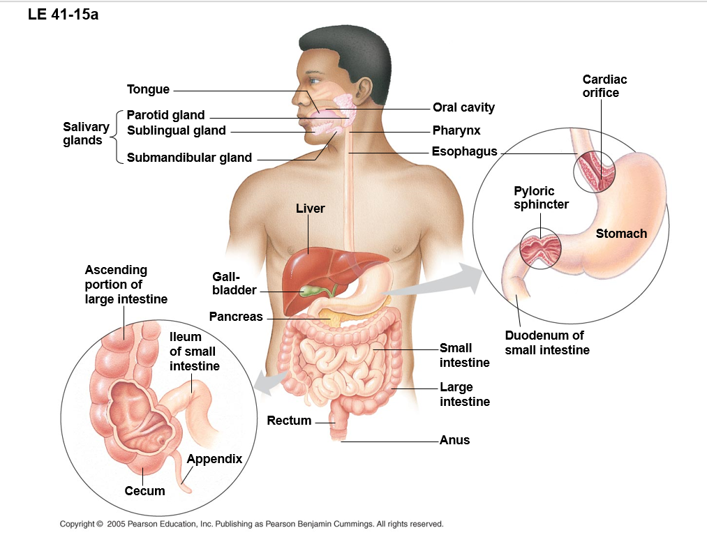
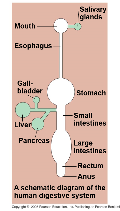
the mammalian digestive system has a alimentary canal and accessory glands that secrete digestive juices through ducts
The accessory glands are: salivary, pancreas, liver, and gallbladder
food is pushed along by peristalsis, which is the rhythmic contractions of muscles in the wall of the canal
sphincters, which close off the tube, regulates the passage of material b/t chambers of the canal — think butt hole
a plasmid in a bacterial cell is accessory DNA. This is for in bad situation, and can be removed and be OK.
The accessory organs in humans ARE NOT (typically) optional
Saliva is of lubricating and beginning the digestion of food
teeth are to break apart food
•In the oral cavity, food is lubricated and digestion begins
•Humans produce more than a liter of saliva each day – may salivate in anticipation
•Saliva – protects the lining of mouth from abrasions, lubricates food, buffers to prevent tooth decay, contains antibacterial agents
•Teeth chew food into smaller particles that are exposed to salivary amylase, initiating breakdown of glucose polymers
Tongue: tastes food, manipulates food while chewing, shapes food into ball called bolus
•The region we call our throat is the pharynx, a junction that opens to both the esophagus and the windpipe (trachea)
•Trachea covered by epiglottis, cartilaginous flap
•This keeps food from going down the “wrong pipe”
•The esophagus conducts food from the pharynx down to the stomach by peristalsis
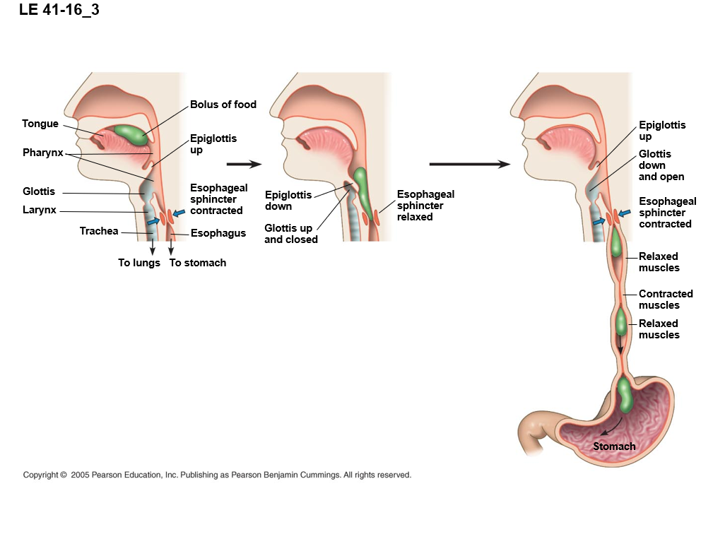
there are skeletal muscles at the upper esophagus, not true for rest of digestive system
Date 3/24/25
GLP makes you feel more full
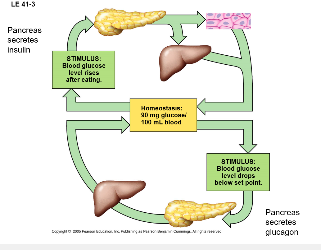
insulin’s responsibility is to reduce your blood sugar
GLP-1 goal to stimulate insulin and to down regulate glucagon to prevent glycogen and fat storage
insulin resistance occurs when the cells sorta loss the receptors and have a harder time responding to the insulin
Dr. Daft thinks the GLP-1 drugs are too good to be true, this is because every thing in medicine has a cost, and there is no clear long term downsides that have been identified yet.
The stomach stores food and secretes gastric juice, consisting of HCl, pepsin, and mucus.
Pepsin is secreted as inactive pepsinogen: pepsin is activated when mixed with HCl in the stomach — begins with hydrolosis of protiens
mucus protects the stomach lining from gastric juice
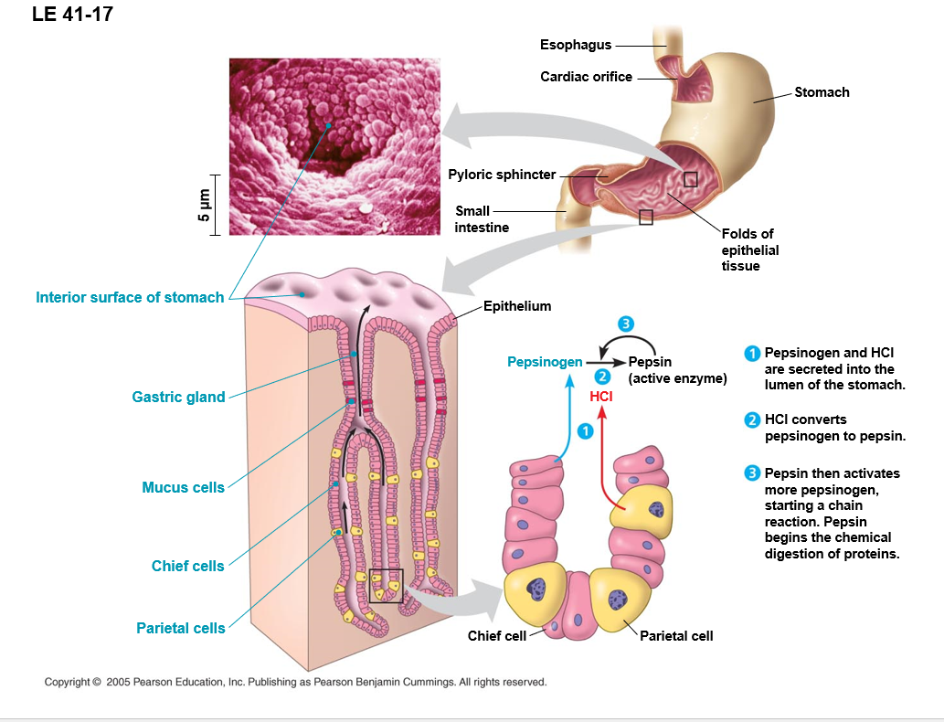
Pepsinogen is needed to get pepsin
stomach pH matters
Enzymes are real pH sensitive
acidity denatures proteins,
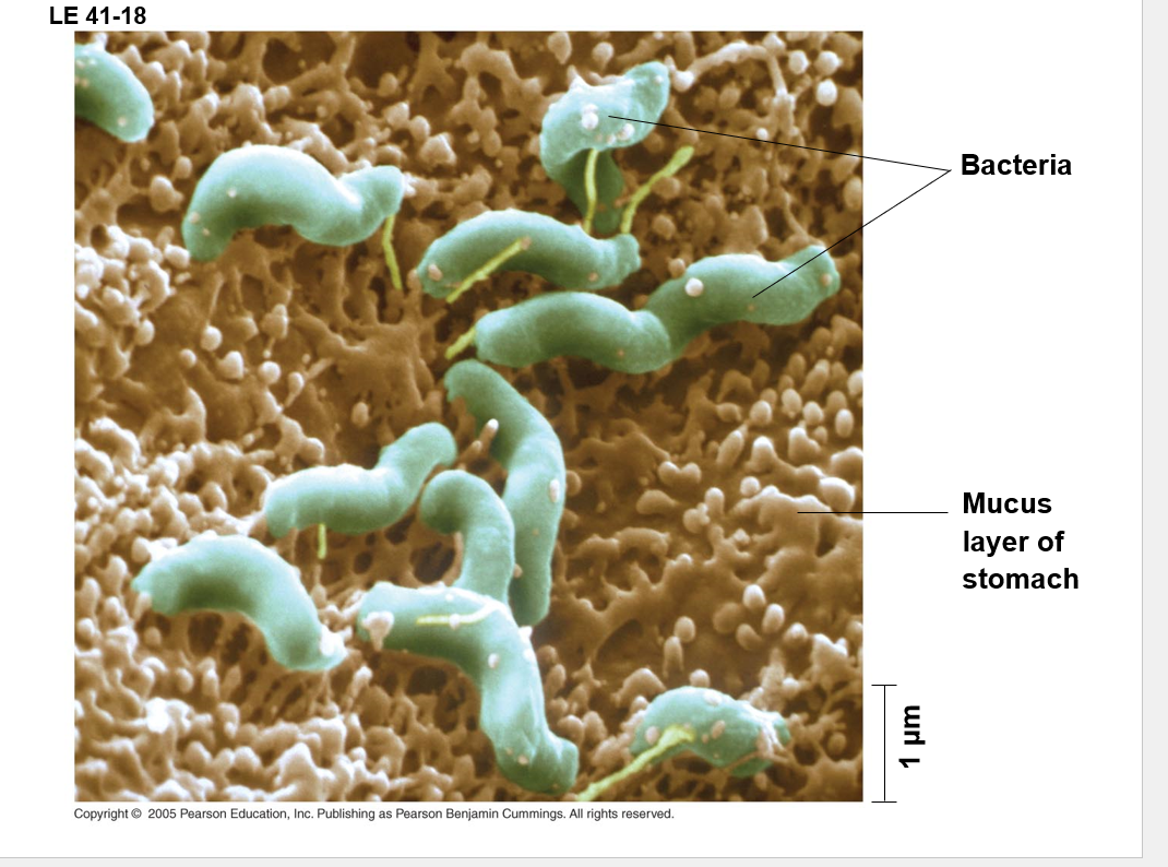
the acids in the stomach kills pathogens, some things survive, but most get killed.
Structural Histology plug from Dr. Daft
Gastric ulsers are lesions in the lining of the stomach. they are mainly cause by H. Pylori
—Destrory protective mucus and cause inflamation of stomach lining
—Acidic gastric jucies can attack the stomach tissue
—In severe ulcers, the erosion can produce a hole in the stomach wall and cause life-threatening internal bleeding and infection
can grow every where. Low or High pH
Chyme= acids mixed with food. What passes through the stomach into the small intestines
Pyloric sphincter is the opening from stomach to Sm. intestines
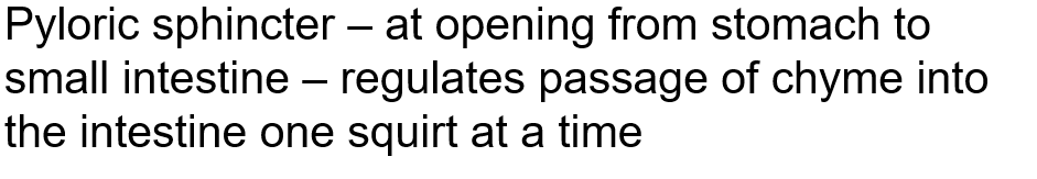
Takes 2-6 hrs for the stomach to empty this way
small intestines are the longest section of the alimentary canal because the surface area is useful for the absorption
Bicarbonate is a buffer that’ll help neutralize acids. This is why the pancreas secretes it, because acid is, surprise surprise kinda hard on tissues.
You can still make bile w/out the gallbladder, it just comes straight from the liver, and doesn’t work quite as well as with the gallbladder.
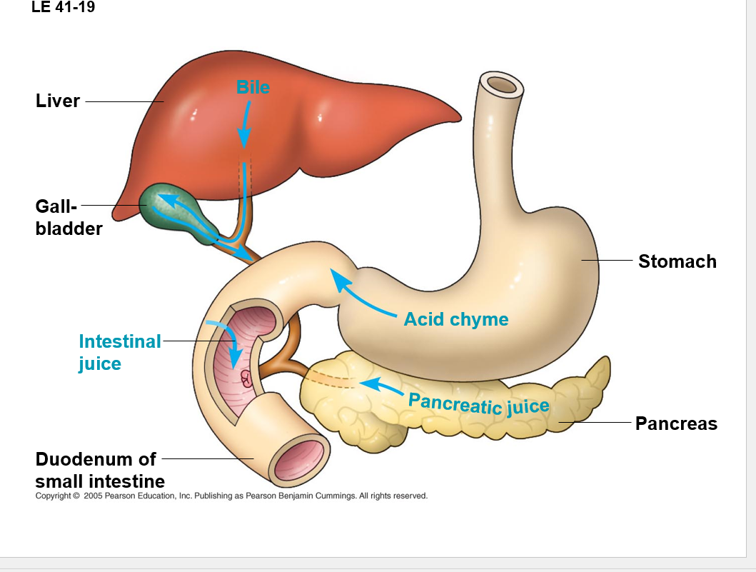
•The pancreas produces proteases, protein-digesting enzymes that are activated after entering the duodenum
–Trypsin and chymotrypsin
•Alkaline rich solution
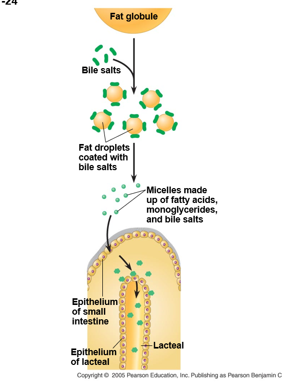
The liver makes bile which absorbs fats and aids and digetstion — stored in gallbadder
—No digestive enzymes but contains bile salts — act as detergents that aid in digestions and absorption of fats
the epithlial lining of the duodenum, called the brush border, produces several digestive enzymes
enzymatic digestion is completed as peristalsis moves the chyme and digestive juices along the small intastines
^^^^^ABOVE ON EXAM^^^^
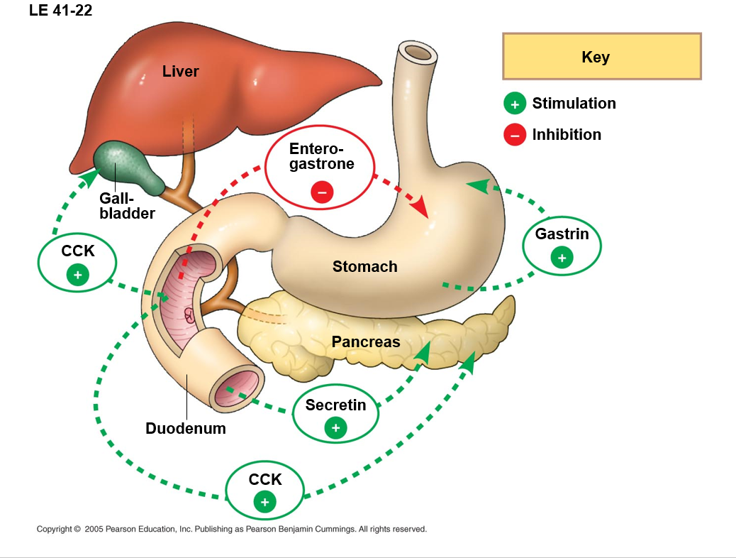
Hormones help coordinate the secretion of digestive juices into the alimentary canal
Amino acid (AA) and fatty acids (FA) in duodenum trigger release of CCK – stimulates release of digestive enzymes from pancreas and bile from gallbladder
Enterogastrone — secreted by duodenum — inhibits peristalsis and acid secretion by stomach — slows (It slows up to give you a chance to digest)
Gastrin — from stomach stimulates the production of gastric juices
Secretion — secreted by duodenum — stimulates the pancreas to release sodium bicarbonate which neutralizes acid chyme from stomach
how long is the duodenum in a human? aprx. the length of a ruler
•The small intestine has a huge surface area (size of tennis court), due to villi and microvilli that are exposed to the intestinal lumen
•The enormous microvillar surface greatly increases the rate of nutrient absorption
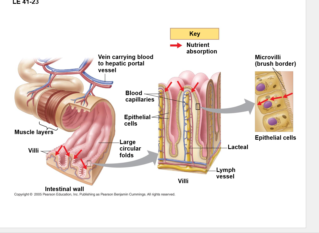
Mico villi are not responsible for the movement ANYTHING in the intestines. They are soly there for surface area and the help of absorption.
WEDNESSDAY/FRIDAY WE PLAY WITH CHALK!!!!
Date:3/26/25
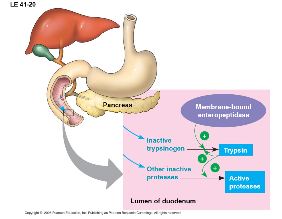
alimentary organs, accessory organs and what’s digested there
INSERT PIC OF CHALK HERE
•Each villus contains a network of blood vessels and a small lymphatic vessel called a lacteal
•Nutrients are absorbed across the intestinal epithelium and then across the unicellular epithelium of the capillaries or lacteals
•Amino acids and sugars pass through the epithelium of the small intestine and enter the bloodstream – active transport
•After glycerol and fatty acids are absorbed by epithelial cells, they are recombined into fats within these cells
•These fats are mixed with cholesterol and coated with protein, forming molecules called chylomicrons, which are transported into lacteals
Lead to blood vessels into the heart
•Blood vessels and veins that carry blood away from the villi all converge into the hepatic portal vein, a blood vessel that leads directly to the liver
•Liver has first access to amino acids and sugars after a meal
•Liver regulates the blood sugar
•From liver, blood travels to the heart, which pumps the blood and the nutrients it contains to all parts of the body
The large intestine or colon is connected to the small intestine
it’s major function is to recover H2O that’s entered the alimentary canal — liters enter anf most reabsorbed (90%)
Waste of the digestive tract the feces become more solid as they move through the colon — diarrhea and constipation occur here
feces pass through the rectum and exit via the anus
The colon houses strains of the bacterium Escherichia coli, some of which provide Vit.s
—Biotin, folic acid, vit.K, and several B vitamins – supplement our dietary intake
•Some bacteria produce methane and hydrogen sulfide gases
•Feces contain masses of bacteria, cellulose and other undigested materials
•Anus has voluntary and involuntary sphincter
Date:3/28/25
what’s being digested where, alementery canal, must know blood flow through heart
Every organism must exchange materials with its environment
exchanges ultimately occur at the cellular level
In unicellular organisms these exchanges occur directly with the enviornment
for most cells making up multicellular organisms, direct exchange with the environment is not possible
•A salmon’s feathery gills are an example of a specialized exchange system in animals
Transport systems connect the orgnas of exchange with the body cells
most complex animals have internal transport systems that circulate fluid
•Diffusion is inadequate – 3 hours to diffuse 1cm
•Circulatory ensures that no substance must diffuse very far to enter or leave a cell
Two types of circulatory systems: open and closed
•In insects, other arthropods, and most molluscs blood bathes the organs directly in an open circulatory system
•There is no distinction between blood and interstitial fluid, and this general body fluid is more correctly called hemolymph
•Hearts pump the hemolymph into an interconnected system of sinuses, spaces surrounding the organs for exchange
•Ostia – pores in heart to draw hemolymph into during heart relaxation
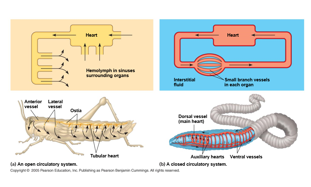
in closed circulary system blood is confined to vessels and is distinct from the interstitial fluid
•Heart pumps into large vessels that branch into smaller ones
•Closed systems are more efficient at transporting circulatory fluids to tissues and cells
Open:
lower E cost — lower hydrostatic pressure
Less E to build and maintain vessels
–Other functions – hydrostatic skeleton in molted molluscs
Closed
More effective in transporting fluids
Humans have four chambered heart
—one of two atria - receive blood returning to heart
—one or two ventricals - chamber that pumps blood out of the heart
•Arteries carry blood to capillaries, the sites of chemical exchange between the blood and interstitial fluid
•Veins return blood from capillaries to the heart
•Human body have a total of about 100,000 km of blood vessels
•In general, the number of blood vessels in a particular organ are correlated with that organ’s metabolic requirements
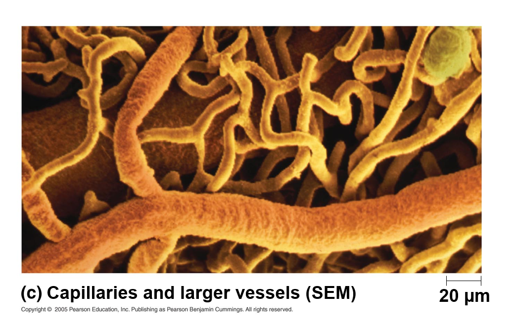
capilaries are only one cell layer thicc that’s to make it easier to trasmit stuff

fish: one chamber, amphibians: 3 chamber, reptiles: 3 chambers but more divided, Mammals and birds: four chamber
heart valves dictate a one way flow of blood through the heart
blood begines its flow with the right ventrical pumping blood to the lungs
in the lungs the blood loads O2 and unloads CO2
•Oxygen-rich blood from the lungs enters the heart at the left atrium and is pumped to the body tissues by the left ventricle
•Blood returns to the heart through the right atrium
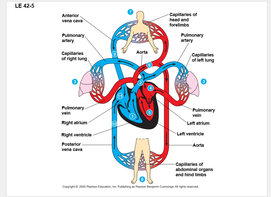
all blood tecnically still has a bit of O2 in it
artery: away from heart
veins: into the heart
MUST KNOW THE HEART STUFF AND BE ABLE TO DRAW IT!!!
right =pulmonary circuit:
left = systemic circuit
a closer look at the mammalian heart provides a better understanding of double circulation
consist mostly of cardiac muscles
•Contraction of atria completes filling of ventricles
•Ventricles pump blood to all body organs
•Valves prevent backflow of blood within the heart
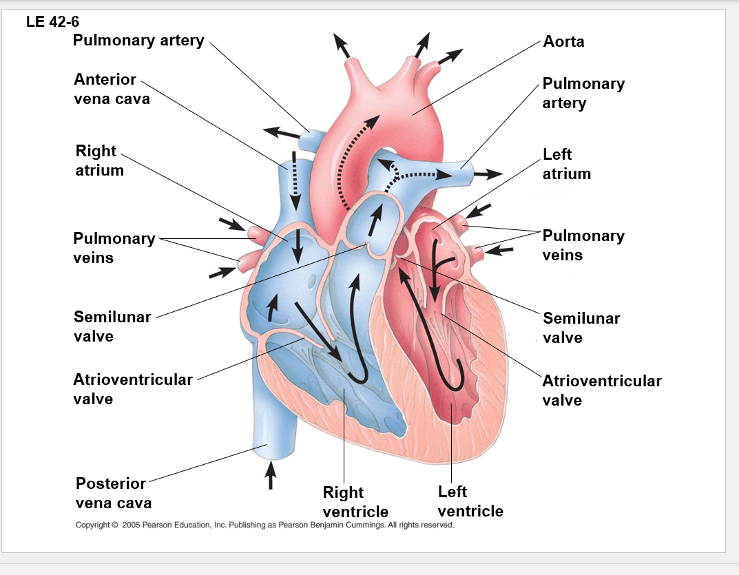
tricuspid valve is better name for atrioventricular valve
outer bit is the cardiac mussel
left ventricle is the most muscular
this is because the right ventricle is just sending it to your lungs, but your left ventricle has to make the blood go all the way around the body
The heart contracts and relaxes in a rhythmic cycle called the cardiac cycle
the contraction of pumping phase is called systole/systolic
the relaxation or filling phase is called diastole/diastolic
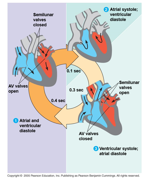
contract-systole
relax-diastole
cardiac output is the volume of blood pumped into the systemic circulation per minute
–Consists of heart rate and stroke volume – amt of blood pumped by the left ventricle each contraction
–About 5.25 L/min – equivalent to the total volume of blood in the human body
–During exercise – pump an amt of blood matching an average person’s body mass every 2-3 minutes
DATE:3/31/25
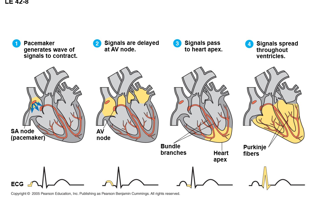
Electrical signal goes from SA, AV, bundle branches(budle of Hiss?), purkinji fibers
the sinoatrial (SA) node or pacemaker sets the rate and timing at which cardiac muscles cells contract — lets your heart beat at aprox. 80bpm
inpulses from the SA node travel to the atruoventricular (AV) node
at the AV node the impulses are delayed and then travel to the Purkinje fibers that make the ventricles contract
the pacemaker in influenced by nerves, hormones, body temp, and excercise
an increase of 1C raises the heart rate by ~10bpm
Exercise – additional oxygen to the body
The physical principles that govern movement of H2O in plumbing systems also influence the functioning of animal circulatory systems
three levels in blood vessels
—Connective tissue - lets vessel stretch and recoil
—Smooth muscle
—Endothelium - single layer of flattened cells that provides a smooth surface that minimizes resistance to the flow of blood
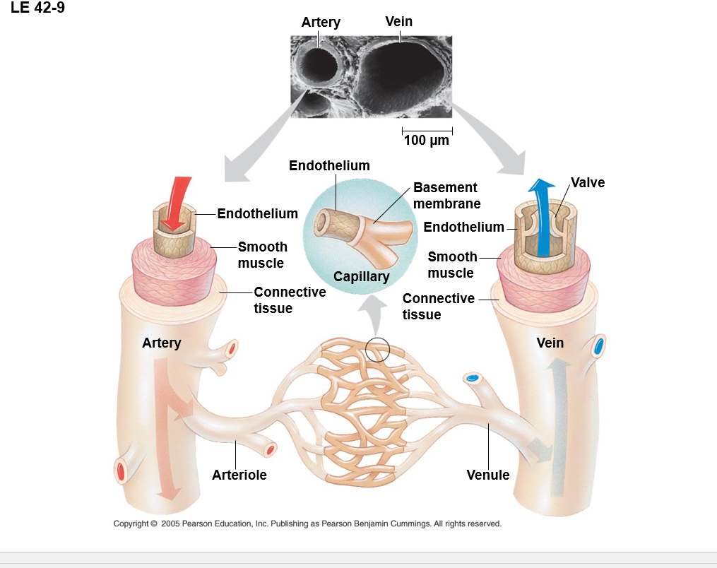
arteries have more smooth muscle, as such they are more uniform in shape
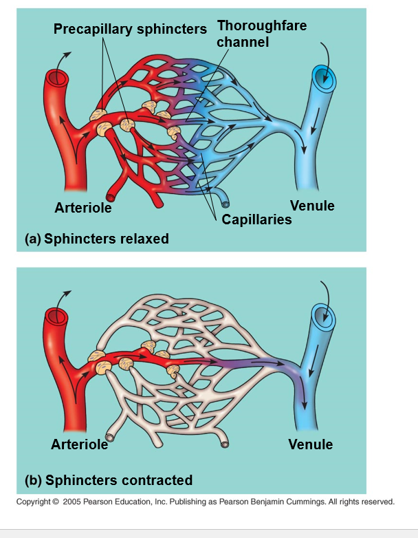
Capillary is where fluid exchange goes down — no layer of smooth muceles
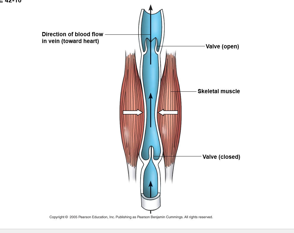
veins have valves to prevent back flow to make it easier to work against gravity, they dont really have smooth muscle — really relies on skeletal muscles to move the blood
if both medium, a vein would have a greater diameter than the artery
if you sit for a long time the skeletal muscles aren’t moving the blood, which can make blood pool, and clot in your veins, creating a DVT, which once you get up/move again can dislodge and go to your heart/lungs/brain and kill you
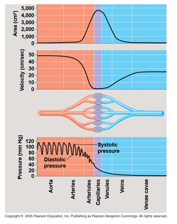
the further the blood gets from the heart the slower it gets; this is to help the capillaries defuse O2/CO2 (figure two right above)
capillaries in major organs are normally filled to capacity
blood supply varies in many other sites
after a meal blood supply to digestive tract
strenuous exercise - blood diverted to skeletal muscles and skin
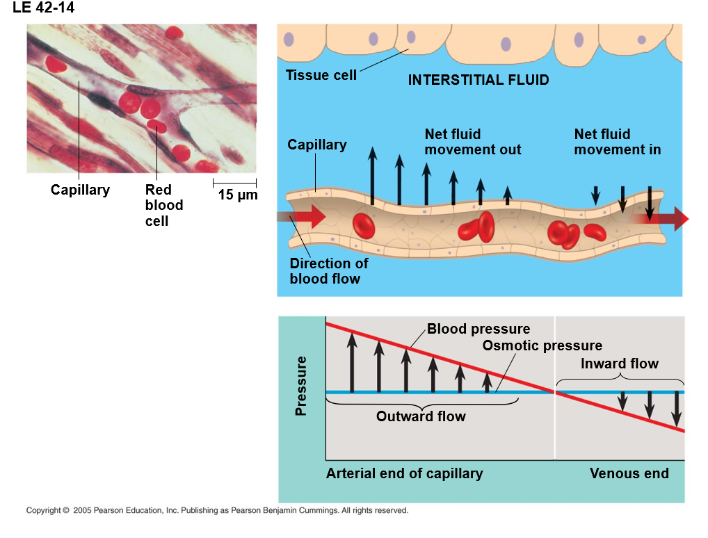
the hydrostatic/systolic pressure has enough force that when we get down to one layer of capillary we are pushing fluid out. this is good, BC the nutrients it receives. proteins, such as Microalbumins, antibodies, and such do not as easily escape. This will create a gradient where there are excess proteins, but not fluids, causing fluids to rush back in.
—even with that there’s a net flow out
—if the water doesn’t come back this can cause edema (swelling/fluid buildup/retaining H2O) (this is typically a sign of heart failure/disease)
The lymphatic system returns fluid to the body from the capillary beds
the system aids in body defense - lymph nodes become swollen
Fluid reenters the circulation directly at the venous end of the capillary bed and indirectly through the lymphatic system
fats travel through the lymphatic system
WBCs live in the lymph nodes, the swelling is because they sense danger and start screaming and multiplying to prepare and defend against it.
In invertibrates with open circulation blood (hemolymph) is not different from interstitial fluid
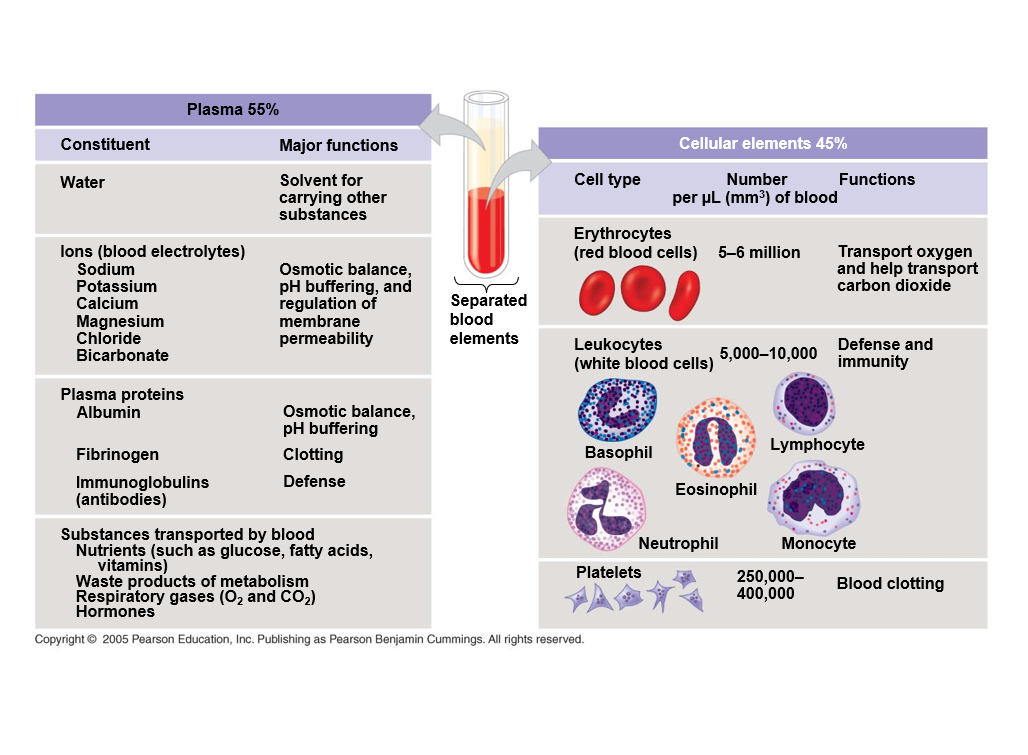
Albumin is a prominent protein in egg whites
the cellular elements of blood wear out and are replaced constantly
Erythrocytes — 3-4 months
erythrocytes, leukocytes, and platelets all develop from a common source, pluripotent stem cells in the red marrow of bones
if tissues do not receive enough O2 the kidney synthesizes and secretes erythropoietin/EPO stimulates erythrocyte production
DATE: 4/2/25
blood is made in red blood marrow
as you get older the red marrow becomes more yellow due to fat
as you age red bone marrow mostly is in hips and ends of long bones
if tissues do not receive enough O2 the kidney synthesizes and secretes erythropoietin/EPO stimulates erythrocyte production
EPO - abused by athletes to increase erythrocyte levels - blood doping
more RBC=more O2 carrying capacity and energy
this can create extra pressure on your heart and cause you to BLEH x-x
Leukemia - person makes too many leukocytes which doesn’t allow red blood cells to be made
These WBCs aren’t really that great
—also not mentioned but sharp decrease in platelets/thrombocytes typically - really fun to run a CBC and be the first to realize someone’s cancer’s back 🙃
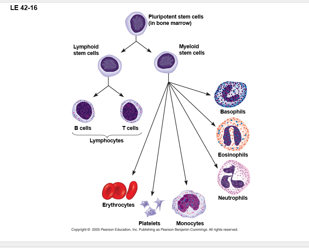
monocytes become macrophages
Basophils and eosinophils are sins when ya’ have allergies
When the endothelium of a blood vessel is damaged, the clotting mechanism begins
a cascade of complex reactions converts fibrinogen to fibrin, forming a clot
Hemophilia - excessive bleeding from minor cuts due to defects in clotting process
these people get platelet transfusions to help with the whole clotting thing
AIDS/HIV effect those with that a good deal
AIDS used to be called the 4Hs - hemophiliacs, Hattians, Heroin, Homosexuals
Blood clots - thrombus
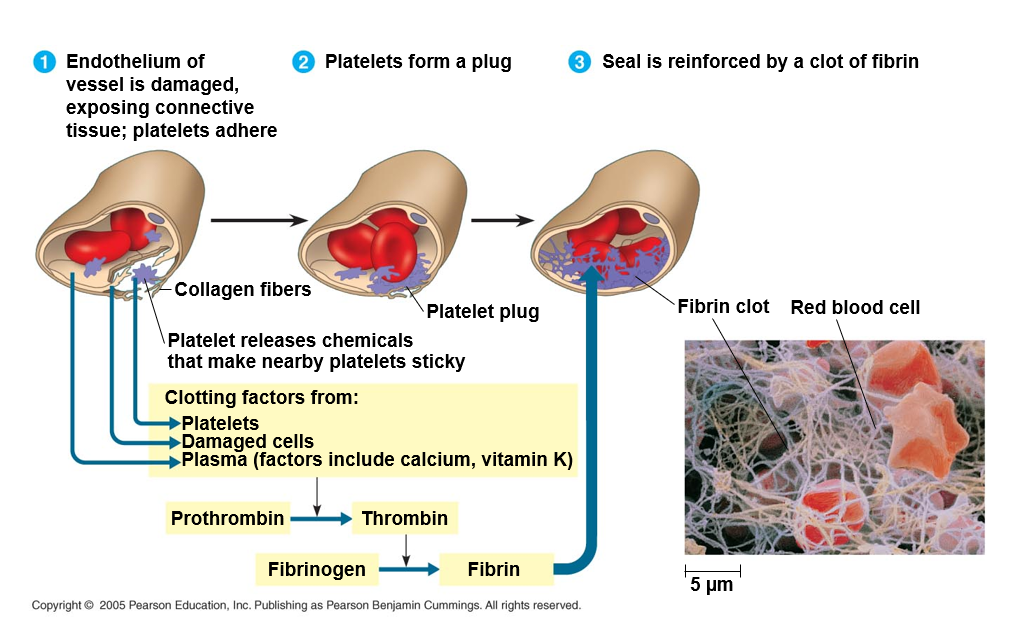
Clotting when damage to endothelial layer the system sees collagen screams because WHY and starts to make clots
Prothrombin, thrombin, fibrinogen, fibrin
Cardiovascular diseases are disorders of the heart and the blood vessels
they account for more that 1/2 the deaths in the United States
•Due to smoking, lack of exercise, a diet rich in animal fat, high concentrations of cholesterol
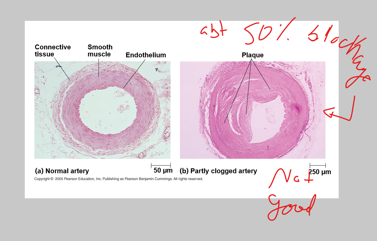
macrophages become foam cells and try to break doesnt cholesterol and that causes them to expand. If they burst that can cause further clotting, which for the reasons of fourign particals is not good. That makes you more apt to clot
plaque build up comes from too much (bad) cholestoral
Hypertension or high blood pressure promotes athrosclerosis and increases the rish of heart attack(MI) and stroke
an MI is the death of cardiac muscle tissue resulting from blockage of one or more coronary arteries
•A stroke is the death of nervous tissue in the brain, usually resulting from rupture or blockage of arteries in the head
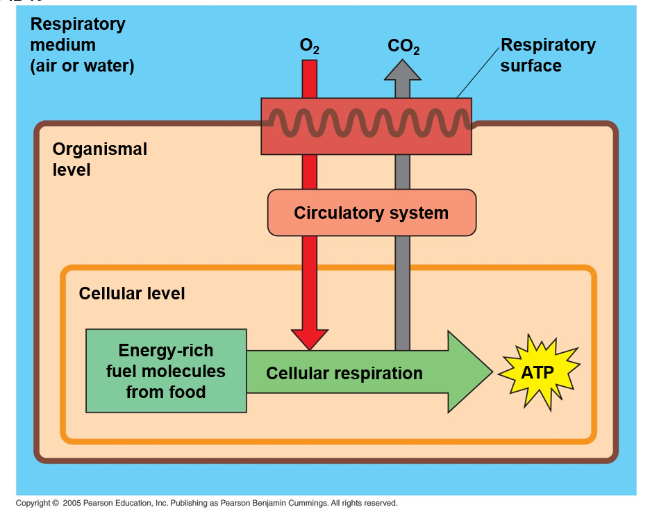
Gas exchange supplies O2 for cellular resperation and desposes of CO2
Spiders. land snails and most terrestrial vertebrates have internal lungs
lungs are restricted to one location
circulatory system must send O2 to rest of body
size and complexity of correlated with an animal’s metabolic rate
a system of branching ducts conveys air to lungs
air inhaled through the nostrils passes through the pharynx into the trachea, bronchi, bronchioles, and dead-end alveoli, where gas exchange occurs
•Nostril – filtered, warmed, humidified, and sampled for odors
•Larynx – moves upward to close epiglottis during eating – voice box
•Trachea – windpipe – C-shaped rings of cartilages maintains shape
•Trachea forks into two bronchi – leading to each lung
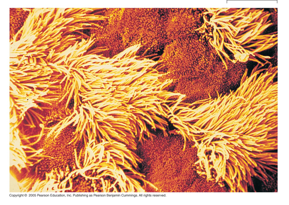
•Bronchioles – fine tubes – covered with cilia and mucus
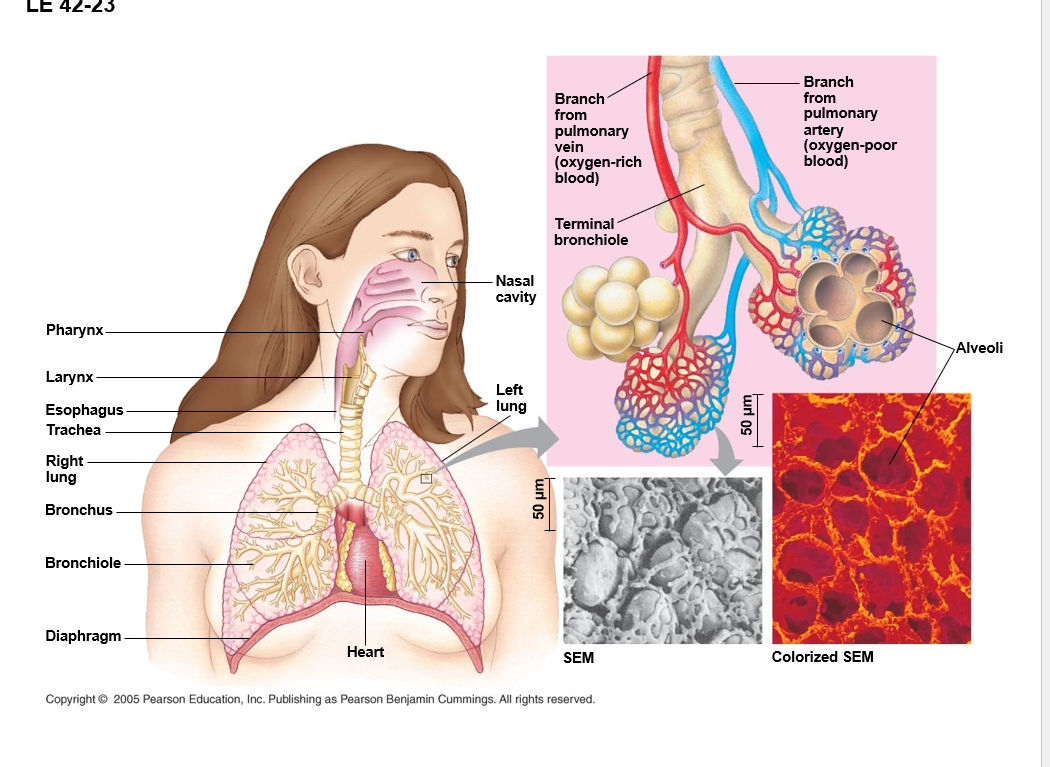
alveoli -air sacks
the process that ventilates the lungs in breathing the alternate inhalation and exhalation of air
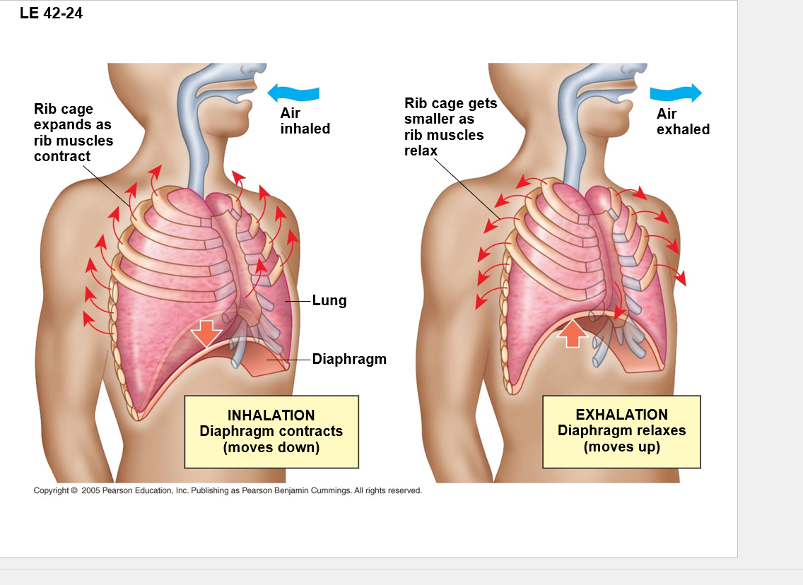
when you breath in you you cause a drop in pressure (below atmosphiric), causing air to rush in to fill the void
When you exhale you decrease pressure and the air rushes back out
•Mammals ventilate their lungs by negative pressure breathing, which pulls air into the lungs
•Lung volume increases as the rib muscles and diaphragm contract
•Tidal volume – volume of air a mammal inhales and exhales with each breath – 500mL
•Vital capacity – maximum tidal volume during forced breathing
•Residual volume – air remaining in lung after forceful exhale—there will ALWAYS be a little left. no matter what you can do with it, it will be there.
In humans the main breathing control centers are in two regions of the brain, the medulla oblongata and the pons
the medulla regulates the rate and depth of breathing in response the pH changes in the cerebrospinal fluid
The medulla adjusts breathing rate and depth to match metabolic demands
Match breathing with circulation – exercise
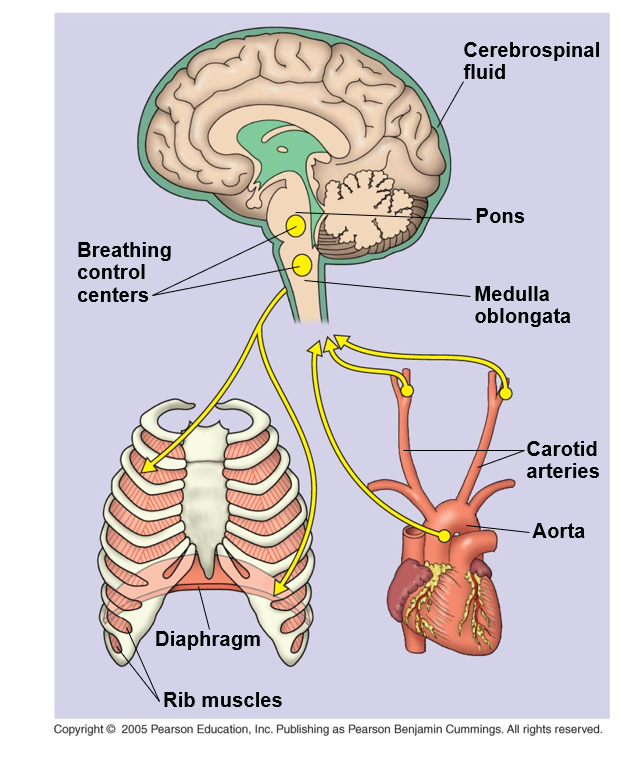
if the medula or pons are damaged you’re sorta just dead, as theres no other part of the brain that can (typically) makes up for it
•The metabolic demands of many organisms require that the blood transport large quantities of O2 and CO2
Gases diffuse down pressure gradients in the lungs and other organs
diffusion of a gas depends on differences in a quantity called partial pressure
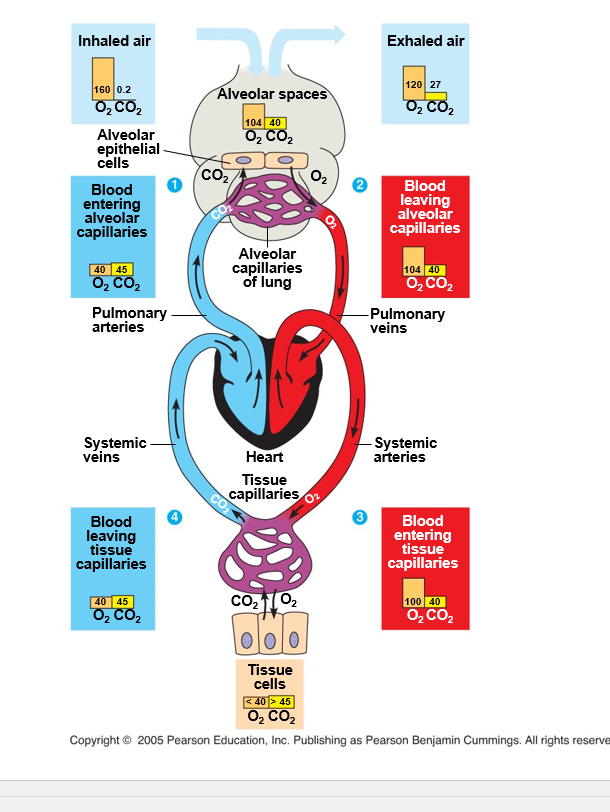
you have two points: the top in heart and lungs, the bottom is the tissue level (where O2 is delivered)
CO2 will diffuse into the alveoli and O2 will diffuse into the blood because of the pressure difference. It wants to be equal
gas exchange is controlled by partial pressure — this is the basic principles of gas diffusion
DATE: 4/4/25
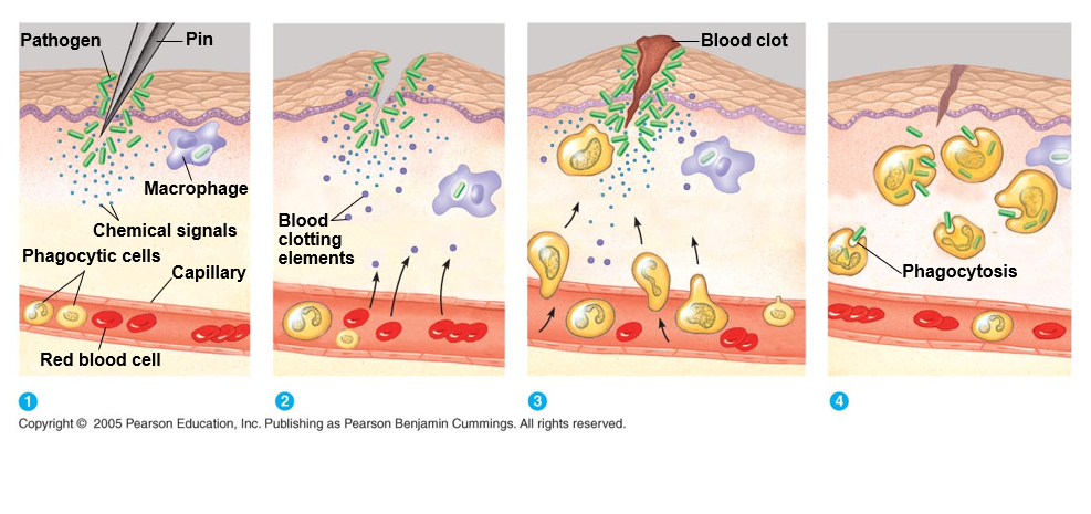
An animal must defend itself from many dangerous pathogens in may encounter
two major kinds of defense have evolved: innate immunity and acquired immunity
acquired immunity: is from vaccines, makes memory so that your body can fight off the infection before you know it or lessen the effects of the infection.
Innate immunity is present before any exposure to pathogens and is effective from the time of birth
it involves nonspecific responses to pathogens
innate immunity consists of external barriers plus internal cellular and chemical defenses
key internal defenses are macrophages and other phagocytic cells
Mucosal membranes are in the respiratory tract, urogenital tract
the salt on you skin keeps stuff from growing
pus is a good sign cause its dead neutrophils, which means your immune system is working
Acquired immunity or adaptive immunity develops after exposure to agents such as microbes toxins or other foreign substances
it involves a very specific response to pathogens
recognition is by WBCs called lymphocytes — includes memory B and T cells
some lymphocytes (B cells) produce antibodies; others destroy infected cells, cancer cells or foreign tissue
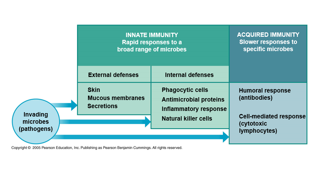
humoral responce = B cells
Cell mediated responce = T cells
Skin and mucous membranes are physical barriers to entry of microorganisms and viruses
mucous membrane cells produce mucus a viscois fluid that traps microbes and other particles
In the trachea, ciliated epithelial cells sweep mucus and any entrapped microbes upward, preventing microbes from entering the lungs
Secretions of the skin and mucous membranes provide an environment hostile to microbes
secretions give the skin a pH between 3 and 5 acidic enough to prevent colonization of many microbes
skin secretions include proteins such as lysozyme which digests bacterial cell walls
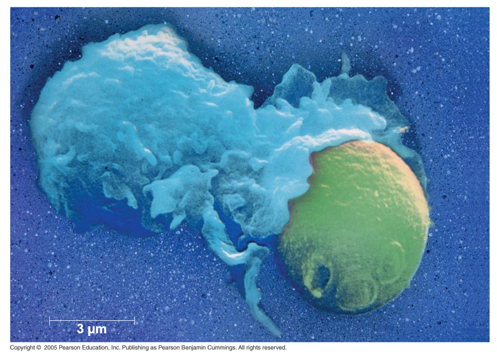
inteernal cellular defenses depend mainly on phagocytosis
WBCs called phagocytes ingest microoragnisms and initiate inflammation
NK cells also play a role in innate defenses by limiting the spread of microbes before the body can mount the acquired immune responce
Phagocytes attach to prey via surface receptors and engulf them forming a vacuole that fuses with a lysosome
microbes destroyed in lysosome
–Nitric oxide may poison the engulfed microbes
–Lysozyme degrade microbial components
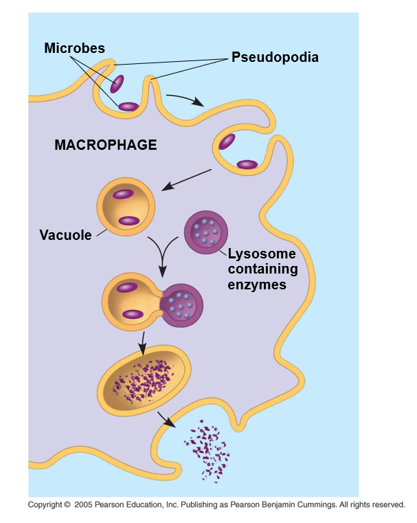
there is something missing in the diagrame, that is that some of the little bits would be presented by special receptor on the macrophage so the immune system can see it and generate a memory and effector responses
endocytosis is the generic name for phagocytosis
probably specifically receptor endo/phagocytosis
if you activate a B and T cell you get an effector for now and memory for later
Neutrophils - most sbundant WBCs engulf and destroy microbes
Macrophages a type of phagocyte migrate through the body and are found in organs of the lymphatic system — belongs to a category called antigen presenting cells - tissue specifc with different names
considered large eaters
can be found in either tissue or circulating in lymphatic system
the lymphatic system defends aganst pathogens
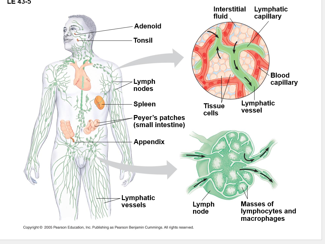
lymph nodes= filters for pathogens
the white dots are germinal centers (masses of lymphocytes and macrophages)
the stuff in the lymph eventually end up in the blood
spleen is sorta an accessory organ to all this, same with Peyer’s patches, tonsils, appendix
you have antimicrobial proteins ~30, making up the complement system which causes lysis of invading cells and helps trigger inflammation
interferons provide innate defense against viruses and help activate macrophages
—inhibit viral reproduction
Date: 4/7/25
digestive, cardio, pulmonary, immune on the test ❤ I love the human body
green stuff = bacteria
once barrier is broken immune system
little white dots from macrophage chemokines to call more cells
the exiting from the blood vessels are diapedesis
they are probs neutrophils
NK cells attack virus infected body cells and cancer cells
they trigger apoptosis in the cells they attack
-if something looks weird they kill it
invertebrets don’t have as complex an immune system
the exoskeloten is the 1st line of defense as the barrer
•Hemolymph contain hemocytes that ingest bacteria or produce antimicrobial peptides to kill it
•Most do not exhibit acquired immunity
•Acquired immunity is the body’s second major kind of defense
•An antigen is a foreign molecule that is recognized by lymphocytes and elicits a response from them
•A lymphocyte and/or antibody recognizes and binds to a small portion of the antigen called an epitope
the epitope is on the bad stuff and the memory is when the WBCs recognize and are able to kill it gooder
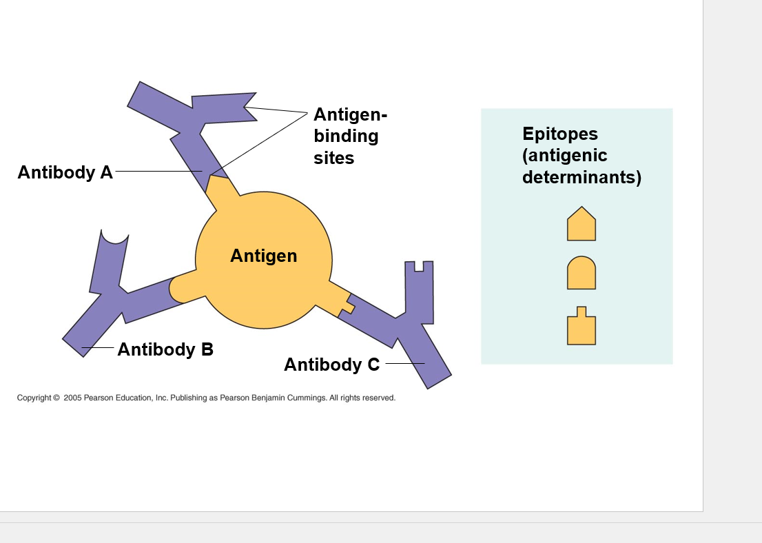
the multipul touch points are helpful for when the pathogen mutates.
when a pathogen recognizes something reacting with it it can down regulate that, and the multipul touch points let it still fight it even when that happens.
not putting all the eggs in one basket basically
Antibodies are made by B-cells — think Cells at work

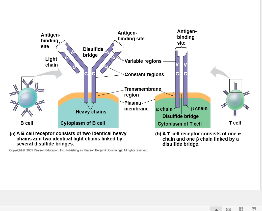
a B cell receptor can detach and that is when it becomes an antibody
a T cell receptor is always attached to the surface of the cell
The ‘V’ is what gives the ability to recognize the stuff
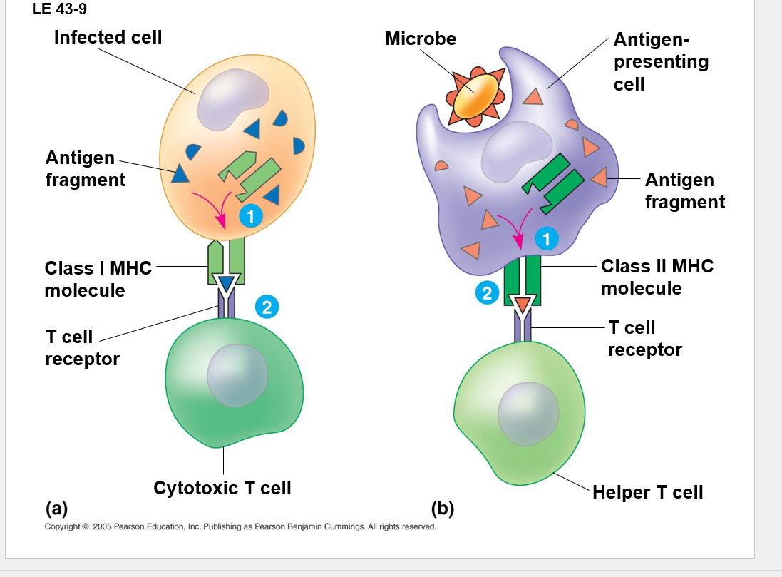
T cells cannot interact with antigens unless presented with it
B cells interact with free floating antigens and can just grab the stuff
CD4-helper: Activates the immune system making effector and memory cells
CD8 - Cytotoxic T Cell: kills the stuff —
What is a class I/II MHC mol.? Major histone compatibility complex there are 2 classes BC there are 2 types that present
almost every cell in your body will be able to present MHC I (not all ofc, but a good amt.)
—It’s not just bad stuff that’s presented on thees mols. when they break down old stuff it can be presented. if its just old stuff from part of a norm. cell the T cell will just scotch along it’s merry way, not if phathogen tho. if that T cell all like: “Yikes, thats bad, DIE!”
MHC II:
macrophges, dedritic, and B cells are really the only ones this is on
it’s these guys jobs to present stuff
this is specifc to only things that EAT/phagocyte - so not in like cardic cells for example
when it eats a bad thing it hypes it up and it gets it gone baby
Macrophages will eat up broken bone and present it too, so it’s not always bad stuff here either
if something gets activated when it’s you you get an autoimmune dissorder
Lymphocytes in thymus develop into T cells
Lymphocytes that remain in bone marrow become B cells
They are both in grade school together, and the T cells move off to school (Thymus University), while the B cells stay at the community (Bone marrow college)
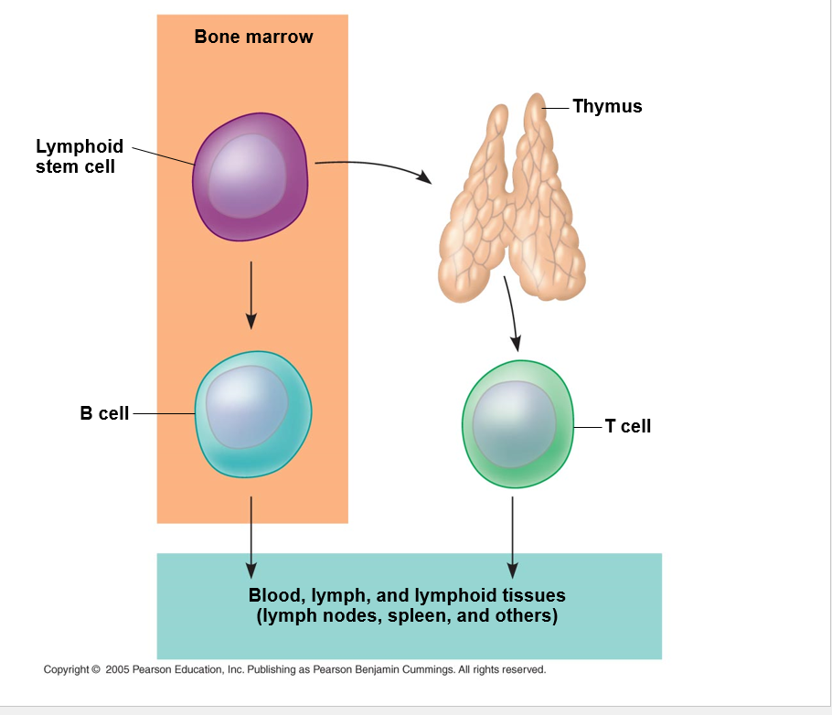
thymus is secondary organ for the lymphatic system — kinda right above the heart
•Each person has 1 million different B cells and 10 million different T cells
•Random, permanent gene rearrangement forms functional genes encoding the B or T cell antigen receptor chains
Possible 1.65X10⁶ combinations
this means circulating in your body there are T and B cells circulating in your body that have receptors for pathogens that may not even exist.
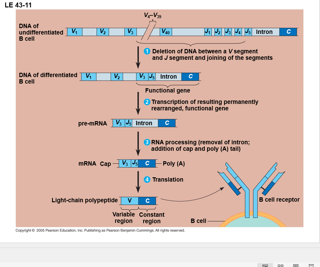
all those V and J regions combine to make the receptor
you can get this stuff all spliced together and that lets you recognize things you’ve never seen before.