5/28 Connective Tissues, Proper Connective Tissue, and Adipose Tissue
Connective Tissue: Definition and Function
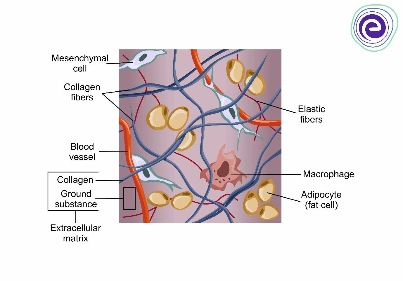
Connective tissue is defined as a highly vascular tissue responsible for connecting other tissues and structures. It is characterized by an abundant extracellular matrix (ECM) composed of protein fibers and ground substance.
Key Functions:
Organ structure: Providing the internal framework for various organs.
Mechanical strength: Offering physical durability to bodily structures.
Space filling: Occupying the voids between more specialized tissues.
Physical and metabolic support: Serving as a medium for the exchange of nutrients and wastes for specialized tissues.
General Structure of Connective Tissue
Connective tissue consists primarily of cells and the extracellular matrix (ECM).
Cells:
These are diverse in shape, function, and origin.
Extracellular Matrix (ECM):
The ECM is the major constituent of connective tissue.
Protein Fibers: Includes collagen, elastic, and reticular fibers.
Ground Substance: This is the amorphous component composed of:
Proteoglycans: These are large molecules composed of sulfated glycosaminoglycans (GAGs) with a protein core that aid in retaining water and provide compressive strength to the tissue.
Multiadhesive glycoproteins: These proteins help anchor the cells to the extracellular matrix and facilitate communication between cells, playing a critical role in tissue repair and development.
Glycosaminoglycans (GAGs): GAGs are often sulfonated, which enhances their negative charge and makes them highly hydrophilic, allowing them to retain water and contribute to the viscoelastic properties of the extracellular matrix.
What are the most important cells in connective tissues?
Fibroblasts
Description:
The most common cell of connective tissue.
Responsible for producing and maintaining the extracellular matrix (ECM).
Functions:
Synthesize and secrete:
Collagen fibers → provide tensile strength.
Elastic fibers (elastin) → provide elasticity and flexibility.
Ground substance → gel-like medium that supports cells and fibers.
Importance:
Provide structural support to connective tissues.
Maintain the extracellular matrix (ECM).
Play a vital role in wound healing and tissue repair by producing new collagen and matrix after injury.
Myofibroblasts:
Specialized fibroblasts involved in wound healing.
Produce collagen and ECM.
Contract the wound to help close it during healing.
Fibroblast = Builds and repairs connective tissue
Myofibroblast = Builds, repairs, and contracts wounds
Adipocytes, which store fat and contribute to energy reserves;
Macrophages, which assist in immune defense and tissue maintenance,
Mast cells, which play a key role in inflammatory responses.
Structural-Functional Features of ECM Components
Collagen Proteins Family: Forms collagen fibers which provide tensile strength to resist pulling, tearing, and stretching.
Elastin Protein: Forms elastic fibers which provide elasticity, allowing structures to return to their original shape after mechanical distortion.
Ground Substance Components:
Hydrophilic Proteoglycans
Glycosaminoglycans (GAG)
Multiadhesive Proteins
Functions: These components contribute to tissue volume, facilitate nutrient/metabolic waste exchange, provide lubrication, and act as a barrier against the penetration of microorganisms.
Fibers of Connective Tissue: Collagen and Reticular Fibers
Collagen is the most abundant protein in the human body, accounting for approximately of its dry weight. This estimate is after you remove the water as humans are 70% of water. These proteins are synthesized and secreted primarily by fibroblasts.
Mechanical Properties: Extremely strong and resistant to normal shearing and tearing forces.
Extracellular Polymerization: After secretion, they undergo polymerization to form different types of collagen fibers.
Notable Localizations:
Connective tissues
Basement membranes
External laminae of nerve and muscle cells
Protein Polymerization → Fiber Formation
Collagen Fibers
Collagen is made of smaller protein units called collagen monomers (tropocollagen molecules).
These monomers assemble (polymerize) outside the cell.
The result is collagen fibers, which are strong, rope-like structures.
Function: Provides tensile strength (resistance to pulling forces) in connective tissues like tendons, skin, and bone matrix.
Actin Fibers (Comparison Example)
Actin exists as globular actin (G-actin) monomers.
These monomers polymerize into filamentous actin (F-actin).
F-actin strands bundle together to form actin filaments (microfilaments).
Function: Important for cell shape, movement, and muscle contraction.
Big Concept Connection
Both collagen and actin follow the same principle:
This process is essential because: Small protein subunits → polymerize → form functional fibers
Individual proteins are too small to provide structure
Fibers give strength, stability, and organization to tissues and cells
Collagen = extracellular strength (outside cells, connective tissue)
Actin = intracellular movement (inside cells, cytoskeleton)
Detailed Classification of Collagen Types

Quick Memory Tip
Type I = Bone, skin, tendons ("strong fibers")
Type II = Cartilage
Type III = Reticular fibers (supportive network in organs and blood vessels)
Type IV = Basement membrane
Characteristics of Specific Collagen Fibers
Type I Collagen (Tension)
Most abundant type of collagen in the human body, providing tensile strength to various tissues such as bones, tendons, and skin.
Appearance: Eosinophilic/Acidophilic; very thick fibers.
Function: resistance to tension
Structure: Fills the ECM; surrounded by ground substance (which appears as unstained areas in microscopy). Striated appearance (dark-light bands) under Transmission Electron Microscopy (TEM).
Biosynthesis: Secreted mainly by fibroblasts.
Common Locations: Ligaments, tendons, organ capsules, dermis, bone, and dentin.
Eosinophilia (acidophilia): strong pink fibers (very obvious)
Type II Collagen (Pressure)
Appearance: Eosinophilic, though less evident than Type I due to the presence of basophilic GAGs. Thicker than some fibers but thinner than Type I. Faint pink in a blue/purple matrix
WHY?
Collagen in the extracellular matrix is rich in proteins, so it tends to bind acidic dyes like eosin, appearing pink under H&E staining. However, this staining is sometimes less intense than in Type I collagen because the matrix also contains basophilic glycosaminoglycans (GAGs), which attract hematoxylin and can partially mask the eosin signal.
It is embedded in a very rich proteoglycan/GAG matrix
GAGs are strongly basophilic (negative) → attract hematoxylin
So the blue-purple ground substance can mask the pink collagen
Eosin (acidic dye) binds to basic/protein-rich structures like collagen, producing a pink appearance (acidophilia).
Hematoxylin (basic dye) binds to acidic components like GAGs, producing a blue-purple color (basophilia).
So the final appearance depends on the balance between protein-rich collagen (eosinophilic) and GAG-rich ground substance (basophilic).
Cytoplasmic proteins are relatively basic and bind eosin, while nucleic acids in the nucleus are negatively charged due to phosphate groups and bind hematoxylin
Structure: Seen in the interterritorial matrix of hyaline cartilage.
Common Locations: Cartilage and the vitreous body of the eye.
Function: Resistance to Pressure
Type III Collagen (Reticular Fibers)
Appearance: Not well seen in routine H&E preparations. Highlights as black in silver impregnation (argyrophilia) and fuchsia with Periodic Acid Schiff (PAS) due to carbohydrate content. BUT it often looks finer and more delicate than Type I collagen.
Type III collagen is best seen with silver stains (reticulin stain) → appears black
So even though it’s eosinophilic in H&E, it’s often identified using silver-staining techniques.
Function: Forms the delicate stroma of expandable organs, lymphoid.
Common Locations: Lymphoid organs (except the thymus), liver, endocrine glands (e.g., adrenal cortex), and bone marrow.
Collagen Synthesis and Polymerization
Intracellular Process
Nucleus: Formation of mRNA for each type of chain.
RER Synthesis: Synthesis of procollagen chains with propeptides at both ends; clipping of the signal peptide.
Hydroxylation: Hydroxylation of specific prolyl and lysyl residues in the ER (Vitamin C dependent).
Steps is dependent on vitamin C, which is crucial for the hydroxylation process, ensuring proper production of collagen formation and stability.
Glycosylation: Attachment of soluble galactosyl and glucosyl sugars to specific hydroxylysyl residues.
Assembly: Assembly of procollagen molecules into a triple helix with nonhelical propeptides.
Transport: Transport of soluble procollagen to the Golgi complex via transfer vesicles.
Packaging: Packaging of soluble procollagen into secretory vesicles.
Exocytosis: Secretory vesicles, assisted by microtubules and microfilaments transport molecules to the cell surface/extracellular space.
Microtubules guide the vesicle from point A to point B
Collagen Synthesis (High-Yield)
Preprocollagen
Made in the rough ER.
Contains a signal peptide.
⬇ signal peptide removed
Procollagen
Hydroxylation of proline and lysine occurs here (requires Vitamin C).
Triple helix forms.
⬇ Golgi processing and secretion
Procollagen is secreted outside the cell
⬇ cleavage of terminal peptides
Tropocollagen
Forms in the extracellular space after procollagen is trimmed.
⬇ cross-linking by lysyl oxidase
Collagen fibrils
⬇
Collagen fibers
Easy Memory Trick
Pro = Produced inside the cell
Tropo = Travels outside the cell
Preprocollagen → Procollagen → Tropocollagen → Collagen Fibrils → Collagen Fibers
A common exam question is:
Q: Which form is secreted from the fibroblast?
A: Procollagen
Q: Which form undergoes extracellular cross-linking?
A: Tropocollagen
Q: What enzyme is required for collagen cross-linking?
A: Lysyl oxidase
Q: What vitamin is required for hydroxylation of proline and lysine?
A: Vitamin C
Extracellular Process
Cleavage: Procollagen peptidases cleave the nonhelical terminal peptides, transforming procollagen into insoluble collagen molecules.
Aggregation: Collagen molecules aggregate to form collagen fibrils.
Reinforcement: Fibrillar structure is reinforced by covalent cross-links catalyzed by the enzyme lysyl oxidase.
Inside the fibroblast:
Rough ER (RER) → synthesizes procollagen
Golgi apparatus → modifies, packages, and prepares procollagen for secretion
Exocytosis → procollagen is released into the extracellular matrix (ECM)
Outside the cell (ECM):
4. Procollagen is cleaved into tropocollagen
5. Tropocollagen molecules assemble into collagen fibrils
6. Fibrils bundle into collagen fibers
7. Lysyl oxidase cross-links the fibers to increase strength
In summary, Collagen Synthesis and Polymerization
Extracellular Processing of Procollagen
Procollagen molecules are released from the cell by exocytosis into the extracellular space.
Once outside the cell, procollagen peptidases remove (cleave) most of the non-helical terminal peptides.
This cleavage converts procollagen into soluble collagen molecules (tropocollagen).
Formation of Collagen Fibers
Soluble collagen molecules aggregate together.
The aggregated collagen molecules organize to form collagen fibrils and collagen fibers.
Key Concept
Synthesis of procollagen occurs inside the cell.
Polymerization and formation of collagen fibers occur outside the cell (in the extracellular matrix).
Therefore:
Intracellular: Procollagen synthesis.
Extracellular: Procollagen processing, collagen aggregation, and collagen fiber formation.
Summary
Procollagen is synthesized inside the cell.
Procollagen is secreted by exocytosis.
Procollagen peptidases cleave terminal peptides.
Soluble collagen molecules are formed.
Collagen molecules aggregate and polymerize.
Collagen fibers are produced in the extracellular space.
Important Exam Point: The polymerization of collagen into fibers occurs outside the cell, whereas procollagen synthesis occurs inside the cell.
Elastic Fibers
Composition: A composite of fibrillin and elastin.
Physical Features: Thinner than Type I collagen as reticular fibers; distention-resistant.
Formation Sequence:
Fibroblasts and smooth muscle cells secrete fibrillin subunits.
Elastin is deposited into the fibrillin scaffold and cross-linked.
Elastin accumulates in the electron-dense center, with fibrillin microfibers visible on the surface.
Staining: Stain poorly with H&E; stain darker than collagen with orcein and aldehyde fuchsin.
Locations:
Sparse networks with collagen bundles.
Elastic lamellae (fenestrated sheets) in blood vessel walls (mainly arteries).
Dermis and mesentery.
Function: Resist tension and deformation; they provide structural support to organs and tissues throughout the body. They function to return to there orginial position.
Secreted: by fibroblasts and smooth muscle cells, meaning
In summary
Meaning: these cells produce and release Type III procollagen, which is then processed outside the cell to form Type III collagen fibers.
Type III Collagen (Reticular Collagen)
Secreted mainly by fibroblasts and smooth muscle cells.
Forms reticular fibers (a delicate network of supporting fibers).
Found in:
Lymph nodes, Spleen, Bone marrow, Liver, Blood vessel walls, Skin, Granulation tissue during wound healing
Process
Fibroblasts or smooth muscle cells synthesize Type III procollagen inside the cell.
Procollagen is secreted into the extracellular space.
Terminal peptides are cleaved by procollagen peptidases.
Type III collagen molecules assemble into reticular fibers outside the cell.
Fibroblast
⬇
Rough ER → synthesize procollagen
⬇
Golgi apparatus → modify and package procollagen
⬇
Exocytosis
⬇
Extracellular Matrix (ECM)
⬇
Procollagen is cleaved to tropocollagen
⬇
Type III collagen molecules assemble into reticular fibers
Ground Substance
Ground substance is a highly hydrated, transparent, and viscous mixture.
The most abundant substance in ground substances if WATERRRR
Components:
1. Proteoglycans: Sulfated GAGs attached to a core protein.
Is a sulfated glycosaminoglycan attached to a core protein
2. Multiadhesive proteins: Large molecules with branched oligosaccharide chains.
3. Glycosaminoglycans (GAGs): Specifically, hyaluronic acid (hyaluronate).
Function: Its viscosity allows for the diffusion of small molecules while acting as a barrier against the penetration of microorganisms (MO).
Cells of the Connective Tissue
Fibroblasts (fibrocytes):
Produce extracellular fibers and ground substance. Most importantly they produce the extracellular matrix.
Origin: Mesenchymal cells
Types:
Active Fibroblasts: Spindle-shaped with intense synthetic activity. Feature abundant eosinophilic cytoplasm, well-developed RER (basophilia), Golgi apparatus, and a large, ovoid, euchromatic nucleus with a prominent nucleolus. Only divides if needed.
Passive Fibrocytes: Quiescent cells, smaller than fibroblasts. Featureless cytoplasm, fewer processes, less RER (cytoplasmic acidophilia), and a small, heterochromatic nucleus.
Myofibroblasts: Involved in wound healing. Contain abundant actin and myosin for contraction. Not identifiable in routine H&E. Features a nucleus with an undulating surface.
Plasma cells:
Origin: B lymphocytes.
Features: Large, ovoid cells with intense cytoplasmic basophilia (RER). Feature a large Golgi apparatus (appearing as a "negative Golgi" area).
Nucleus: Eccentric and spherical with a "Clockface" appearance (alternating heterochromatin and euchromatin).
Function: Produce and secrete antibodies.
Activation: The activation and differentiation of P lymphocytes result in a plasma cell that is actually in charge of the production of antibodies. Changes in the structure and function of the immunoglobulin will be secreted as antibodies, which play a crucial role in the immune response by identifying and neutralizing pathogens.
Role of the Rough ER in Plasma Cells
B lymphocyte activates and differentiates into a plasma cell.
Plasma cells need to produce huge amounts of antibodies (proteins).
The rough ER, which is covered with ribosomes, synthesizes the antibody protein chains.
The newly made antibodies enter the lumen of the rough ER, where they begin folding and assembly.
The antibodies are then transported to the Golgi apparatus for further processing and packaging.
Finally, the antibodies are secreted from the cell by exocytosis.
Why Plasma Cells Have So Much Rough ER
Antibodies are proteins.
Cells that secrete large amounts of protein have abundant rough ER.
Under a microscope, plasma cells appear basophilic because they contain extensive rough ER rich in ribosomal RNA.
Lymphocytes:
Various immune and defense functions.
Leukocytes:
Important Vocabulary
Leukocytes = all white blood cells (WBCs).
Be careful not to confuse a specific type of leukocyte (such as an eosinophil) with the entire group.
Leukocyte = the broad category (all white blood cells).
Specific types are = Lymphocyte, neutrophil, eosinophil, basophil, and monocyte
Analogy
Fruit = leukocyte
Apple = lymphocyte
Orange = neutrophil
Banana = eosinophil
Types of Leukocytes
Granulocytes
Neutrophils – first responders to bacterial infections
Neutrophils leukocytes: phagocytosis of bacteria, Elimination of bacteria
Eosinophils – involved in parasite defense and allergic reactions
Eosinophilic leukocytes: Modulate allergic/vasoactive reactions and defend against parasites.
Basophils – release histamine and participate in allergic responses
Agranulocytes
Lymphocytes
B lymphocytes (produce antibodies through plasma cells)
T lymphocytes
Natural killer (NK) cells
Monocytes
Differentiate into macrophages in tissues, so Macrophages derive from monocytes and are essential for phagocytosis.
Perform phagocytosis
Wandering cells
Origin: circulating blood cells
Migrate from blood to connective tissues
Mostly during inflammation
Most functions in CT during a few hours
Undergo apoptosis
Some are antigen-presenting cells APCs
Macrophages:
Phagocytosis of ECM components, debris, and microbes; antigen processing and presentation; secretion of growth factors.
Origin: Monocytes.
Features: Eccentric, oval, or kidney-shaped nucleus. Abundant lysosomes and regular irregular surfaces varying in size & shape. Acidophilic cytoplasm.
Function: Abundant in sites of inflammation: Turnover protein, apoptotic cells, tissue debris, and other particulate material.
Types: Include both white and brown adipose tissue, which serve in energy storage and insulation.
Location: Found throughout the body, particularly beneath the skin, around internal organs, and in bone marrow.
Mononuclear Phagocyte System (MPS):
Macrophage-derived, long-living cells.
Distributed in stroma of most organs.
Functions:
✓Debris removal
✓Uptake, processing and presentation of antigens
✓Activation of lymphocytes
Monocytes-to-macrophage differentiation:
✓Increased cell size
✓Increased protein sysnthesis
✓Increased number of GA and lysosomes
paste chat
Steps
1. Monocytes Become Macrophages
Monocytes circulate in the blood.
When they leave the bloodstream and enter tissues, they differentiate into macrophages.
Monocyte (blood) → Macrophage (tissue)
2. Macrophages Perform Phagocytosis
Phagocytosis means "cell eating."
Macrophages:
Engulf bacteria, viruses, dead cells, and debris.
Digest them inside lysosomes.
3. Macrophages Present Antigens
After a macrophage eats a pathogen:
It breaks the pathog
en into small pieces (antigens).
It displays some of those antigen fragments on its surface using MHC Class II molecules.
4. What Is MHC Class II?
MHC Class II is like a display case on the macrophage's surface.
The macrophage is essentially saying: "Look what I found! This piece came from a bacterium." The antigen is placed in the MHC II molecule so other immune cells can inspect it.
5. T Cells Recognize the Antigen
Specifically, Helper T cells (CD4+ T cells) recognize antigens presented on MHC Class II.
When a Helper T cell recognizes the antigen:
It becomes activated.
It releases cytokines.
It helps activate B cells and other immune cells.
This strengthens the immune response.
Monocyte → Macrophage → Phagocytosis → Antigen Processing → Antigen Display on MHC II → Helper T Cell Activation → Stronger Immune Response
Mast cells and basophilic leukocytes:
Produce pharmacologically active molecules (e.g., histamine).
Structure: Oval or irregular cells filled with basophilic granules (metachromasia) that often obscure the nucleus. Occasional mitochondria, Little RER, few Golgi apparatus, and a sparse cytoplasm contribute to their unique functionality in immune responses.
Origin: Hematopoietic stem cells in bone marrow.
Types: 1:26
Perivascular: Near blood vessels in skin and mesenteries.
Mucosal: In digestive and respiratory mucosa.
Hypersensitivity Mechanism:
First exposure to Antigen (Ag): IgE from plasma cells binds to mast cell receptors.
Second exposure: Cross-linkage of IgE receptors occurs when Ag binds to IgE.
Activation of adenylate cyclase and proteins phosphorylation.
enters the cell, triggering exocytosis of granules.
Phospholipases act on membrane phospholipids to release leukotrienes.
First Exposure (Sensitization Phase)
This sequence describes mast cell activation during a Type I (immediate) hypersensitivity reaction, such as allergies, hay fever, asthma, or anaphylaxis.
An antigen (allergen) enters the body.
B lymphocytes differentiate into plasma cells and produce IgE antibodies.
The IgE antibodies bind to Fcε receptors on the surface of mast cells.
The person is now sensitized, but symptoms usually do not occur yet.
Second Exposure (Activation Phase)
The same antigen enters the body again.
The antigen binds to and cross-links adjacent IgE molecules on the mast cell surface.
This cross-linking activates intracellular signaling pathways, including:
Activation of adenylate cyclase
Protein phosphorylation cascades
These signaling events increase intracellular calcium.
Calcium-Dependent Degranulation
Calcium ions enter the mast cell. Ca^{2+}
The increase in intracellular calcium triggers exocytosis (degranulation).
Granules release preformed mediators such as:
Histamine
Heparin
Proteases
Synthesis of New Mediators
Phospholipases act on membrane phospholipids.
This leads to the production of arachidonic acid metabolites, including:
Leukotrienes (cause bronchoconstriction, increased mucus production, and increased vascular permeability)
Prostaglandins
Quick Flow Chart
1st exposure
Antigen → Plasma cells produce IgE → IgE binds mast cells2nd exposure
Antigen → Cross-links IgE → Mast cell activation → ↑ Ca²⁺ → Degranulation → Histamine release + Leukotriene production → Allergy symptoms
High-Yield Exam Point
Histamine is a preformed mediator stored in mast cell granules and released immediately.
Leukotrienes are newly synthesized mediators produced after mast cell activation and are important causes of prolonged bronchoconstriction in asthma.
Adipocytes:
Origin: Mesenchymal cells.
Structure: Large cells containing lipid droplets (triglycerides).
Function: Triglyceride storage, regulation of energy metabolism, response to hormone/nerve stimuli, endocrine organ (releasing regulatory substances), and thermal insulation. Storage of energy and heat production, Storage of neutral fats.
Brown Adipose Tissue (BAT)
Structure
Small cells and various lipid droplets.
Brown adipocytes are multilocular, meaning they contain many small lipid droplets rather than one large droplet.
Brown adipose tissue is highly vascularized, with numerous capillaries surrounding the adipocytes.
Lobules are better delineated than those of white adipose tissue
Components
External lamina of Type IV collagen.
Rich blood supply.
Darker Appearance
Abundant mitochondria with cytochrome pigment between the lipid droplets.
Large number of capillaries.
They contain numerous mitochondria, which give the tissue its characteristic brown color.
Function
Release heat and warm blood.
The main function of brown adipose tissue is non-shivering thermogenesis (heat production without muscle shivering).
Metabolism
Relatively long-term energy storage.
Location
Newborns
2%–5% of newborn body weight is in the neck, back, and shoulders.
It reduces during childhood and adolescence.
Adults
Around the kidneys.
Around the adrenal glands.
Around the aorta.
In the mediastinum.
White Adipose Tissue (WAT)
Structure
Single one large lipid droplet in the cytoplasm (unilocular).
"Signet-ring" appearance (nucleus and organelles displaced to the periphery).
Spherical cells when isolated and polyhedral cells when in tissues.
Large cells.
Components
Clear appearance.
External lamina of Type IV collagen.
Rich blood supply.
Function: Fat Storage and Large cells,
Metabolism: Relatively long-term energy storage
Connective Tissue Support
Septa of connective tissue carry blood vessels and nerves and form incomplete lobules.
The connective tissue surrounding adipocytes contains fibroblasts and macrophages.
It provides structural support for individual adipocytes and helps bind them together into adipose tissue.
Formation of Adipose Tissue
Adipocytes are individual fat cells.
When many adipocytes cluster together in large groups (aggregates), they form adipose tissue, which is a specialized type of connective tissue.
Easy Way to Remember
One adipocyte = one fat cell
Many adipocytes grouped together = adipose tissue
Amount in the Body
Adipose tissue accounts for 15%–20% of body weight in men (and a higher percentage in women).
Stain:
Oil Red O and Sudan stains are commonly used on frozen tissue sections because routine tissue processing with alcohol and xylene removes most lipids. Most common.
Sudan Black B stains phospholipids, neutral fats, and some lipoproteins.
Osmium tetroxide both fixes and stains lipids black and is often used in electron microscopy.
Histogenesis of Adipose Tissue
Adipose tissue develops from mesenchymal stem cells and skeletal myogenic progenitor cells. Under the influence of the transcription factor PPARγ/RXR (the "master switch" for adipocyte development) and regulatory factors such as PRDM16 and PGC-1, these precursor cells differentiate into lipoblasts (preadipocytes). As they mature, they progress through early, intermediate, and late stages of development, eventually becoming mature white adipocytes (white fat cells) or brown adipocytes (brown fat cells). Under certain conditions, white and brown adipocytes can transdifferentiate, meaning they can convert from one type to the other.
Classification of Connective Tissue
Connective Tissue Proper:
Loose (Areolar) Connective Tissue
Composition: Roughly equal parts of cells (mostly fibroblasts), fibers (mostly collagen), and ground substance.
Properties: Delicate consistency, flexible, little resistance to stress.
Locations: Papillary dermis, lamina propria of mucosae; always found underneath epithelial membranes.
Dense Irregular Connective Tissue
Composition: Abundant Type I collagen fibers in a random, interwoven (3D) network; fewer cells; less ground substance.
Properties: Resists stress from all directions.
Locations: Reticular (deep) dermis, organ capsules.
Dense Regular Connective Tissue
Composition: Parallel alignment of Type I collagen fibers with fibroblasts (tendinocytes) in between. Very little ground substance and very few blood vessels.
Properties: Great resistance to prolonged or repeated stress from the same direction.
Specialized cells: Tendinocytes feature cytoplasmic folds that envelop portions of the fibers.
Embryonic Connective Tissue:
Mesenchyme
Composition: Primarily in the embryo. Small spindle-shaped cells with processes contacting neighbors. Viscous ground substance with very sparse fibers.
Function: Stem cells that give origin to different cell types.
Mucoid (Mucous) Tissue
Composition: Abundant ground substance (hyaluronate), sparse collagen, and scattered fibroblasts/mesenchymal stem cells.
Properties: Gelatinous consistency.
Locations: Umbilical cord (Wharton’s jelly), vitreous chamber of the eye, and pulp cavity of young teeth.
Specialized Connective Tissues:
Reticular Tissue
Composition: Abundant Type III collagen forming a delicate network supported by modified fibroblasts called reticulocytes.
Locations: Bone marrow, lymph nodes, and spleen (except the thymus).
Reticular dermis is not made of reticular connective tissues; it’s made from irregular dense connective tissue, which provides strength and elasticity to the skin.
Blood, Bone, Cartilage, Adipose, Reticular.
Questions & Discussion
Q: Which of the following is a distinctive characteristic of connective tissue?
a) Scarce ECM
b) Abundant blood capillaries
c) More cells than ECM
d) Highly specialized tissue
e) Few varieties
(Note: Connective tissue is highly vascular and typically has more ECM than cells, which refutes 'a' and 'c').
Q: What type of cell of the connective tissue reacts to allergens?
a) Mast cells
b) Plasma cells
c) Macrophages
d) Fibroblasts
e) Adipocytes
Q: According to its cellular population, what type of connective tissue is usually associated with inflammatory reactions?
a) Dense regular connective tissue
b) Dense irregular connective tissue
c) Reticular tissue
d) Adipose tissue
e) Loose (areolar) connective tissue