Lecture 11: Sensory Neurons
3 Broad Nervous System Function
Sensory Input
Integration
Motor output
The Peripheral Nervous System Sensory Afferent Information is Received by Sensory Receptors.
Sensory Receptors Can be classified by:
Location
Structure
Function
Sensory Receptor Stimulation initiates a localized depolarization- aka- Receptor Potential
The Receptor Potential can generate an action potential if it reaches threshold
The exception are the Rods and Cones of the Retina
Sensory receptors can also be divided into two classes based on how they adapt to continuous stimulation
Tonic Receptors- Slowly adapting R that Continue to fire as long as the stimulus is present
ex) Baroreceptors and Proprioceptors
Phasic Receptors- Rapidly adapting R that fire when they first receive the stimulus, but cease firing if the strength of the stimulus remains constant. Once a stimulus reaches a steady intensity, these receptors adapt and turn off
Ex) Olfactory Receptors
Sensory Receptors by Function
Mechanoreceptors
Skin Receptors- Specialized R for Light Touch, Deep Touch, Texture, Stretch… etc
Proprioceptors- Found in Muscles and Tendons- Help the Cerebellum determine where our body is in space
Baroreceptors – Sense changes in Arterial Pressure- Found in the Carotid Sinus and Aortic Arch
Thermoreceptors
Cold Receptors- More of these in the Mammal Body
Warm Receptors
Chemoreceptors
Taste- Gustatory Epithelial Cells- aka Taste Buds
Smell- Olfactory Sensory Neurons- Olfactory Epithelium
Osmolality- Detect Osmolarity Changes in the blood
Carbon Dioxide Receptors- Detect Carbon Dioxide concentration changes- located in the Medulla and the Aortic/Carotid bodies
Photoreceptors
Light R- Rods and Cones in the Retina (eye)
Nociceptors
Noxious Stimuli R- Detect Pain and Itch- Free Nerve Endings in the Skin, Muscle, Joints and Organs. Are classified by the type of Nerve Fiber
Sensory Receptors located in the Skin Mechanoreceptors, Thermoreceptors and Nociceptors
Mechanoreceptors
Meissner’s Corpuscle: Touch
Pacinian Corpuscle: Vibration
Ruffini Organ: Stretch
Merkel Disks: Steady Pressure and Texture
Free Nerve Endings: Pain, Itch, Cold and Warm
Thermoreceptors
Receptor Structure: Free Nerve Endings
These use cation channels called: Transient Receptor Vanilloid 1 Channels (TRPV1)
There are distinct receptors for Cold and Warm → One receptor can NOT detect both
Locations are:
Peripheral (skin)- Sense surface temperatures; Highest concentration in the face and ears
Central (Viscera, Spinal Cord and Hypothalamus) Monitor the body’s core Temp Cold Receptors are found in higher concentrations in the skin than Warm R- This is why we are more sensitive to cold
Osmoreceptors
Classified as either Central or Peripheral based on location
Central Osmoreceptors → Primarily found in the Anterior Hypothalamus
This specialized area of the hypothalamus is a No Blood-Brain-Barrier Zone to allow communication about Osmolality status
These Osmoreceptors contain 2 specialized channels:
An osmotically activated ion channel called “Transient Receptor Potential Vanilloid Channel” or TRPV1
A receptor that is sensitive to Angiotensin II called “Angiotensin Receptor Type 1” or AT1R
When Serum Osmolality increases above the threshold limit, the TRPV1 channel stimulates the thirst response of the hypothalamus.
Variations in blood volume as a result of increased osmolarity triggers the kidneys to release Renin (which increases Angiotensin II levels) which regulates ADH release from the Hypothalamus-Posterior Pituitary axis. The AT1R channel is stimulated by an increase in Angiotensin level
Carbon Dioxide Chemoreceptors
1) Central Chemoreceptors-
Located mainly in the Medulla but also other areas of the brain that contain respiratory nuclei
Detect rising CO2 levels indirectly, by detecting increases in H+ ions in the CSF
CO2 crosses the Blood-Brain-Barrier rapidly, causing a fall in pH of the CSF
(WHY?)→ Carbonic Anhydrase Pathway that causes a release of H+ ions
It is the H+ ions in the CSF that directly stimulate the central chemoreceptors and increases ventilation rates
They are responsible for most of the Respiratory response to change in CO2
2) Peripheral Chemoreceptors-
Located in the Aortic Arch and Carotid Bodies
Respond to levels of Arterial Oxygen
They can also respond to high CO2 and low pH, but to a lesser extent
The carotid bodies respond to arterial hypoxia by sending signals to the medulla to increase breathing rate and depth
The carotid bodies are more sensitive to changes in CO2 and pH than the aortic bodies
The aortic bodies are connected to the cardiovascular centers of the medulla and when they detect hypoxia (PO2 < 60 mmHg) they signal the medulla to increase breathing rate and depth
Specialized Peripheral Chemoreceptors
1. Olfactory ChemoR
These R respond to a Ligand called an Odorant
These are GPCR that are found on the cell membranes of the CILIA of the Olfactory R Neurons
The Signal from the Odorant molecule is transferred to an electrical signal by the opening of Chemically gated Na/Ca channels
Transduction of olfactory information occurs in the olfactory epithelium
Binding of the Odorant activates the GPCR that contains a special Alpha Subunit called G alpha “olf”
Olfaction has 2 Main Components
Odorant- Induced signal Transduction Pathway
Olfactory Adaptation or Odor Fatigue
Ca2+ in this pathway: Know what calcium and calmodulin are doing in these pathways!!
Gustatory Pathway
Each taste cell is specialized to detect only one type of Ligand.
Taste Receptor Cells (TRC) are present in oval clusters
On the tongue called Taste Buds
Different TRCs are shaded in different colors
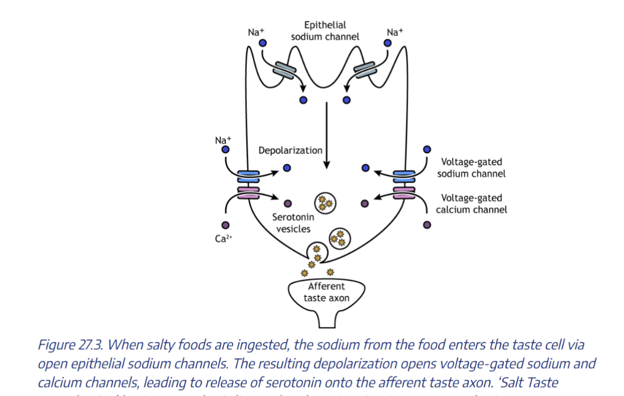
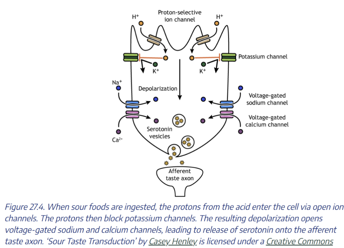
Taste ligands create Ca 2+ signals that release Serotonin or ATP
Sodium Taste Receptor Cell
Type III Taste Receptor Cell
Type II Receptor Cell
Sweet, Umami or Bitter Taste Receptor Cell
These all fall under the classification of a Type II Receptor Cell
What differentiates them is the G-Protein Coupled Receptor that is present.
Sweet TRC have T1R1 GPCR
Umami TRC have T1R1 GPCR
Bitter TRC have T2R GPCR
All three signals work by activating cAMP and PLC- which opens VR K Channels and causes IP3 to trigger Ca release- Ca release then triggers ATP release which activates the Neuron underneath
Phototransduction and the Retina
The Retina is the sensory organ of the eye
Neurons of the Retina are organized into 5 layers
1) Photoreceptors
2) Bipolar Cells
3) Ganglion Cells
4) Amacrine Cells
5) Horizontal Cells
Sensory information passes from the photoreceptors to
the bipolar neurons then the ganglion cells whose axons
form the Optic Nerve
Photoreceptors Rods and Cones of the Retina
Photoreceptors are the neurons that convert light energy into electrical signals
Rods
Function well in low light
Used in Night Vision
Black and White Vision
Cones
High-acuity Vision
Color Vision