BIOC 3001
1. What is biochemistry?
Biochemistry is the study of the chemical substances and chemical processes that occur within living organisms.
The basic idea of the chapter is:
Living organisms → cells → biomolecules → atoms → subatomic particles
Despite the enormous diversity of organisms, the same fundamental chemical principles apply across life.
SPONCH
Most biologically important molecules are built primarily from:
S = sulfur
P = phosphorus
O = oxygen
N = nitrogen
C = carbon
H = hydrogen
The lecture emphasizes that these elements are especially useful biologically because they can form the bonds needed to construct biomolecules.
A major theme is that living systems need both stability and flexibility. Molecules must be stable enough to exist but their interactions also need to be breakable/rearrangeable so biochemical processes can occur.
Define biomolecule and macromolecule and distinguish organic/biological molecules from inorganic molecules
Biomolecule
A biomolecule is a molecule present in living organisms.
This includes large molecules such as:
proteins
carbohydrates
lipids
nucleic acids
but also smaller biological molecules such as metabolites and natural products.
Macromolecule
A macromolecule is a very large molecule, commonly formed by combining smaller subunits called monomers.
The lecture describes macromolecules as typically containing thousands of atoms or more.
Important distinction
Not every biomolecule is a macromolecule.
For example:
Glucose → biomolecule, but relatively small.
Protein → biomolecule AND macromolecule.
Organic vs. inorganic
For this course, a useful distinction is:
Organic molecules
generally carbon-based biological molecules
examples: glucose, proteins, lipids, DNA
Inorganic molecules
examples include water and inorganic ions/salts
can still be extremely important biologically even though they aren't part of the four major organic biomolecule classes
Know the four major classes of biomolecules
The four major classes are:
Class | Major building blocks/features | Important functions | Examples |
|---|---|---|---|
Proteins | Amino acids | Enzymes, receptors/signaling, structure | Enzymes, receptors |
Carbohydrates | Sugars such as glucose | Fuel, energy storage, cell recognition | Glucose, glycogen, starch |
Lipids | Hydrophobic/amphipathic molecules | Energy storage, membranes, signaling | Phospholipids |
Nucleic acids | Nucleotides | Genetic information, gene expression | DNA, RNA |
These are the four classes your professor explicitly identifies.
Proteins
Building blocks
Amino acids
Approximately 20 common amino acids are used to build proteins.
Amino acids are joined through peptide bonds.
General amino-acid structure:
H
|
H3N+ — C — COO−
|
R
The R group/side chain distinguishes one amino acid from another.
Protein functions
Proteins can function as:
enzymes
receptors
signaling molecules
structural components
Proteins are therefore extremely versatile biomolecules.
Carbohydrates
The lecture describes carbohydrates as both fuel and informational molecules.
Important building block
Glucose
Energy storage
Animals:
glucose → glycogen
Plants:
glucose → starch
Functions
Carbohydrates function in:
energy/fuel
energy storage
signaling
cell-cell recognition
Carbohydrates can also form linear or branched chains.
Lipids
Lipids are particularly important for:
fuel storage
signaling
biological membranes
The lecture emphasizes the distinction between:
Hydrophilic
→ interacts with/dissolves in water
versus
Hydrophobic
→ does not dissolve readily in water.
Phospholipids have both:
Hydrophilic head
O
|
|
Hydrophobic tails
||
||
That property allows them to form biological membranes.
Why membranes matter
Membranes allow compartmentalization.
Different biochemical reactions can therefore occur in different regions of the cell.
Nucleic Acids
Building blocks
Nucleotides
Nucleotides are joined by phosphodiester bonds.
Two major types
DNA — deoxyribonucleic acid
Bases:
A, T, G, C
Usually double-stranded.
RNA — ribonucleic acid
Bases:
A, U, G, C
Usually single-stranded.
Functions
DNA/RNA:
store genetic information
maintain genetic information
allow expression of genetic information
Individual nucleotides can also function in:
energy
signaling
cofactors
ATP is an important example.
Central Dogma
The lecture introduces:
DNA → RNA → Protein
More specifically:
DNA
↓ transcription
RNA
↓ translation
Protein
DNA can also undergo:
DNA → DNA
replication
The key point is that DNA does not directly specify amino-acid assembly. Information is first transferred to RNA, which serves as the template used to make proteins.
Categorize novel biomolecules from their structures
You should learn to recognize characteristic structural clues.
Protein/amino acid clues
Look for:
amino group
carboxyl group
central α-carbon
peptide bonds
Typical peptide linkage:
–C(=O)–NH–
Carbohydrate clues
Look for:
many –OH groups
carbon backbone
often ring structures
approximately carbon + hydrogen + oxygen
Glucose is the classic example.
Lipid clues
Look for:
long hydrocarbon regions
relatively few polar groups
fatty-acid chains
hydrophobic character
Phospholipids have:
polar head + nonpolar tails
Nucleic-acid clues
Look for:
phosphate + sugar + nitrogenous base
A nucleotide contains all three.
A nucleic-acid polymer has a repeating sugar-phosphate backbone.
Know common valences
These are extremely useful for determining whether a molecular structure makes sense.
Atom | Typical number of bonds |
|---|---|
H | 1 |
O | 2 |
N | 3 |
C | 4 |
A useful mnemonic:
HONC 1234
H = 1
O = 2
N = 3
C = 4
For example, carbon wants four total bonds:
    H
    |
H — C — H
    |
    H
Oxygen commonly forms two:
H — O — HRank biological objects according to size and complexity
The chapter's size comparison progresses from small chemical species to increasingly complex biological structures.
A useful ranking is:
subatomic particle < atom < small molecule < building block/monomer < macromolecule < macromolecular complex < organelle < cell
For example:
electron
↓
carbon atom
↓
water
↓
glucose/amino acid
↓
protein/DNA
↓
ribosome
↓
mitochondrion
↓
cell
Chapter 2 — Water, Bonding & Buffers
This chapter is considerably more chemistry-heavy.
There are four major themes:
Water → chemical bonds → acids/bases → buffers
Describe the chemical structure of water
Water:
H₂O
δ−
O
/ \
δ+H Hδ+
Water contains polar covalent bonds.
Oxygen attracts the shared electrons more strongly than hydrogen.
Therefore:
O → partial negative charge (δ−)
H → partial positive charge (δ+)
The molecule therefore has a dipole.
The lecture connects this directly to electronegativity and molecular shape.
Relate water's structure to its unique properties
This relationship is crucial:
electronegativity + molecular shape
↓
polar O–H bonds
↓
water has a dipole
↓
hydrogen bonding
↓
water's unusual properties
The lecture identifies properties such as:
high boiling point
high melting point
surface tension
solvation ability
as consequences largely associated with hydrogen bonding.
Water can also form hydrogen bonds with functional groups on biomolecules, and hydrogen bonding plus hydrophobic interactions contribute strongly to the 3D structures of biomolecules.
Diagram the ionization of water
Water can ionize.
A useful representation is:
2 H₂O ⇌ H₃O⁺ + OH⁻
Often simplified in biochemical equations to:
H₂O ⇌ H⁺ + OH⁻
The important concept is that a proton can be transferred between water molecules, producing acidic and basic species.
Relate energy to breaking and forming chemical bonds
This is particularly testable because the lecture gives a mnemonic:
BREAK-IN / MAKE-OUT
Breaking bonds → energy goes IN
Making bonds → energy comes OUT
The lecture describes bond breaking as requiring energy input and bond formation as releasing energy.
So:
Break bond → energy required
Make bond → energy released
Know the six major types of chemical bonding/interactions
Your professor divides them into:
Covalent
Nonpolar covalent
Polar covalent
Noncovalent
Ionic/electrostatic
Hydrogen bonds
Van der Waals interactions
Hydrophobic interactions
Covalent bonds
Atoms share electrons.
Nonpolar covalent
Electrons are approximately equally shared.
Usually occurs when atoms have similar electronegativity.
Polar covalent
Electrons are unequally shared.
Creates partial charges:
δ+ and δ−
Water contains polar covalent bonds.
Strength
Covalent bonds are generally much stronger than individual noncovalent interactions.
Ionic/electrostatic interactions
Large differences in electronegativity can result in electron transfer and the creation of charged ions.
Opposite charges attract: + ← attraction → −
Hydrogen Bonds
A hydrogen covalently attached to an electronegative atom can be attracted to another electronegative atom, particularly oxygen or nitrogen.
For example:
O—H ··· O
↑
H bond
Individual hydrogen bonds are relatively weak.
But:
many weak H-bonds together → major biological effect
They are important for stabilizing biological structures.
Van der Waals Interactions
These arise from temporary fluctuations in electron distribution.
Individually:
very weak
Collectively:
potentially substantial
There is an optimal distance between interacting atoms called the van der Waals contact distance. Too far apart gives little attraction; too close causes electron-cloud repulsion.
Hydrophobic Interactions
Hydrophobic = "water fearing."
Nonpolar molecules do not interact favorably with water.
Therefore, nonpolar groups tend to cluster together in aqueous environments.
This phenomenon is enormously important for:
protein structure
membrane formation
The lecture specifically emphasizes hydrophobic interactions as particularly important for proteins in aqueous environments and essential to membrane formation.
Explain how bonds/interactions contribute to protein structure
Protein structures depend on a combination of interactions.
Covalent bonds
Provide strong chemical connections, including the peptide-bonded backbone.
Hydrogen bonds
Help stabilize particular arrangements of protein structure.
Ionic/electrostatic interactions
Attraction between oppositely charged groups.
Hydrophobic interactions
Hydrophobic groups tend to cluster away from water.
Van der Waals interactions
Help stabilize atoms that are packed at appropriate distances.
The key concept is:
One noncovalent interaction may be weak, but many interactions acting together can strongly stabilize a protein.
This is particularly clear for hydrogen bonds, which the lecture describes as relatively weak individually but important because of their abundance.
Define equilibrium, dissociation, association, and acid dissociation constants
Consider:
HA ⇌ H⁺ + A⁻
Equilibrium constant
Represents the ratio between different chemical species at equilibrium.
Dissociation constant
Measures the equilibrium favoring separated/dissociated species relative to associated species.
For:
HA ⇌ H⁺ + A⁻
a dissociation expression is:
Kd=[H+][A-]/[HA]
Association constant
Essentially the inverse:
Kaassociation = [HA]/[H+][A-]
The lecture emphasizes that association and dissociation constants are inverses.
Acid Dissociation Constant — Ka
For an acid:

Ka describes the tendency of an acid to dissociate and release its proton.
Larger Ka
More dissociation.
Smaller Ka
Less dissociation.
pH and pKa
pH

Higher [H⁺]:
→ lower pH
Lower [H⁺]:
→ higher pH
pKa

Therefore:
Ka and pKa move in opposite directions.
Large Ka → small pKa
Small Ka → large pKa
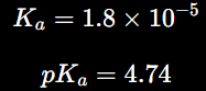
The lecture uses acetic acid as an example:
The Most Important pH/pKa Rule
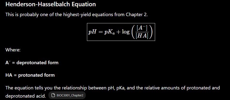
This is one of the most useful rules to memorize for this chapter.
pH = pKa
50% protonated / 50% deprotonated
The amino-acid examples on the slides illustrate this directly.
pH < pKa
More protonated.
pH > pKa
More deprotonated.
Memorize:
LOW pH → HOLD H⁺
HIGH pH → RELEASE H⁺
Example: carboxyl group
Consider:

If:
pKa = 2
and:
pH = 2
then:
50% COOH / 50% COO⁻
At pH 7, pH is well above the pKa, so the carboxyl group is predominantly deprotonated: COO⁻.
The lecture therefore describes carboxyl groups as acidic and typically negatively charged around pH 7.
Example: amino group

Suppose:
pKa = 9
At:
pH = 9
approximately:
50% NH₃⁺ / 50% NH₂
At pH 7:
pH < pKa
therefore the amino group is predominantly:
NH₃⁺
The lecture consequently describes the amino group as basic and positively charged around pH 7.



Extremely Useful Ratio Table
pH relative to pKa | A⁻ : HA | Interpretation |
|---|---|---|
pKa − 2 | 1:100 | ~99% protonated |
pKa − 1 | 1:10 | ~91% protonated |
pKa | 1:1 | 50/50 |
pKa + 1 | 10:1 | ~91% deprotonated |
pKa + 2 | 100:1 | ~99% deprotonated |
State the useful pH range of a biological buffer
A buffer contains:
weak acid HA + conjugate base A⁻
The useful buffering range is approximately:


Why are buffers biologically important?
Proteins are highly sensitive to pH.
The lecture gives the chain:
change pH
↓
change protein charge
↓
change noncovalent interactions
↓
change protein shape
↓
change protein function
This is particularly important for enzymes.
The lecture shows three examples with different optimal pH ranges:
pepsin
fumarase
lysozyme
This illustrates why biological systems need to maintain appropriate local pH conditions.
Functional Groups Worth Recognizing

If you're preparing for an exam, I would know these cold:
Water is polar because of electronegativity + molecular geometry.
Water molecules form hydrogen bonds.
Breaking bonds requires energy; forming bonds releases energy.
Covalent interactions: polar + nonpolar.
Noncovalent interactions: ionic, H-bonds, van der Waals, hydrophobic.
Many individually weak interactions can strongly stabilize biomolecules.
pH=−log[H+]\displaystyle pH=-\log[H^+]
pKa=−logKa\displaystyle pK_a=-\log K_a
Large Ka = small pKa.
pH < pKa → protonated form favored.
pH > pKa → deprotonated form favored.
pH = pKa → 50/50.
pH=pKa+log([A−]/[HA])\displaystyle pH=pK_a+\log([A^-]/[HA])
Best buffering range = pKa ± 1.
Changes in pH can alter protein charge → interactions → structure → function.
Amino Acids
1. Draw a generic amino acid and label its five key components
Amino acids are the building blocks of proteins. All 20 common amino acids share the same basic backbone, and differ in their R group/side chain.
At approximately neutral pH, the generic structure is:
H
|
H3N+—Cα—COO−
|
R
The five key parts are:
α-carbon
α-amino group: usually
NH3+α-carboxyl group: usually
COO−α-hydrogen
R group / side chain
The R group is the only part that differs among the 20 standard amino acids.
Amino acids and chirality
If the α-carbon is attached to four different groups, the amino acid is chiral.
That produces two mirror-image forms:
L-amino acid
D-amino acid
The lecture emphasizes that proteins use L-amino acids.
One important exception is glycine, because its R group is another H, so its α-carbon is not bonded to four different groups.
Define “zwitterion” and explain its relevance
A zwitterion is a molecule that has both a formal positive charge and a formal negative charge, while having no overall net charge.
At approximately neutral pH, a typical amino acid looks like:
NH3+
|
H — Cα — COO−
|
R
So:
amino group = +1
carboxyl group = −1
total = 0
The lecture defines amino acids lacking charged R groups as zwitterions at neutral pH.
Why this matters biologically
Amino-acid charge depends strongly on pH.
As pH changes:
amino groups can gain or lose H⁺
carboxyl groups can gain or lose H⁺
some R groups can also ionize
This changes the amino acid's electrical charge and therefore affects:
electrostatic interactions
protein folding
protein solubility
enzyme activity
Cationic, zwitterionic, and anionic forms
The lecture summarizes the progression as:
Low pH
Lots of H⁺ available.
NH3+
|
H — C — COOH
|
R
Net charge:
+1
This is the cationic form.
Near neutral pH
NH3+
|
H — C — COO−
|
R
Net charge:
0
This is the zwitterion.
High pH
NH2
|
H — C — COO−
|
R
Net charge:
−1
This is the anionic form.
Isoelectric point — pI
The isoelectric point, pI, is the pH where an amino acid has:
net charge= 0
For amino acids without ionizable side chains, this usually corresponds to the zwitterionic form.
Important:
pI is not necessarily pH 7.
It depends on the amino acid's ionizable groups.
Estimate the molecular weight of a protein
This objective is very testable.
The average molecular mass of an amino-acid residue in a protein is approximately:
110 Da\boxed{110\text{ Da}}
The lecture highlights an example around 137 kDa → ~110 kDa average amino-acid reasoning and uses amino-acid molecular masses to establish this approximation.
The quick protein estimate is:
Protein MW≈(number of amino acids)(110 Da)
Example
A protein has 300 amino acids.
300×110=33,000 Da=33 kDa
So:
300-aa protein≈33 kDa
Reverse calculation
A protein is 55 kDa.
55,000/110=500
So it contains approximately:
500 amino acids
Estimate amino- and carboxyl-group charge from pH
This uses the Chapter 2 rule:
pH < pKa
More protonated
pH > pKa
More deprotonated
The Chapter 3 lecture gives approximate average values:
pKa(α-COOH)≈2.2
and
pKa(α-NH3+)≈9.4
Carboxyl group
COOH ⇌ COO− + H+
Below pH 2.2
Mostly:
COOH
Charge:
0
Above pH 2.2
Mostly:
COO⁻
Charge:
−1
Amino group
NH3+ ⇌ NH2 + H+
Below pH 9.4
Mostly:
NH₃⁺
Charge:
+1
Above pH 9.4
Mostly:
NH₂
Charge:
0
At physiological/neutral pH
For a typical amino acid around pH 7:
pH>2.2
so:
COO⁻
and:
pH<9.4
so:
NH₃⁺
Thus:
NH3+ — CH(R) — COO−
Know the 20 common amino acids
Your professor uses this classification scheme: simple, aliphatic, cyclic, aromatic, hydroxyl, amide, acidic, basic, and sulfhydryl.
Class | Amino acid | 3-letter | 1-letter |
|---|---|---|---|
Simple | Glycine | Gly | G |
Simple | Alanine | Ala | A |
Aliphatic | Valine | Val | V |
Aliphatic | Leucine | Leu | L |
Aliphatic | Isoleucine | Ile | I |
Aliphatic | Methionine | Met | M |
Cyclic | Proline | Pro | P |
Aromatic | Phenylalanine | Phe | F |
Aromatic | Tyrosine | Tyr | Y |
Aromatic | Tryptophan | Trp | W |
Hydroxyl | Serine | Ser | S |
Hydroxyl | Threonine | Thr | T |
Amide | Asparagine | Asn | N |
Amide | Glutamine | Gln | Q |
Acidic | Aspartate | Asp | D |
Acidic | Glutamate | Glu | E |
Basic | Lysine | Lys | K |
Basic | Arginine | Arg | R |
Basic | Histidine | His | H |
Sulfhydryl | Cysteine | Cys | C |
A good strategy from the last slide is to learn them by category, rather than trying to memorize all 20 randomly.
Simple amino acids — G, A
Glycine — Gly, G
Side chain:
R = H
Glycine is extremely small and flexible.
The lecture says glycine can adopt conformations unavailable to other amino acids and is more flexible than other amino acids.
Because of its small size, glycine can occur:
inside proteins
outside proteins
in tightly packed regions
Alanine — Ala, A
Side chain:
R = CH3
Alanine is also small.
The lecture notes that alanine is often useful as a substitution when researchers want minimal structural or functional disturbance.
Aliphatic amino acids — V, L, I, M
These are:
Valine
Leucine
Isoleucine
Methionine
They contain primarily nonpolar hydrocarbon-like side chains.
The lecture describes them as:
nonpolar
hydrophobic
often found in protein cores
important for protein folding
Valine — V
CH3
\
R = CH
/
CH3
Leucine — L
R = CH2—CH(CH3)2
Isoleucine — I
R = CH(CH3)—CH2—CH3
Valine, leucine, and isoleucine are branched-chain amino acids.
Methionine — M
Contains sulfur:
R = CH2—CH2—S—CH3
But this sulfur is present in a thioether.
The lecture specifically warns not to confuse methionine with cysteine: methionine's sulfur is not highly reactive like cysteine's thiol.
Cyclic amino acid — Proline
Proline — Pro, P
Proline's side chain loops back and attaches to the amino nitrogen.
This gives it a rigid cyclic structure.
Consequences:
restricts rotation
disrupts some secondary protein structures
frequently appears at turns/bends
A very good association is:
Proline = protein bend/turn
Aromatic amino acids — F, Y, W
These contain aromatic ring systems.
Phenylalanine — Phe, F
Contains a benzene-like phenyl group.
Generally:
hydrophobic
nonpolar
Tyrosine — Tyr, Y
Similar to phenylalanine but contains an:
-OH group
Therefore tyrosine can participate in hydrogen bonding and is more polar than phenylalanine.
Its side-chain pKa is approximately:
10
Tryptophan — Trp, W
Contains an indole ring.
The lecture describes indole as a fused six-membered benzene ring plus five-membered nitrogen-containing ring.
UV absorption
Tyrosine and tryptophan absorb light around: 280 nm
Tryptophan contributes the most strongly.
This becomes important when measuring protein concentration.
Hydroxyl amino acids — S, T
Serine — Ser, S
R = CH2OH
Threonine — Thr, T
Contains a hydroxyl-containing side chain.
These amino acids are:
polar
uncharged
hydrophilic
capable of hydrogen bonding
The lecture associates them with protein surface hydration.
Their hydroxyl groups have high pKa values, so they are generally not ionized under ordinary biological conditions.
Amide amino acids — N, Q
Asparagine — Asn, N
R = CH2—CONH2
Glutamine — Gln, Q
R = CH2—CH2—CONH2
These contain terminal carboxamide groups.
They are:
polar
hydrophilic
uncharged
non-ionizable under normal conditions
capable of hydrogen bonding
A useful relationship:
Asparagine corresponds structurally to aspartate
and
Glutamine corresponds structurally to glutamate
except amides are uncharged.
Acidic amino acids — D, E
Aspartate — Asp, D
Side chain contains:
-CH2-COO−
Glutamate — Glu, E
Side chain:
-CH2-CH2-COO−
Their R-group pKa is approximately:
4
Therefore, at physiological pH: pH≈7 > pKa≈4
so they are predominantly deprotonated.
Side-chain charge: -1
They can participate in:
electrostatic interactions
hydrogen bonding
nucleophilic reactions
Basic amino acids — K, R, H
Lysine — Lys, K
Lysine has a terminal amino group.
Its side-chain pKa is approximately: 10.5
Therefore around pH 7:
pH<pKa
so it remains protonated and positively charged: +1
The lecture places lysine frequently on protein surfaces and associates it with electrostatic interactions.
Arginine — Arg, R
Contains a guanidinium group.
Side-chain pKa: 12
Therefore it is strongly positively charged around neutral pH.
Arginine can participate in:
electrostatic interactions
hydrogen bonding
interactions with nucleic acids
some hydrophobic interactions along its carbon chain
The lecture also notes that arginine is commonly found on protein surfaces.
Histidine — His, H
Histidine is particularly important.
It contains an imidazole ring.
pKa: 6
Because physiological pH is relatively close to this pKa, histidine can readily change protonation state.
That allows histidine to:
donate H⁺
accept H⁺
This is why it frequently appears in enzyme active sites.
A strong association:
Histidine = acid/base catalysis
Sulfhydryl amino acid — Cysteine
Cysteine — Cys, C
Side chain:
R = CH2—SH
Contains a sulfhydryl/thiol group.
Side-chain pKa: 8.5
The thiol group is highly chemically reactive.
Cysteine can undergo oxidation.
Two cysteines can form a:
disulfide bond
Cys—SH + HS—Cys
↓ oxidation
Cys—S—S—Cys
The linked product is called:
cystine
The lecture stresses that disulfide bonds can be critically important for protein structure.
Seven amino acids with important side-chain pKa values
The lecture highlights that seven amino acids have biologically relevant ionizable side chains.
Amino acid | Side-chain pKa | Approximate charge near pH 7 |
|---|---|---|
Aspartate, D | ~3.7–4 | −1 |
Glutamate, E | ~4.2 | −1 |
Histidine, H | ~6 | often partly protonated |
Cysteine, C | ~8.5 | mostly 0 |
Tyrosine, Y | ~10 | mostly 0 |
Lysine, K | ~10.5 | +1 |
Arginine, R | ~12 | +1 |
For this course, the most important shortcut is:
At pH 7
Negative
D
E
Positive
K
R
Special
H, because its pKa is close to physiological pH
Usually neutral but ionizable
C
Y
Predict where an amino acid will be located in a protein
This is one of the most conceptually important objectives.
The key question is:
Is the side chain hydrophobic or hydrophilic?
For a typical soluble protein in water:
Hydrophobic side chains
Prefer the protein interior/core.
Examples:
V
L
I
M
F
Why?
They avoid contact with water.
The lecture explicitly says aliphatic amino acids help promote folding and are often found in protein cores.
Polar/charged side chains
Often appear on the:
protein surface
Examples:
S
T
N
Q
D
E
K
R
These interact favorably with water through:
hydrogen bonding
ion-dipole interactions
electrostatic interactions
For example, the lecture specifically describes lysine and arginine as commonly present on protein surfaces.
Aromatic residues
F, Y, W can be somewhat more complicated.
The lecture notes that aromatic side chains are relatively nonpolar and may be partially buried on the protein surface.
Tyrosine is more surface-compatible because its OH can hydrogen bond.
Glycine and Proline are structural exceptions
Glycine
Very small + flexible.
Likely in:
tight spaces
flexible regions
unusual conformations
Proline
Rigid.
Likely in:
turns
bends
and can disrupt regular secondary structure.
Cysteine has a structural role
Cysteines may appear at locations where two portions of the protein need to be covalently linked.
Two cysteine residues:
Cys−SH+HS−Cys
can form:
Cys−S−S−Cys
This stabilizes protein structure.
A practical protein-location rule
For a soluble protein, imagine:
WATER
D K S N R Q
\ /
\ /
---------
| V L I F |
| M W | ← hydrophobic core
| V L |
---------
/ \
T E K
WATER
So:
Inside
Mostly hydrophobic residues
V L I M F W
Surface
Mostly polar/charged residues
S T N Q D E K R
Special cases
G → flexibility/tight spaces
P → turns/bends
C → disulfides
H → enzyme active sites
This is a very useful exam framework.
1. Amino acids are joined by peptide bonds
A peptide bond is a special type of covalent amide bond formed between:
the carboxyl group of one amino acid
the amino group of another amino acid
Formation occurs through a condensation reaction, releasing water.
Conceptually:
AA1—COOH + H2N—AA2
↓
AA1—CO—NH—AA2 + H2O
↑
peptide bond
The reverse process is hydrolysis, in which water is used to break the peptide bond.
2. Polypeptides have directionality
Every polypeptide has two different ends:
N-terminus
The end containing the free amino group.
C-terminus
The end containing the free carboxyl group.
Protein sequences are always written:
N-terminus→C-terminus
Your lecture shows the five-residue peptide YGGFL and emphasizes that peptides are read from N → C, with the N-terminus conventionally shown on the left.
So if you see:
YGGFL
you read it:
Tyr → Gly → Gly → Phe → Leu
from N-terminus to C-terminus.
3. The peptide backbone repeats
The backbone follows the repeating pattern:
N−Cα−C−N−Cα−C−N−Cα−C...
The R groups project away from this backbone.
That means proteins essentially contain:
Backbone → common structural framework
R groups → determine many of the protein's chemical properties
Because most α-amino and α-carboxyl groups become incorporated into peptide bonds, the lecture emphasizes that the side chains become major determinants of the chemical properties of the protein.
Peptide bonds are planar
This is one of the most important structural properties to know.
The peptide bond has partial double-bond character because of resonance.
Conceptually:
O O−
|| |
— C — N — ↔ — C = N+ —
| |
H H
Because of this resonance:
rotation around the peptide C–N bond is restricted
the peptide bond is essentially planar
six atoms lie approximately in one plane
the peptide bond itself is uncharged
neighboring α-carbons generally adopt the trans configuration
Important distinction
A polypeptide is therefore:
Flexible, but conformationally restricted.
It isn't a completely floppy chain.
Phi (φ) and Psi (ψ) angles allow flexibility
Although rotation around the peptide C–N bond is restricted, rotation can occur around two other backbone bonds.
Phi — φ
Rotation around:
N−Cα
Psi — ψ
Rotation around:
Cα−C
Your professor describes these as the two effective degrees of freedom in the peptide backbone.
Together:
ϕ+ψ
determine the path taken by the polypeptide chain.
Not every φ/ψ combination is physically possible because atoms would collide with one another.
Allowed combinations are represented using a:
Ramachandran plot
Name and Describe the Four Levels of Protein Structure
This hierarchy is extremely important.
Level | What it describes | Major stabilizing feature |
|---|---|---|
Primary | Amino-acid sequence | Peptide bonds |
Secondary | Local backbone folding | Backbone H-bonds |
Tertiary | Overall 3D structure of one polypeptide | Side-chain interactions |
Quaternary | Association of multiple polypeptide chains | Mostly noncovalent interactions |
1. Primary Structure
Primary structure is simply the:
linear amino-acid sequence
For example:
N—Met—Gly—Ser—Leu—Val—CThe amino acids are connected through peptide bonds.
Genes ultimately specify the unique amino-acid sequence of proteins.
Think:
Primary = sequence
2. Secondary Structure
Secondary structure describes local folding of the polypeptide backbone.
It is produced primarily through:
hydrogen bonding between backbone groups
Specifically:
C=O⋯H−N
The carbonyl oxygen acts as a hydrogen-bond acceptor and the amide hydrogen participates as a donor.
Your lecture divides secondary structure into:
α-helices
β-sheets
turns and loops
The two main repeating structures you especially need to know are:
α-helix and β-sheet
3. Tertiary Structure
Tertiary structure is the:
overall 3D folded structure of one polypeptide chain
The lecture describes it as the three-dimensional, folded, biologically active conformation of a single polypeptide.
Unlike secondary structure, tertiary structure depends heavily on interactions involving R groups.
These include:
hydrophobic interactions
hydrogen bonds
ionic interactions
disulfide bonds
4. Quaternary Structure
Quaternary structure occurs when:
two or more separate polypeptide chains associate\boxed{\text{two or more separate polypeptide chains associate}}
Each individual chain is called a:
subunit
The lecture states that quaternary structure is held together primarily by noncovalent interactions and is stabilized by the same types of forces involved in tertiary structure.
Example: Hemoglobin
Hemoglobin contains four subunits:
α2β2
Two α subunits + two β subunits.
Easy memory trick
1° = Sequence
2° = Sections of regular folding
3° = Three-dimensional single chain
4° = Four-ish/multiple chains together
Identify the Two Main Secondary Structures and Their Bonds
A. α-Helix
The α-helix is a coiled structure stabilized by hydrogen bonds within the same polypeptide chain.
The R groups point: Outward away from the central helix
Conceptually:
R R
\ /
\ /
/ \
/ \
/ \
/ \
↑
α-helical
backbone
R groups point outward
α-helix hydrogen bonding
The important rule is:
i→i+4
The C=O of one residue hydrogen-bonds with the N–H of a residue four positions later.
For example:
Residue 1 C=O ··· H-N Residue 5
Residue 2 C=O ··· H-N Residue 6
Residue 3 C=O ··· H-N Residue 7
These repeated hydrogen bonds stabilize the helix.
The lecture also notes that α-helices are usually right-handed.
Proline and α-helices
Proline is generally unfavorable within an α-helix because its rigid cyclic structure interferes with normal helix geometry.
Remember:
Proline = helix disruptor
β-Sheet
β-sheets consist of individual:
β-strands
positioned next to one another.
Hydrogen bonds form between:
C=O
of one strand and:
N−H
of an adjacent strand.
Unlike an α-helix, the interacting sections can be far apart in the primary amino-acid sequence.
For example:
Residues 10–15
→→→→→→
|||||| H bonds
←←←←←←
Residues 80–85
Residues 10–15 and 80–85 might be far apart in primary structure but become neighbors after protein folding.
Parallel vs. Antiparallel β-Sheets
There are two arrangements.
Parallel
Both strands run in the same N → C direction:
N → → → C
H bonds
N → → → C
Antiparallel
The strands run in opposite directions:
N → → → C
H bonds
C ← ← ← N
α-Helix vs. β-Sheet
Feature | α-Helix | β-Sheet |
|---|---|---|
Shape | Coil/helix | Extended sheet |
H-bonds | Within helix | Between neighboring strands |
Main interaction | Backbone C=O···H–N | Backbone C=O···H–N |
Important pattern | i → i+4 | Adjacent β-strands |
R groups | Point outward | Project from sheet |
Variants | Usually right-handed | Parallel/antiparallel |
Critical exam point
Both α-helices and β-sheets are stabilized primarily by:
backbone hydrogen bonds
Not by hydrogen bonding between R groups.
Turns and Loops
Proteins also need structures that connect α-helices and β-sheets.
β-turns
Turns allow the polypeptide chain to reverse direction.
Your lecture emphasizes:
Proline is commonly found in turns.
Glycine is also common.
Asp and Asn can occur frequently.
a hydrogen bond can form between residue i and i+3.
This makes sense based on Chapter 3:
Proline → rigid → encourages bends
Glycine → tiny and flexible → fits tight turns
Loops
Loops connect other secondary structures but have more variable sequences.
If a loop has an important biological function, such as interacting with another protein, its sequence may be conserved
Predict an Amino Acid's Location in a Protein
For a typical soluble protein surrounded by water, the major principle is:
hydrophobic inside; hydrophilic outside
The lecture states that hydrophobic R groups tend to orient inward, while polar or ionized groups tend to orient outward toward water.
Hydrophobic amino acids → protein core
Nonpolar side chains dislike interacting with water.
Examples include:
Leu
Ile
Val
Met
Phe
So:
WATER
↓
┌─────────────┐
│ polar │
│ V L I │
│ M F │ ← hydrophobic core
│ polar │
└─────────────┘
↑
WATER
Your professor specifically gives Leu and Phe as examples of residues normally oriented inward.
Polar/charged amino acids → protein surface
These interact favorably with water.
Examples:
Polar uncharged
Ser
Thr
Asn
Gln
Negatively charged
Asp
Glu
Positively charged
Lys
Arg
Your professor specifically gives Ser, Gln, and Arg as examples of outward-facing residues.
Important exceptions
This is a general rule, not an absolute rule.
A charged or polar residue can occur inside a protein if it has an important structural or functional role, such as:
enzyme catalysis
substrate binding
hydrogen bonding
ionic interactions
Glycine is also described in the lecture as being able to tolerate either aqueous or nonaqueous environments.
Explain the Forces Stabilizing Proteins
Your professor identifies four major forces stabilizing tertiary structure:
1. Hydrophobic interactions2. Hydrogen bonds3. Ionic interactions4. Disulfide bridges
1. Hydrophobic Interactions — Most Important
The lecture specifically labels the hydrophobic interaction as the most important force in tertiary folding.
Nonpolar R groups cluster together away from water.
For example:
Water Water Water Water
┌─────────┐
│ Leu Val │
│ Phe Ile │ ← hydrophobic core
│ Met Leu │
└─────────┘
Water Water Water Water
This drives formation of compact globular protein structures.
2. Hydrogen Bonds
Hydrogen bonds can form between polar groups.
Common participants include:
Ser
Thr
Asn
Gln
Tyr
Hydrogen bonding also occurs extensively between the backbone C=O and N–H groups, especially in secondary structure.
One H-bond is weak compared with a covalent bond, but many together can substantially stabilize a protein.
3. Ionic/Electrostatic Interactions
Oppositely charged R groups attract.
For example:
Asp−⋯Lys+
or
Glu−⋯Arg+
These can stabilize particular regions of the folded protein.
Changing pH can change side-chain protonation and therefore disrupt these interactions—which is one reason protein structure can be sensitive to pH.
4. Disulfide Bonds
These are different from the previous three because they are:
covalent bonds
Two cysteine side chains can undergo oxidation:
Cys−SH+HS−Cys
↓
Cys−S−S−Cys
The linked pair is called:
cystine
Your professor identifies these cysteine-cysteine disulfide bridges as one of the forces capable of stabilizing tertiary protein structure.
Hemoglobin: Cooperativity & Allosterism
Big Picture
The main biological problem is that cells require O₂ for respiration, but oxygen has low solubility in aqueous environments and simple diffusion is inadequate for large organisms. Oxygen-carrier proteins solve this problem.
Two major proteins in this chapter are:
Myoglobin → stores/binds O₂ in muscle
Hemoglobin → transports O₂ from lungs → tissues
The most important concept is that hemoglobin isn't simply an oxygen-binding protein. Its four subunits communicate with one another, allowing hemoglobin to change its oxygen affinity depending on conditions.
Explain the Biochemical Differences Between Hemoglobin and Myoglobin
Myoglobin
Myoglobin is located in the cytosol of muscle cells, especially red muscle tissue. It facilitates oxygen transport within muscle and serves an oxygen-binding/storage role.
Structure
Myoglobin is:
a single polypeptide
approximately 18 kDa
predominantly/all α-helical
contains one heme
binds a maximum of one O₂ molecule
Because it consists of one polypeptide, its highest level of organization is tertiary structure.
Hemoglobin
Hemoglobin is found in red blood cells and transports oxygen from the lungs to peripheral tissues. The lecture also emphasizes its role in transporting H⁺ and CO₂.
Adult hemoglobin contains four subunits:
α2β2
Therefore, hemoglobin has quaternary structure.
Each subunit contains one heme group and can bind one O₂.
Therefore:
4 hemes→4 O2
per hemoglobin tetramer.
Feature | Myoglobin | Hemoglobin |
|---|---|---|
Major location | Muscle-cell cytosol | Red blood cells |
Major role | O₂ binding/storage in muscle | O₂ transport |
Polypeptides | 1 | 4 |
Structure | Tertiary | Quaternary |
Adult subunits | One chain | α₂β₂ |
Heme groups | 1 | 4 |
Maximum O₂ | 1 | 4 |
O₂-binding curve | Hyperbolic | Sigmoidal |
Cooperativity | No | Yes |
Allosteric regulation | Not emphasized here | Yes |
the overall fold of myoglobin resembles an individual hemoglobin globin subunit, but hemoglobin combines four globin subunits into a quaternary complex.
Why Their Differences Matter
The two proteins have different jobs, so they need different oxygen-binding behavior.
Myoglobin
Myoglobin binds O₂ strongly and stores/facilitates O₂ in muscle.
Because it only has one O₂-binding site, there are no other subunits or binding sites with which to communicate.
Therefore:
Myoglobin does NOT exhibit cooperativity\boxed{\text{Myoglobin does NOT exhibit cooperativity}}
Its O₂-binding curve is hyperbolic.
Hemoglobin
Hemoglobin needs to do something more complicated:
bind O₂ in lungs AND release O₂ in tissues\boxed{\text{bind O₂ in lungs AND release O₂ in tissues}}
Its four subunits interact, producing cooperative oxygen binding.
Therefore its curve is:
sigmoidal
The lecture's graph on page 15 shows why this matters: across the illustrated lung-to-tissue pO₂ change, myoglobin releases only about 7%, whereas hemoglobin releases about 66%. Thus, cooperativity dramatically enhances oxygen delivery.
Key idea
Myoglobin is designed to hold O₂; hemoglobin is designed to pick it up and efficiently give it away.
Describe the Heme Group and What Changes When O₂ Binds
Both hemoglobin and myoglobin bind oxygen using a:
heme group
Globins contain heme in a hydrophobic pocket created by the folded protein.
Structure of Heme
Heme consists of:
1. Protoporphyrin
An organic ring system.
2. Ferrous iron
A central:
Fe2+
The Fe²⁺ sits in the center of the porphyrin and interacts with four nitrogen atoms.
However, Fe²⁺ can form a total of six coordination interactions.
Think of it as:
6th site
↑
Fe
/|\
/ | \
four porphyrin N
|
↓
5th site
The Six Coordination Sites
Sites 1–4
Four nitrogen atoms of the porphyrin ring coordinate Fe²⁺.
Site 5 — Proximal Histidine
The fifth site is occupied by the imidazole group of a:
proximal histidine
This histidine connects the heme iron to the globin protein.
Site 6 — O₂
The sixth coordination site binds:
O2
The bound oxygen also interacts with another histidine called the:
distal histidine
Your lecture's diagram on page 12 distinguishes these clearly: the proximal histidine directly coordinates Fe²⁺, while O₂ occupies the sixth coordination site and interacts with the distal histidine.
What Happens When O₂ Binds?
This tiny molecular event triggers a much larger protein structural change.
Without O₂
The Fe²⁺ is positioned slightly outside the plane of the porphyrin ring.
O₂ binds
O₂ occupies the sixth coordination site.
↓
Fe²⁺ moves
The iron moves:
into the plane of the heme
↓
Proximal histidine moves
Because the proximal His is attached to Fe²⁺, it moves too.
↓
α-helix moves
The proximal histidine is part of an α-helix, so movement is transmitted into the protein.
↓
Subunits move
This structural change is communicated to the other hemoglobin subunits.
↓
Hemoglobin changes conformation
T→R
Your lecture describes this structural pathway directly.
Cooperativity, T/R States, and Allosterism
First: What Is Allosterism?
Your lecture defines allosterism as binding of a molecule at one site that causes a conformational change altering protein activity.
In simple terms:
binding at one location affects behavior somewhere else
What Is Cooperativity?
Cooperativity is a particular form/consequence of allosteric behavior in which:
binding at one site changes affinity at other sites
Hemoglobin exhibits positive cooperativity.
That means:
Binding of one O₂ makes subsequent O₂ binding easier.
And as hemoglobin transitions toward its high-affinity state, the remaining sites increasingly favor O₂ binding.
T State vs. R State
Hemoglobin exists dynamically between two important conformational states.
T State — Tense
T=lower O2 affinity
Associated with:
deoxyhemoglobin
weaker O₂ binding
O₂ release
extensive αβ dimer interfaces
R State — Relaxed
R=higher O2 affinity
Associated with:
oxyhemoglobin
stronger O₂ binding
subunit rearrangement
During the T → R transition, the two αβ dimers rotate relative to one another.
Why Cooperativity Produces a Sigmoidal Curve
Myoglobin has one O₂-binding site:
hyperbolic curve
Hemoglobin's interacting sites produce:
sigmoidal/S-shaped curve
At relatively low O₂:
Hemoglobin favors lower-affinity behavior.
As O₂ begins binding:
O2→conformational change
which promotes additional O₂ binding.
Thus the curve rises steeply through its cooperative region.
At high pO₂:
Hemoglobin becomes highly saturated.
The lecture specifically identifies the sigmoidal shape as a hallmark of cooperativity arising from interactions between distinct O₂-binding sites
Why Cooperativity Is Biologically Useful
Think about the two environments hemoglobin encounters.
Lungs
High pO₂
↓
O₂ binding is favored
↓
R state favored
↓
Hemoglobin becomes highly saturated
Tissues
Lower pO₂
↓
O₂ dissociates
↓
T state becomes more favorable
↓
More O₂ can be released
So:
Cooperativity improves O₂ loading in lungs and unloading in tissues
That is exactly what a transport protein needs.
How 2,3-BPG, CO₂ and H⁺ Affect Hemoglobin
Hemoglobin doesn't respond only to O₂.
Its oxygen affinity is regulated by allosteric effectors.
Your lecture identifies:
2,3-BPG
CO₂
H⁺
and also Cl⁻
as allosteric inhibitors that stabilize the T form.
The central rule is:
T state↑→O2 affinity↓→O2 release↑
1. 2,3-BPG
2,3-bisphosphoglycerate (2,3-BPG) is an important allosteric effector of hemoglobin.
The lecture reports that erythrocytes contain roughly:
5 mM 2,3-BPG at sea level
How does it work?
2,3-BPG binds at the interface of the hemoglobin subunits and makes contacts with the β-subunits.
↓
It stabilizes:
T state
↓
Hemoglobin's O₂ affinity decreases.
↓
O₂ dissociation increases.
So memorize:
2,3-BPG↑→T↑→O2 affinity↓→O2 release↑
On an O₂-binding curve, the graph shows this as a shift toward higher pO₂ for a given fractional saturation.
H⁺ and CO₂ — The Bohr Effect
Actively respiring tissues produce:
CO2
and conditions associated with increased:
H+
The lecture states that both enhance oxygen release from hemoglobin at the tissue level. This phenomenon is called the:
Bohr effect
Why This Is Useful
Consider an actively working muscle.
The muscle needs lots of oxygen for cellular respiration.
At the same time, active metabolism produces CO₂.
Therefore:
Active tissue
↓
CO₂ ↑ / H⁺ ↑
↓
T state stabilized
↓
Hb O₂ affinity ↓
↓
O₂ released
↓
O₂ delivered to active tissue
This creates a very useful physiological relationship:
The tissues producing metabolic signals associated with high activity encourage hemoglobin to release more oxygen exactly where oxygen is needed.
CO₂, H⁺ and pH
The diagram connects CO₂ with the carbonic acid/bicarbonate system:
CO2+H2O⇌H2CO3⇌HCO3−+H+
and identifies carbonic anhydrase in this process.
Therefore, increasing CO₂ is associated with increased H⁺ and decreased pH in the RBC environment shown.
The graph on the same page compares approximately:
pH=7.4
with
pH=7.2
The lower-pH condition promotes greater O₂ release over the illustrated tissue-to-lung range.
All Three Effectors Together
Effector | Effect on T state | O₂ affinity | O₂ release |
|---|---|---|---|
2,3-BPG ↑ | ↑ | ↓ | ↑ |
CO₂ ↑ | ↑ | ↓ | ↑ |
H⁺ ↑ / pH ↓ | ↑ | ↓ | ↑ |
So the master rule is:
2,3-BPG, CO2, H+→T state→↓O2 affinity→↑O2 release
Molecular Recognition, Allosterism, and Cooperativity
The beginning of the chapter gives three definitions worth distinguishing.
Molecular recognition
Specific interaction/binding between molecules through noncovalent interactions.
Examples include:
receptor + ligand
protein + protein
enzyme + substrate
enzyme + regulator
enzyme + inhibitor
Allosterism
Binding at one location produces a conformational change that affects protein behavior elsewhere.
Cooperativity
Binding at one site alters binding affinity at additional sites.
So:
Molecular recognition → allosteric structural change → cooperative behavior
in the context of hemoglobin.
Enzymes
1. General Properties of Enzymes
An enzyme is a biological catalyst: it increases the rate of a chemical reaction without itself being consumed.
Your lecture identifies several important properties:
Most enzymes are proteins.
Enzyme names commonly end in -ase.
They accelerate reactions under relatively mild/physiological conditions.
They are highly specific for their reactions.
Substrates bind to a particular region called the active site.
Enzymes can be regulated.
Enzymes are not used up or permanently changed by the reaction.
Some RNA molecules also possess catalytic activity; these are ribozymes.
The general reaction can be represented:

where:
E = enzyme
S = substrate
ES = enzyme-substrate complex
P = product
Notice that E appears on both sides. The enzyme participates in the reaction but is regenerated.
2. Why Are Enzymes Excellent Biological Catalysts?
Cells need chemical reactions to happen at useful rates under physiological conditions.
Enzymes solve this problem primarily by: lowering activation energy
enzymes increase reaction rates while not changing the reaction's equilibrium constant or overall energetics.
Enzymes are particularly effective because they combine:
Specificity + catalytic power + mild reaction conditions + regulation.
Their specificity comes from substrate recognition involving:
noncovalent interactions
three-dimensional shape
charge
3. Enzyme Cofactors
Some enzymes require additional nonprotein components to function.

The slides further distinguish:
Prosthetic group: remains unchanged during the reaction.
Cosubstrate: must subsequently be regenerated.
4. ΔG and Reaction Favorability
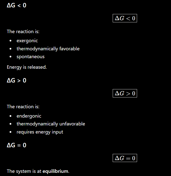
A central distinction in this chapter is between thermodynamics and kinetics.


7. Interpreting an Energy Diagram
X-axis
Reaction progress
It represents progression from:
S→P
It is not simply time.
Y-axis
Free energy\boxed{\text{Free energy}}
Reactant/substrate
The starting energetic state.
Product
The final energetic state.
The difference between them is:
ΔG\boxed{\Delta G}
Peak
The top of the curve represents the: transition state
The energy required to reach it from substrate is:
ΔG‡
or activation energy.
The enzyme provides a pathway with a smaller activation-energy barrier, while the initial and final energy levels remain unchanged.
8. Transition State
The transition state is a temporary, extremely unstable molecular arrangement.
It is:
no longer fully substrate, but not yet product.
The energy necessary to reach it is the activation energy. Enzymes facilitate formation of the transition state and therefore decrease the activation-energy barrier.
A useful reaction sequence is:

where X‡X is the transition state.
9. Transition-State Analogs
Transition states are unstable and cannot normally be isolated.
However, scientists can create molecules that resemble them:
transition-state analogs
These can bind strongly to enzymes and inhibit them.
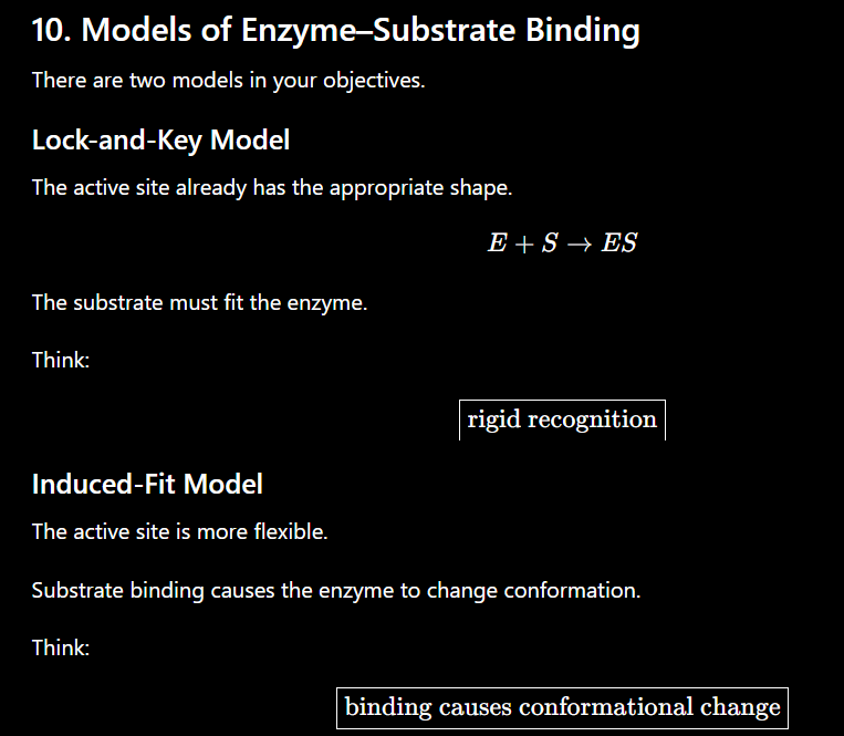
11. Key Characteristics of Active Sites
This is another section I would memorize almost word-for-word for an exam.
An active site:
Is a three-dimensional cleft or crevice.
Can contain amino acids that are far apart in the primary sequence but brought together by protein folding.
Occupies only a small portion of total enzyme volume.
Creates a unique microenvironment.
Interacts with substrate through multiple weak interactions.
Determines enzyme specificity through its molecular architecture.
Stabilizes the transition state.
ENZYME KINETICS
How can we quantitatively measure enzyme behavior?
The answer is enzyme kinetics, particularly Michaelis–Menten kinetics.
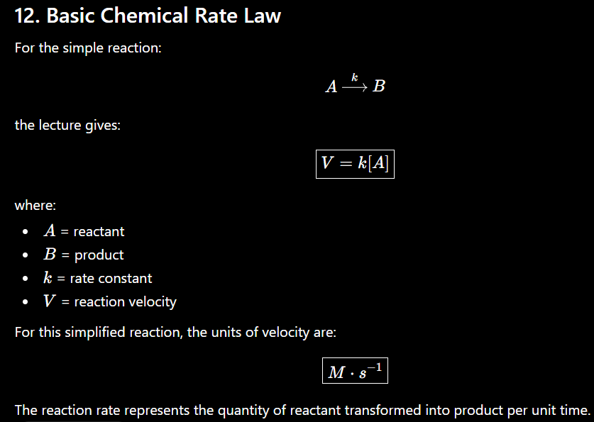
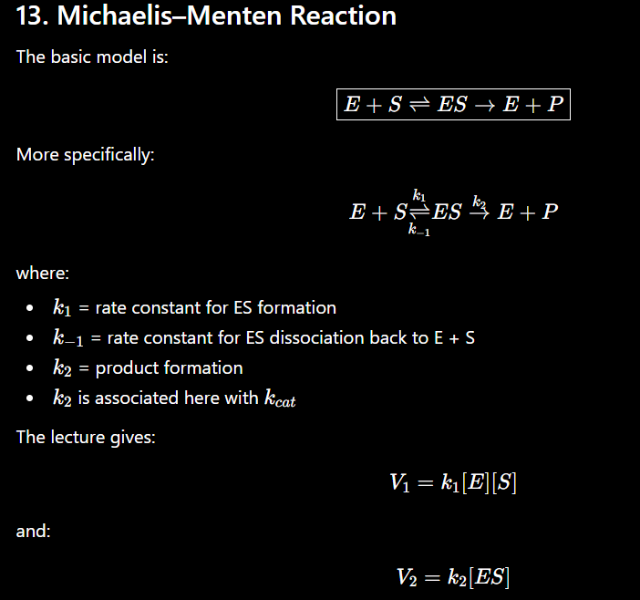
14. Three Michaelis–Menten Assumptions
Know these three:


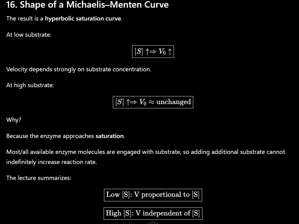
17. Michaelis–Menten Equation
This equation needs to be memorized:

Where:
V0= initial velocity
Vmax = maximum velocity
[S]= substrate concentration
KM = Michaelis constant


So to find KM from a graph:
Find Vmax
↓
Divide by 2
↓
Find Vmax/2 on y-axis
↓
Move to the curve
↓
Move down to x-axis
↓
That [S] = KM
21. Interpreting KM
The lecture uses KM
as a quantity relating enzyme–substrate affinity.
Within that framing:
Lower KM
Half-maximal velocity is reached at a lower substrate concentration.
This is associated with tighter/greater substrate binding affinity.
Higher KM
More substrate is required to reach half-maximal velocity.
This is associated with weaker/lower substrate binding affinity.
A useful shorthand for this course is:


23. Catalytic Efficiency
Another major equation:
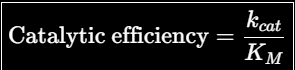
This combines two aspects of enzyme performance emphasized in the lecture:
kcat → ability to catalyze the reaction
KM → substrate-binding behavior
Therefore, a large:
kcat/KM
can result from:
large kcat
small KM
or both
24. Comparing Two Enzymes
Suppose:
Enzyme | KM | kcat |
|---|---|---|
A | 1 mM | 100 s⁻¹ |
B | 10 mM | 20 s⁻¹ |
Catalytic efficiencies:
A=100/1=100 B = 20/10 = 2
So A has the larger kcat/KM
For exam problems, don't look at KM or kcat alone when the question specifically asks about catalytic efficiency.


Kinetic Mechanisms & Inhibitors
1. Big Picture: Enzyme Inhibition
An enzyme inhibitor decreases enzyme activity.
Your chapter divides inhibition into two major categories:
Reversible inhibition involves noncovalent association and dissociation between inhibitor and enzyme. The three types you need to know are:

Irreversible inhibition generally involves covalent attachment that permanently inactivates the enzyme.
The single most important table to memorize from this chapter is:
Inhibition | What inhibitor binds | Apparent KM | Apparent Vmax |
|---|---|---|---|
Competitive | E only | ↑ | unchanged |
Uncompetitive | ES only | ↓ | ↓ |
Noncompetitive | E or ES | unchanged | ↓ |

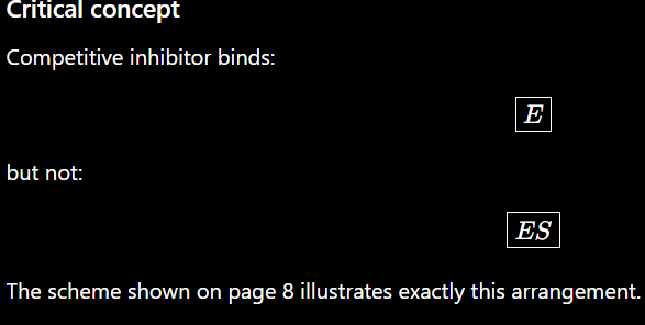
2. Competitive Inhibition
What happens?
A competitive inhibitor competes with the substrate for the enzyme's active site.
The inhibitor often resembles the substrate sufficiently to fit into the active site.
Your slides describe it as the inhibitor essentially "wasting" the enzyme's time while occupying the active site.
Cartoon
Substrate
↓
┌───────────┐
│ ACTIVE │
│ SITE │
│ ENZYME │
└───────────┘
↑
Inhibitor
S and I compete for the SAME site.
Therefore, substrate and inhibitor cannot occupy the active site simultaneously.

S
↓
E ⇌ ES → E + P
↕
EI
↑
I


5. Competitive Inhibition — Michaelis–Menten Plot
Without inhibitor:
Velocity
│ ───────── Vmax
│ ___/
│ __/
│ __/
│ __/
│__/
└────────────────────────── [S]
With competitive inhibitor:
Velocity
│ ───────── SAME Vmax
│ ___/
│ ___/
│ __/ no inhibitor
│ _/
│ _/ competitive inhibitor
│/
└────────────────────────── [S]
The curve shifts effectively to the right because more substrate is required.
So recognize:


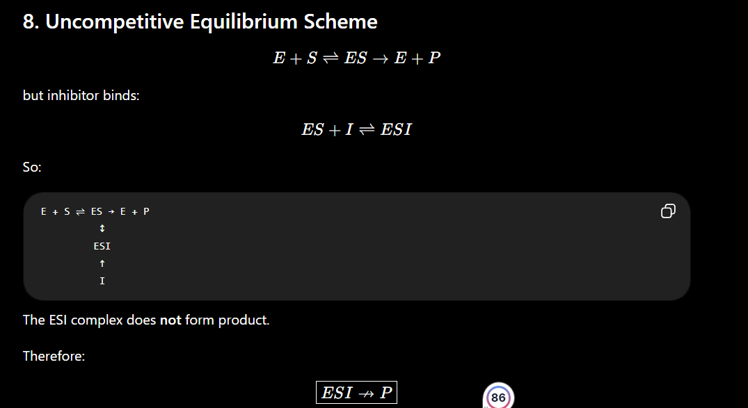
7. Uncompetitive Inhibition
This one is very different.
An uncompetitive inhibitor binds only after the substrate has already bound.
Therefore, it binds:
ES
NOT free enzyme.
Your slides define uncompetitive inhibition specifically as inhibitor binding only to the enzyme-substrate complex.
Cartoon
STEP 1:
Substrate binds enzyme
E + S → ES
STEP 2:
Inhibitor
↓
┌───────────┐
│ S already │
│ bound │
│ ENZYME │
└───────────┘
ESI
Product cannot form

10. Uncompetitive — Michaelis–Menten Plot
The inhibited curve has:
lower Vmax
lower KM
Velocity
│
│ ─────── No inhibitor
│ ___/
│ __/
│ __/
│ /
│ /────────── + inhibitor
│ _/
│/
└────────────────────────── [S]
So recognize:


Cartoon
Substrate
↓
┌───────────┐
I → │ │
│ ENZYME │
└───────────┘
↑
Inhibitor binds
elsewhere
Substrate and inhibitor do not need to compete for the same site.


15. Noncompetitive — Michaelis–Menten Plot
The key feature is:

Velocity
│
│ ───────── No inhibitor
│ ___/
│ __/
│ __/
│ /──────── Lower Vmax
│ _/
│/
└──────────────────────── [S]
↑
Same KM

17. How to Identify the Three Types from Graphs
This is probably one of the most testable parts of the chapter.
Michaelis–Menten
Observation | Inhibitor |
|---|---|
Same VmaxV_{max}, ↑ KMK_M | Competitive |
↓ VmaxV_{max}, ↓ KMK_M | Uncompetitive |
↓ VmaxV_{max}, same KMK_M | Noncompetitive |
Lineweaver–Burk
Pattern | Inhibitor |
|---|---|
Lines meet at y-axis | Competitive |
Lines are parallel | Uncompetitive |
Lines meet at x-axis | Noncompetitive |
Quick memory trick:
COMPETITIVE
→ Same Vmax
→ Y-axis intersection
UNCOMPETITIVE
→ Both decrease
→ Parallel lines
NONCOMPETITIVE
→ Same KM
→ X-axis intersection
18. Can Increasing Substrate Overcome the Inhibition?
Another useful comparison:
Type | More substrate overcomes inhibition? |
|---|---|
Competitive | ✅ Yes |
Uncompetitive | ❌ No |
Noncompetitive | ❌ No |
Competitive inhibition can be overcome because S and I compete for the same active site. Your slides explicitly contrast this with uncompetitive and noncompetitive inhibition, which cannot be overcome with excess substrate.
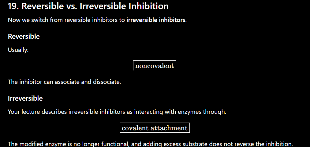
Conceptually: E + I → E—I
↑
covalent bond
Once modified: enzyme inactive
20. Example of Irreversible Inhibition: Aspirin
Aspirin irreversibly inhibits cyclooxygenase (COX) by adding an acetyl group to a serine residue in the catalytic pocket.
That creates a covalent modification that reduces enzyme activity.
The important general principle is:
covalent modification → loss of functional enzyme

22. Serine Proteases
A protease catalyzes the cleavage of peptide bonds.
Chymotrypsin is a:
serine protease
The lecture states that chymotrypsin cleaves proteins on the carboxyl side of aromatic and large hydrophobic amino acids.
Its catalytic activity depends on three amino acids known collectively as the:
catalytic triad
23. Catalytic Triad
You should memorize:

Ser195
Serine becomes the major nucleophile.
Its oxygen ultimately attacks the peptide carbonyl.
His57
Histidine acts as an acid/base catalyst.
Initially, it removes a proton from Ser195.
That converts the serine hydroxyl:
Ser−OH
into a much stronger nucleophile:

Asp102
Aspartate helps orient and stabilize histidine and makes histidine a better proton acceptor.
Your lecture describes the sequence as:
Asp-102→His-57→Ser-195
with histidine removing Ser-195's proton and the resulting alkoxide attacking the substrate peptide bond.
24. Chymotrypsin Mechanism — Big Picture
Mechanism:
Substrate binding
Serine nucleophilic attack on peptide carbonyl
Collapse of tetrahedral intermediate
Release of amine component
Water binding
Water nucleophilic attack on acyl-enzyme intermediate
Collapse of second tetrahedral intermediate
Release of carboxylic acid component

25. Chymotrypsin Mechanism — Step by Step
Step 1 — Substrate Binding
The peptide substrate binds in the active site.
The peptide bond that will be cleaved is positioned next to Ser195.
Step 2 — Activate Ser195
His57 removes the proton from the hydroxyl group of Ser195.
Ser—OH + His
↓
Ser—O⁻ + His—H⁺
This creates the highly reactive: alkoxide ion
Asp102 assists by orienting His57 and making it a better proton acceptor.
26. Step 3 — Nucleophilic Attack
Now:
Ser−O−
attacks the carbonyl carbon of the peptide bond.
Electron movement conceptually:
O
||
R — C — NH — R'
↑
Ser—O⁻
The nucleophile attacks: carbonyl carbon
while the carbonyl π\pi electrons move toward oxygen.
This produces a: tetrahedral intermeadiate
Step 4 — Collapse of First Tetrahedral Intermediate
The tetrahedral intermediate collapses.
The carbonyl reforms and the peptide bond is broken.
His57 participates in proton transfer to facilitate departure of the amine-containing product.
The first product leaves.
However, part of the substrate remains covalently attached to Ser195.
This gives the: acyl - enyme intermeadiate
Step 5 — Water Enters
Now:
H2O
binds to the active site.
His57 acts again as an acid/base catalyst.
It removes a proton from water.
Conceptually:
H2O→OH
This produces a stronger nucleophile.
Step 6 — Water Attacks
The activated water-derived nucleophile attacks the carbonyl carbon of the acyl-enzyme intermediate.
Again:
O
||
Enzyme—O—C—R
↑
OH⁻
This produces a second tetrahedral intermediate.
The oxyanion hole again helps stabilize the negatively charged oxygen.
Step 7 — Second Tetrahedral Intermediate Collapses
The carbonyl reforms.
This breaks the covalent bond between:
substrate and Ser195
Ser195 is regenerated.
Step 8 — Second Product Leaves
The carboxylic acid-containing product leaves.
The enzyme returns to its original state:
E+S→E+P1+P2
Therefore, chymotrypsin has hydrolyzed a peptide bond while regenerating its catalytic triad.
Chymotrypsin Mechanism as One Flow
For studying, I'd memorize it this way:
SUBSTRATE BINDS
↓
His removes H⁺ from Ser195
↓
Ser-O⁻ forms
↓
Ser attacks peptide carbonyl
↓
1st tetrahedral intermediate
↓
Intermediate collapses
↓
Peptide bond breaks
↓
1st product leaves
↓
ACYL-ENZYME
↓
Water enters
↓
His activates water
↓
Water attacks carbonyl
↓
2nd tetrahedral intermediate
↓
Intermediate collapses
↓
Ser-substrate bond breaks
↓
2nd product leaves
↓
ENZYME REGENERATED
Feature | Competitive | Uncompetitive | Noncompetitive |
|---|---|---|---|
Binds E? | Yes | No | Yes |
Binds ES? | No | Yes | Yes |
Active-site competition? | Yes | No | No |
KMK_M | ↑ | ↓ | Same |
VmaxV_{max} | Same | ↓ | ↓ |
Overcome with ↑[S]? | Yes | No | No |
LB pattern | Meet at Y-axis | Parallel | Meet at X-axis |