Split brain research
Aim:
To discover whether other neural processes may also be the subject to hemispheric laterization
Procedure:
- Sperry studied 11 patients who had undergone a commissurotomy to control frequent and sever epileptic seizures
- the main communication line between the two hemispheres were removed
- this allowed Sperry and colleagues to determine which hemispheres were specialised for certain functions, and whether the hemispheres performed tasks independently
I
- patients would have to either describe what they saw (image/object/word), describe what they could feel (object in hands) or draw from a word in front of them
- this information would be shown to either the left or right visual field and therefore processed by only one hemisphere at a time
- in a normal brain the corpus callosum would have shared the information with the other hemisphere, giving a complete picture of the visual world
- presenting information to just one hemisphere meant it could not be shared in split-brain patients, as the corpus callosum is removed
The type of tasks:
- describe what you see
- tactile test
- drawing task
Findings:
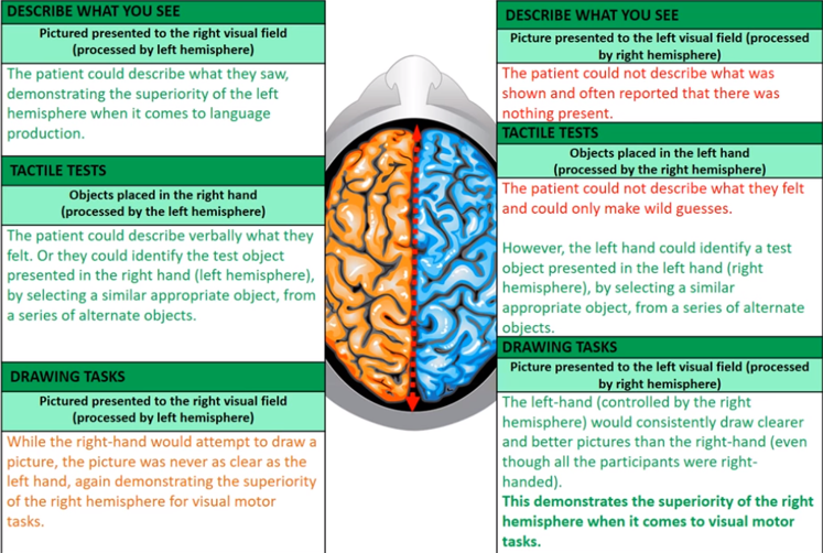
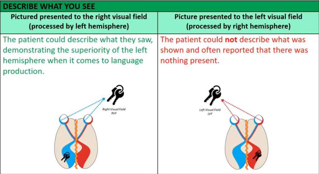
Describe what you see:
- when a picture of an object was shown to a patients right visual field (RVF). they could easily describe what they’d seen
- but they couldn’t when the same object was shown in the left visual field (LVF) and often said there was nothing there
I
Because the language centre is in the left hemisphere so when the image is presented in the left visual field, it is processed in the right hemisphere which has no language centre, and the corpus callosum is absent so the information is not shared by the left hemisphere
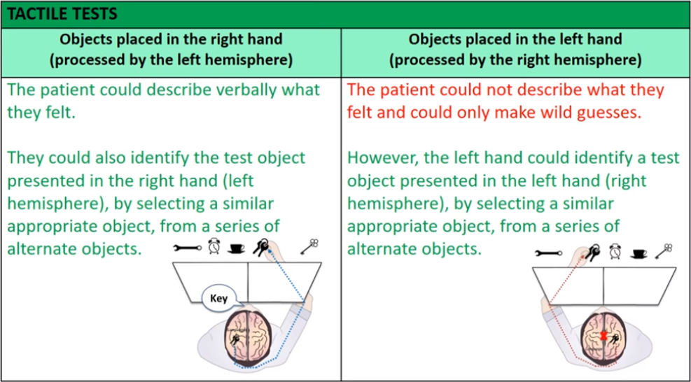
Tactile test:
- although patients could not attach verbal labels to their LVF, using their left hand (and therefore RH), they could select a matching object from a grab-bag closely associated with the object shown
- for example, if a picture of a cigarette was shown to the LVF, they could not verbalise it was a cigarette, but they would grab an ashtray, showing they had understood what the object was using the RH
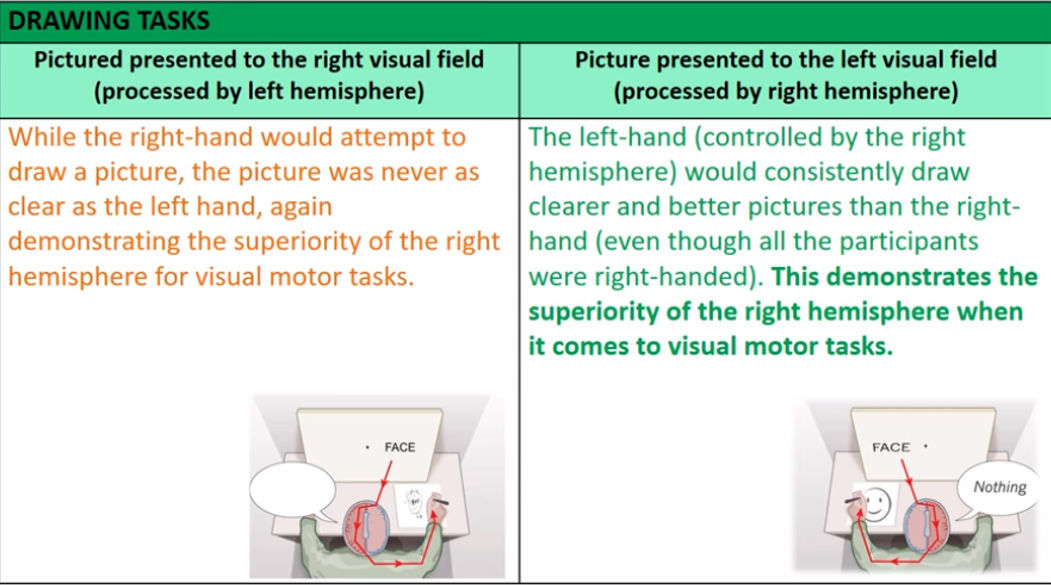
Drawing Task:
- patients had to draw an image of the word projected onto either the right or left visual field
- left-hand (RH) would draw clearer images than right hand (even though patients were all right handed)> RH = superior with visual-motor tasks
- right-hand (LH) would draw less clear images