Brain structure and function
Brain structure and function
Part 1
CNS – brain and spinal cord
Peripheral nervous system PNS – all parts of the NS outside the CNS
PNS split into:
somatic system – links spine and body and sense organs, controls voluntary behaviour – external stimuli.
Autonomic system – serves internal organs and glands, controls automatic functions – internal stimuli.
Sympathetic nervous system – part of autonomic – fight or flight – body arousal, emergency system, activating energy resources.
Parasympathetic nervous system – part of autonomic – rest and digest – relaxes body, active after emotional event, conserves energy.
Sympathetic response starts with release of acetylcholine, this activates sympathetic adrenal-medullary system to release epinephrine (adrenalin) and norepinephrine. -> stress response. AMS works in synchrony with the hypothalamic pituitary-adrenaline axis (HPA) – part of hormonal stress response.
Somatic – sensory input and movement control – transferring sensory information and motor responses to and from CNS. – nerves connect to skin, sensory organs and all skeletal muscles – processes sensory info – voluntary movement and reflexes.
Reflexes – most simple response – sensory receptor – sensory neuron transmits signal via afferent nerve fibres to PNS – dorsal horn of the spinal cord decodes signal – signal travels via ventral horn to efferent nerve fibres to effectors/ muscles – effector responds by contracting.
CNS
Spinal cord – conduit for motor information and sensory information – a centre for coordinating certain reflexes.
anatomy – grey matter (inner layer) body of neurons – white matter (outer layer) axels of neurons. – dorsal horn, a site where afferent/ somatic and autonomic nerves fibres synapse – ventral horn, a site where efferent nerve fibres synapse
function – mainly motor – look on slides (slide 15)
referencing location
Lateral – towards the side of the brain, away from the middle.
Medial – towards middle, away from sides.
Ipsilateral – on the same side.
Contralateral – opposite side.
Anterior/ rostral – in front of; towards face; front of the brain but for spine on the top.
Posterior/ caudal – behind; towards back; for spine on the bottom.
Superior/ dorsal - above; top of head; for spine on the back.
Inferior/ ventral – below; towards feet; for spine front of spinal cord.
Part 2
The brain
Development from birth to adulthood – only 2% of body weight but gets 16% of blood supply – 10x as much blood as muscle tissue – intricate web of blood vessels serving the brain – supplied with oxygen and nutrients.
Gross divisions – left and right hemispheres – build up from grey matter (cell bodies) and white matter (axons) – Cerebellum, brain stem, cerebral cortex.
Fine divisions - 
Brain stem:
Medulla oblongata – lower part of brain stem – transition between spinal cord and brain – origin of some cranial nerves – function, controls vital functions such as heart rate, breathing, blood pressure and vomiting. – damage is fatal.
Pons – bridge from the upper part of the brain stem – connects rest of the brain to cerebellum – bridges spinal cord and brain – function, controls muscle movement, and carries sensory-motor information from PNS, arousal and automatic functions, also involved in sleep and wake cycle.
Midbrain
Tectum (roof) – superior colliculi, forms part of the visual system, controls eye movements – inferior colliculi, forma part of auditory pathway, controls pitch perception, startle response.
Tegmentum (floor)
– periaqueductal grey, specific movement sequences, pain regulation, reproductive and maternal behaviours
– reticular formation, sleep, arousal, attention, muscle tone
– red nuclei, gait, crawling, fine hand movement
– substantia nigra (black matter), dopamine release, relays to basal ganglia, motor planning, eye movement, reward seeking, learning, addiction.
All play roll in motor control
Cerebellum
“small brain” located at back of brain – stem attached to pons by cerebellar peduncles – contains more neurons than cortex – function, motor movement coordination, balance and equilibrium, automated movement sequences, fine movement, muscle tone, also automatic language processing (e.g. talking, writing)
Thalamus
Lies at top of the brainstem – many thalamic nuclei – central relay centre of the brain – function, sensory pathways (but not olfaction/ smell), motor pathways, and cortico-cortical loops, fundamental roll in attention, also involved in consciousness/ sleep and alertness.
Hypothalamus
Bellow thalamus – smaller structure – contains mamillary bodies and projects to pituitary gland – function, controls ANS, roll in homeostasis, regulation of hormones, regulated basic behaviours (e.g. fight or flight)
Limbic system
Hippocampus, amygdala, fornix, cingulum, mamillary bodies, olfactory bulb – thalamus and hypothalamus have large input – amygdala, central for emotional response, fear conditioning, responds to emotionally salient stimuli and attaches emotion to memories – hippocampus, learning and long-term memory storage, can produce new brain cells.
Basal Ganglia
Three main structures – globus palidus, caudate nucleus, putamen – connected with brainstem and thalamus, as well as cortex – function, voluntary high order motor movement, procedural learning, habit learning, eye movement.
Cerebral Cortex
Outer layer of neural tissue – folded providing greater surface area
Split into hemispheres, major grooves/ fissures – secondary groove/ sulcus/ sulci – bulges/ gyrus/ gyri
Inter-hemispheric fissure – splits left and right
Lateral fissure – splits front and back
Central sulcus
These identify boundaries for lobes (more detail on slide)
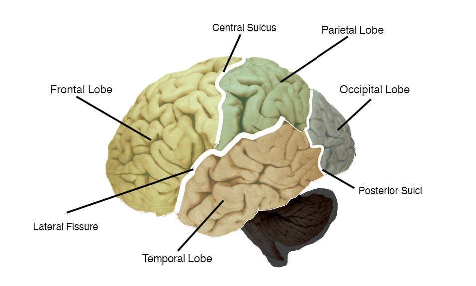
Occipital lobes – processing visual information – calcarine fissure is here, point of primary visual cortex – higher-order vision further processed by visual association cortices.
Parietal lobes – somatosensory cortex and association cortex – processing physical sensory information – differentiate things like temperature and pain – position location and movement of body identifies.
Temporal lobes – superior temporal cortex (Heschel’s gyrus) seat of primary auditory cortex – hearing – Wernicke’s area control language and speech comprehension, damaged leads to not understanding speech – inferior parts organise integrate visual sensory input – crucial roll in sematic memory/ cognition.
Frontal lobes – last region to evolve – posterior proportion contains pre-motor and motor cortices (control motor function) – anteriorly, involved in executive function (planning reasoning, decision making, working memory ext.), attention, social cognition and emotion regulation, language production (Broca’s area) and semantic control.
Inside the hemispheres
Cortical surface also stretches to medial surface. Two major structures – anterior cingulate cortex, involved in emotion processing (mainly sadness, implicated in depression) and self-referential process – corpus callosum, white matter bundle which joins the hemispheres and allows communication between them.
Ventricles
Four interconnected cavities filled with cerebrospinal fluid. Lateral, third, fourth and cerebral aqueduct. Function, protect brain, support against gravity, enables chemical stability and provides nutrients.
Meninges
A protective sheet wrapping around whole CNS – three layers – dura matter, holds it together – arachnoid, cushions it (- subarachnoid space, filled with CSF) – pia mater, allows nourishment.