Week 7- Renal Disorders
Renal System Overview
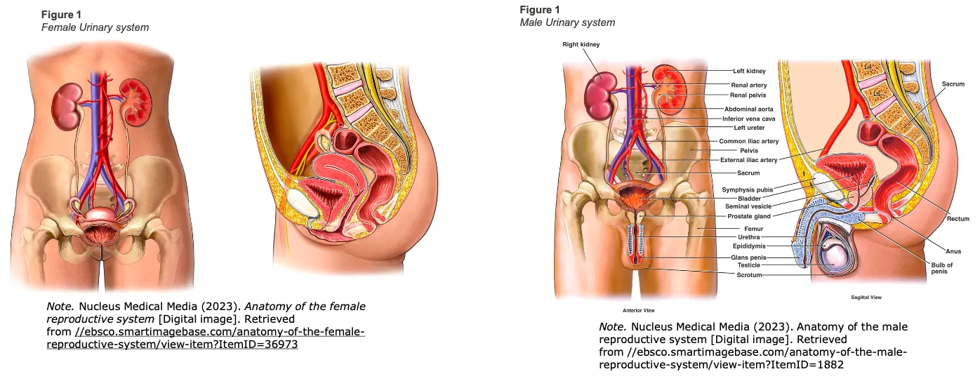
Components of the Renal System
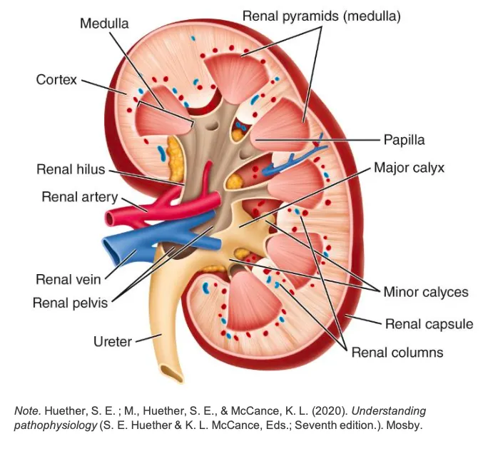
Kidneys
Ureters
Bladder
Urethra
Prostate Gland (in males)
Blood Flow and Purpose
Blood flows from the kidneys through multiple arteries surrounding the nephrons, aiding in the reabsorption of ions before returning deoxygenated blood to the inferior vena cava.
Functions of the Kidneys
Removal of waste from the body.
Regulation of:
Blood pressure
Blood volume
Blood glucose
Blood osmolarity
Nephron Structure
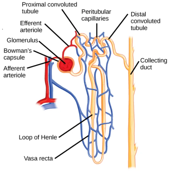
Comprised of:
Renal Corpuscles:
Glomerulus
Bowman’s Capsule
Renal Tubules:
Proximal Convoluted Tubule
Loop of Henle
Distal Convoluted Tubule
Collecting Duct
Glomerular Filtration Rate (GFR)
Definition: The volume of blood filtered per minute by the glomeruli.
Normal GFR: approximately .
Influenced significantly by blood pressure.
Renal System Anatomy and Physiology
Key Organs
Kidneys
Ureters
Bladder
Urethra
Prostate Gland (in males)
Functions of the Kidneys
Regulating Ion Concentration
Regulating Blood pH Levels
Regulating Blood Volume
Regulating Blood Pressure
Managing Blood Osmolarity
Hormone Production
Excreting Waste and Foreign Substances
Nephrons
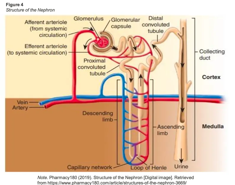
Filtration
Glomerular Capsule
Filtration of plasma, including glucose.
Contains the Glomerulus, surrounded by the Bowman’s capsule, located in the renal cortex.
Renal Tubule Functions
Proximal Convoluted Tubule
Function:
Reabsorption of 80% of electrolytes, water, glucose, amino acids, and bicarbonate.
Secretion of hydrogen ions and creatinine.
Location: Renal cortex
Loop of Henle
Function:
Reabsorption of sodium and chloride in the ascending limb, water in the descending loop.
Location: Inner renal medulla
Distal Convoluted Tubule
Function:
Secretion of potassium, hydrogen, and ammonia.
Reabsorption of water influenced by antidiuretic hormone and bicarbonate.
Regulation of sodium and potassium by aldosterone.
Location: Renal cortex
Glomerular Filtration Rate (GFR)
Definition: The volume of blood filtered per minute by the glomeruli.
Normal GFR:
Factors Influencing GFR:
Net filtration pressure
Total surface area for filtration
Filtration membrane permeability
Tests to Assess GFR:
Creatinine Clearance: An assessment method involving a 24-hour urine collection based on body surface area.
Serum Creatinine Levels: Normal ranges are:
Males:
Females:
Renal Assessment
Concept: A combination of subjective and objective patient information.
Purpose: To analyze kidney function and diagnose issues.
Urinalysis
Objective Measures:
Vital Signs
Inspection
Palpation
Percussion
Auscultation
Urine Analysis: Color, clarity, odour
Weight
Subjective Measures:
Frequency and urgency
Nocturia: Urinating once or more per night
Dysuria: Painful, burning sensation during urination
Hesitancy: Difficulty urinating
Incontinence: Leakage of urine
History of urinary conditions
Significance of Urinalysis:
Establishes baseline information and aids in diagnosis
Optimal Timing: Morning sample best for reliability
Components of Urinalysis
Chemical and Microscopic Findings:
Specific Gravity
pH level
Protein levels
Leucocytes
Nitrites
Blood
Ketones
Glucose
Bilirubin
Urobilinogen
Abnormal Findings and Their Significance
Hematuria: Blood in urine indicates potential kidney injury or disease.
Color Changes:
Dark, smoky color indicates excessive bilirubin.
Odor: Ammonia-like odour can indicate infection (UTI).
Cloudiness: Linked with urinary tract infections (UTI).
pH Levels: Can indicate diabetes insipidus or dehydration.
Renal Serology and Procedures
Serum Levels
Serum Urea: A byproduct of protein metabolism, normal range: .
Creatinine: Removed by kidneys, reliable indicator of GFR, normal range: .
eGFR: Estimates kidney filtering capability, normal: <.
Blood Urea Nitrogen (BUN): Normal range: .
Diagnostic Imaging and Procedures
Renal Ultrasound: Evaluates kidneys.
Bladder Scan: Determines bladder volume.
Cystoscopy: Visualizes the bladder.
Intravenous Pyelogram: Imaging of kidney structures.
Age-related Changes
Impacts of Aging on the Renal System:
Decreased renal tissue
Decreased function of Loop of Henle
Weakening urinary sphincter
Decrease in nephrons and renal blood vessels
Decreased bladder capacity and sensory receptors
Urinary Tract Infection (UTI)
Types:
Upper UTIs (pyelonephritis): Kidney level infection.
Lower UTIs (cystitis, urethritis): Bladder or urethra infection.
Terms | Definition | Location |
|---|---|---|
Urinary Tract Infection | The presence of bacteria in one or more | - |
Pyelonephritis | Inflammation of the parenchyma and | Upper tract infection |
Cystitis | Is inflammation of the bladder, which is most | Lower tract infection |
Urethritis | Is inflammation of the urethra | Lower tract infection |
Urosepsis | Is a urinary tract infection that has spread | Systemic infection |
Risk Factors
Anatomical Factors: Women are at higher risk due to shorter urethra distance to the anus.
Functional Disorders: Constipation, voiding dysfunction can increase risk.
Immune system conditions: Diabetes, HIV increases susceptibility.
Pathophysiology of UTI
General Cause: Bacteria, primarily E. coli, enter through the urethra → ascends to the bladder. → bacteria adheres to urothelium → bacteria multiply → causes infection + inflammation → WBCs + Cytokines + other pro-inflammatory markers reach the site of infection
Cystitis occurs = irritation and swelling of bladder
pyelonephritis = inflammation of kidneys (if infection spreads)
Symptoms of a UTI:
Hesitancy: Difficulty starting urination
Dysuria: Painful or difficult urination
Frequency and Urgency: Urine loss and strong urge to void
Clinical Manifestations:
Symptoms | Description |
|---|---|
Hesitancy | Difficulty starting urination |
Intermittency | Interruption of urine flow while voiding |
Post-void dribbling | Urine loss after completion of voiding |
Urinary retention or incomplete emptying | Inability to empty urine from bladder caused by atonic bladder or obstruction of urethra |
Dysuria | Painful or difficult urination |
Urinary frequency | > 8 times in 24 hours |
Urgency | Sudden, strong or intense desire to void immediately. Commonly accompanied by frequency |
Incontinence | Involuntary or accidental urine loss or leakage |
Nocturia | Awakened by the urge to void two or more times during the night |
Nocturnal enuresis | Passing urine while sleeping (bedwetting) |
Diagnosis and Management
Medications:
Antibiotics (e.g., Trimethoprim; Nitrofurantoin)
Trimethoprim
MOA: Selectively interferes with bacterial biosynthesis of the nucleic acids and the proteins of the organism
Broad spectrum antibiotic
Contraindicated in pregnancy
Nitrofurantoin
MOA: Bacteriostatic and bactericidal as interferes with the bacterial enzymes
Broad spectrum affecting both gram-positive and gram-negative bacteria
Can be used in caution in pregnancy
First generation cercosporin
MOA: Bactericidal as inhibit cell wall synthesis
i.e. Cephalexin
Can be used with caution in pregnancy
Broad spectrum mainly used for gram-positive bacteria
Other medication
Antipyretics
Analgesia
Avoid unnecessary cauterization
Sepsis
If patient is acutely unwell then fluid resuscitation would be appropriate management to stabilize the patient
Patient education
Instructions on antibiotic therapy
Drinking more fluids
Encourage urination once feel the urge
Urination after intercourse
Cleansing of genitals
Avoiding constipation
Avoidance of spermicides
Acute Kidney Injury (AKI)
The sudden impaired functioning of the kidneys.
Pathophysiology
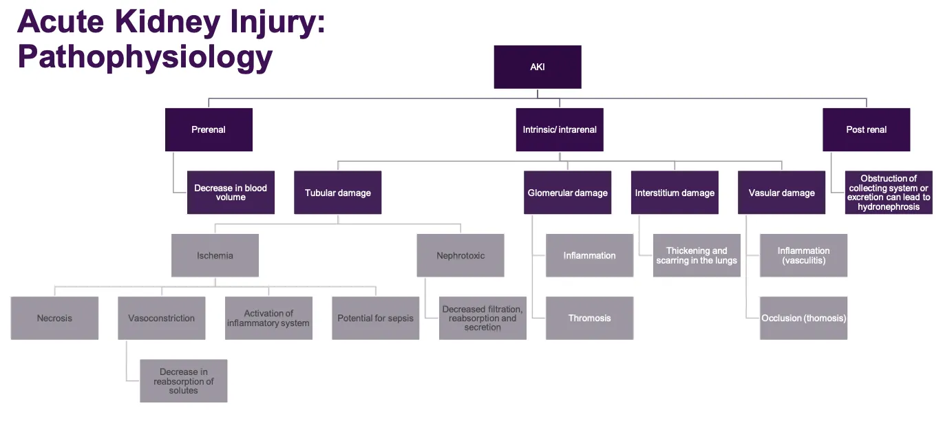
Categories of AKI:
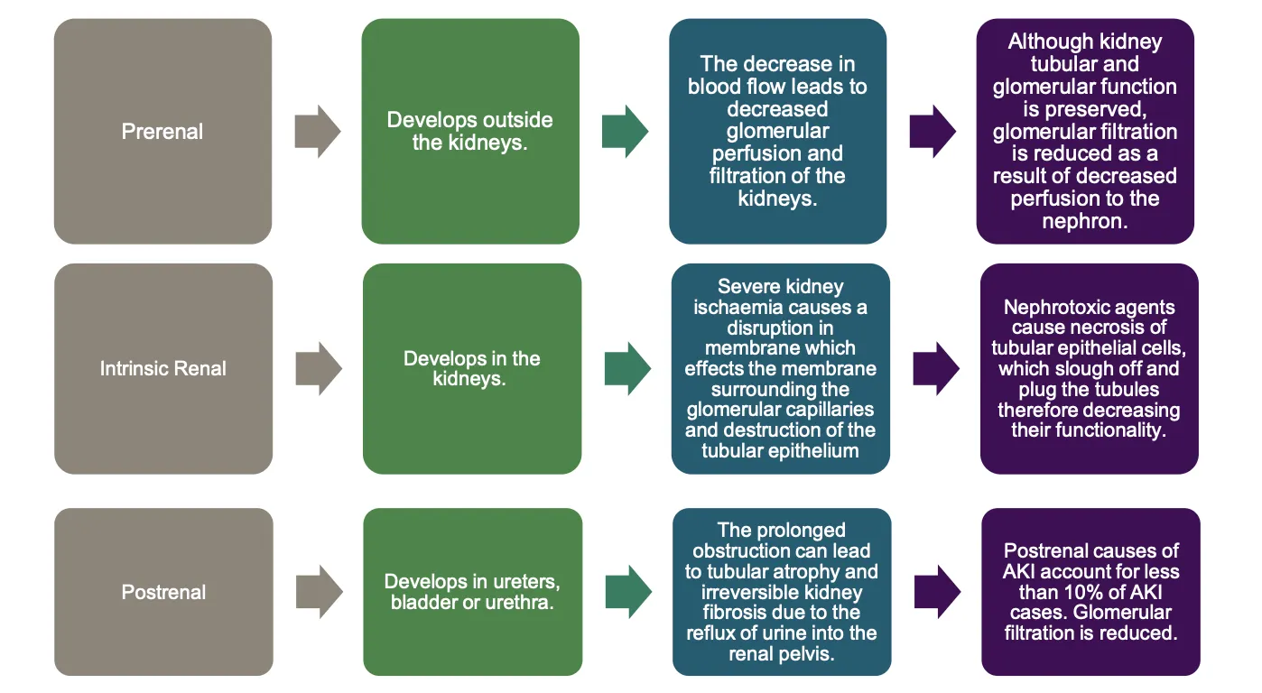
Prerenal: Caused by factors outside the kidney (e.g., dehydration, hemorrhage).
Intrinsic/Intrarenal: Due to direct damage to kidneys (e.g., nephrotoxins).
Postrenal: Due to obstructions (e.g., benign prostatic hyperplasia).
Clinical Features
Oliguria
Hypovolemia
Fluid retention
Severe acidosis
Decreased sodium levels
Increased potassium levels
Increased urea and creatine levels
Neurological changes
Risk Factors:
Hypertension, hypercholesterolemia, infection/sepsis, nephrotoxic agents.
Clinical Phases (RIFLE Classification)
RIFLE | GFR | Urine Output |
|---|---|---|
Risk | Increased creatinine x 1.5 | Dcreased Urine ouput <0.5mg/g/h x 6 |
Injury | Increased creatinine x 2 | Dcreased Urine ouput <0.5mg/kg/h x 12 |
Failure | Increased creatinine x 3 | Dcreased Urine ouput <0.5mg/kg/h x 12 or anuria |
Loss | Loss of kidney function >4 weeks | |
End stage kidney disease | End stage kidney disease >3 months |
Stages of AKI
Phase | Feature | Important Considerations |
|---|---|---|
Initial | Time from when the initial kidney injury takes place to when a reduction in renal function occurs. | Recognition of risk – identification of hypotensive episodes and nephrotoxic agents etc. |
Oliguric | Reduced urine output (less than 400 ml/day) – usually occurs 1-7 days after kidney injury and lasts 10-14 days. | Findings may include fluid overload, hyponatraemia, hyperkalaemia, metabolic acidosis and elevated creatinine and urea and other uraemic symptoms. |
Diuretic | Urine production increases but concentrating ability is not yet recovered. | Due to fluid loss patient has a risk of hypovolaemia, hypotension, hypernatraemia and hypokalaemia. |
Recovery | Occurs when the kidneys regain function and return to normal. | Close follow up and rehabilitation post-discharge. |
Patient Management
General Monitoring: Vital signs, fluid intake/output, lab values.
Management Strategies:
Dietary restrictions, IV fluid therapy, medication administration, hemodialysis as a last resort.
Prerenal: Provide Intravenous therapy to increase blood volume and perfusion to kidneys
Intrinsic renal: Remove the cause of damage for example nephrotoxic medication or treat the infection such as glomerulonephritis
Postrenal: Remove the obstruction through surgery or by providing urinary catheter
Nursing Management Priorities
Pain management, monitoring for changes in vital signs, monitoring at-risk patients.
Chronic Kidney Disease (CKD)
Pathophysiology:
Progressive, irreversible decline in kidney function.
Signs include increased workload on remaining nephrons leading to damage.
Duration: More than 3 months with eGFR <.
Nursing Diagnoses for CKD
Fluid Volume Excess: Due to oliguria characterized by edema, weight gain, and hypertension.
Fatigue: Related to uremia and anemia, marked by pallor and low hemoglobin levels.