13.10 Sliding filament model
In order to contract and cause movement, the actin and myosin filaments within the myofibrils have to slide past each other, muscle contraction is usually described using the sliding filament model.
Sliding filament model
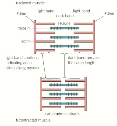
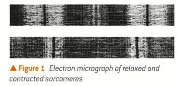
During contraction, the myosin filaments pull the actin filaments inwards and towards the centre of the sarcomere. Resulting in
the light band becoming narrower
the Z lines moving closer together, shortening the sarcomere
the H-zone becoming narrower
The dark band remains the same width, as the myosin filaments themselves have not been shortened, but now overlap the actin filaments by greater amount.
The simultaneous contraction of lots of sarcomeres means that the myofibrils and muscle fibres contract. This results in enough force to pull on a bone and cause movement, when sarcomeres return to their original length the muscle relaxes.
Structure of myosin
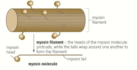
Myosin filaments have globular heads that are hinged which allows them to move back and forwards.
On the head is a binding site for each of actin and ATP
The tails of several hundred myosin molecules are aligned together to form the myosin filament.
Structure of actin
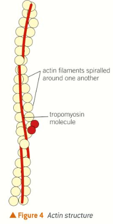
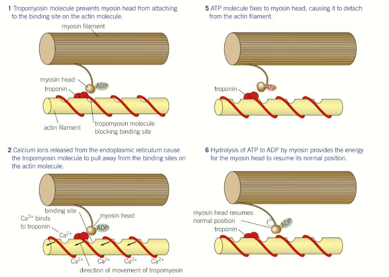
Actin filaments have binding sites for myosin heads, these are called actin-myosin binding sites. However these are often blocked by the presence of another protein called tropomyosin which is held in place by the protein troponin.
When a muscle is in a resting state (relaxed) the actin-myosin sites are blocked by tropomyosin. The myosin heads can therefore not bind to the actin, and the filaments cannot slide past each other.
When a muscle is stimulated to contract, the myosin heads form bonds with actin filaments known as actin-myosin cross-bridges.
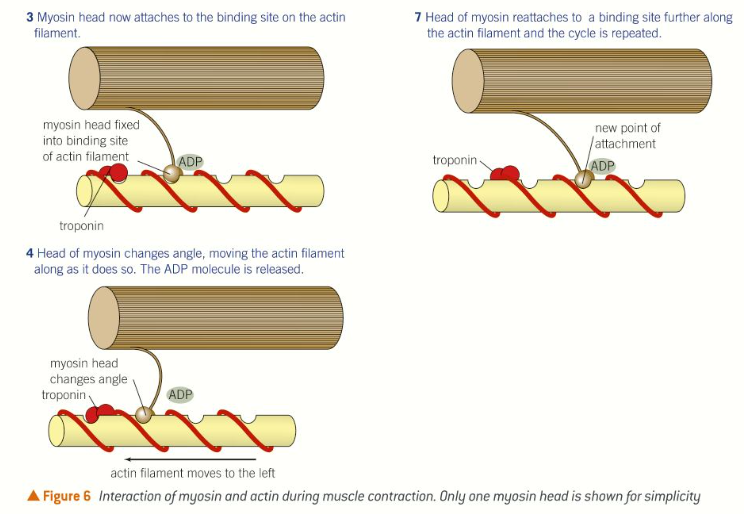
The myosin heads then flex (change angle) in unison, pulling the actin filament along the myosin filament.
The myosin then detaches from the actin and its head returns to its original angle, using ATP.The myosin then reattaches further along the actin filament and the process occurs again, This is repeated up to 100 times per second.
How muscle contraction occurs
Neuromuscular junction
Muscle contraction is triggered when an action potential arrives at a neuromuscular junction - this is the point where a motor neuron and a skeletal muscle fibre meet.
There are many neuromuscular junctions along the length of a muscle to ensure that all the muscle fibres contract simultaneously.
If only one existed, the muscle fibres would not contract together therefore the contraction of the muscle would not be as powerful, it would also be much slower as wave of contraction would have to travel across the muscle to stimulate the individual fibres to contract.
All the muscle fibres supplied by a single motor neurone are known as a motor unit - the fibre act as a single unit. If a strong force is needed, a large number of motor units are stimulated, whereas only a small number are stimulated if a small force is required.
When an action potential reaches the neuromuscular junction, it stimulates calcium ion channels to open.
Calcium ions then diffuse from the synapse into the the synaptic knob, where they cause vesicles to fuse with the presynaptic membrane.
Acetylcholine is released into the synaptic cleft by exocytosis and diffuses across the synapse. It binds to the receptors on the postsynaptic membrane (sarcolemma) opening sodium ion channels and resulting in depolarization.
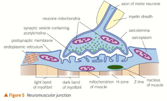
Acetylcholine is the broken down by acetylcholinesterase into choline and ethanoic acid. This prevents the muscle being overstimulated. Choline and ethanoic acid diffuse back into the neurone, where they are recombined into acetylcholine, using energy provided by mitochondria.
Sarcoplasm
The depolarization of the sarcolemma travels deeps into the muscle fibre by spreading though the T-tubules. These are in contact with the sarcoplasmic reticulum. The sarcoplasmic reticulum contains stored calcium ions which it actively absorbs from the sarcoplasm.
When the action potential reaches the sarcoplasmic reticulum, it stimulates calcium ion channels to open. The calcium ions diffuse back down their concentration gradient flooding the sarcoplasm with calcium ions.
The calcium ions bind to troponin causing it to change shape. This pulls on the tropomyosin moving it away from the actin-myosin binding sites on the actin filament. Now that the binding sites have been exposed the myosin heads binds to the actin filament forming an actin-myosin cross-bridge.
Once attached to the actin filament the myosin heads flexes, pulling the actin filament along. The molecule of ADP bound to the myosin head is released. An ATP molecule can now bind to the myosin head, this causes the head to detach from the actin filament.
The calcium ions present in the sarcoplasm also activate the ATPase activity of myosin. This hydrolyses ATP to ADP and phosphate, releasing energy which the myosin head uses to return to its original position.
The myosin head can now attach itself to another actin-myosin binding site further along the actin filament and the cycle in repeated, The cycle continues as long as the muscle remains stimulated. During the period of simulation many actin-myosin bridge form and break rapidly, pulling the actin filament along. This shortens the sarcomere and causes the muscle to contract.
Energy supply during muscle contraction
Muscle contraction requires large quantities of energy. This is provided by the hydrolysis of ATP into ADP and phosphate. The energy is required for the movement of the myosin heads and to enable to sarcoplasmic reticulum to actively reabsorb calcium ions from the sarcoplasm. There are 3 main ways that ATP is generated.
aerobic respiration
anaerobic respiration
creatine phosphate
Aerobic respiration
Most of the ATP used by muscle cells is generated from ADP during oxidative phosphorylation. This chemical reaction takes place inside the mitochondria which are plentiful in muscles. However this can only occur in the presence of oxygen, therefore used for long period of low-intensity exercise.
Anaerobic respiration
In very active muscle, oxygen is used up more quickly than the blood supply can replace it, therefore ATP has to be generated anaerobically.
ATP is made by glycolysis but as no oxygen is present, the pyruvate which is also produced is converted into lactate (lactic acid) This can quickly build up in the muscles resulting in muscle fatigue. Anaerobic respiration is used for short periods of high intensity exercise like sprinting.
Creatine Phosphate
Creatine phosphate is shored in muscle. To form ATP, ADP has to be phosphorylated. Creatine phosphate acts as a reserve supply of phosphate, which is availably immediately to combine with ADP, reforming ATP. This system generates ATP rapidly but the store is used up quickly.
As a result, this is used for short bursts of vigorous exercise like tennis serve, when the muscle is replaced the creatine phosphate stores is replenished using phosphate from ATP.
Monitoring muscle activity with sensors
Sensors can be used to monitor the electrical activity in a muscle. These can be used to measure the strength of a muscle contraction or to track muscle fatigue levels.
Muscle fatigue is a long-lasting reduction of the ability to contract and exert force. It is normally localized and occurs after prolonged,, strong muscle activity. Occasionally this can be beneficial through promoting muscle growth, however usually harmless. Serious injuring is most likely to occur when the level of fatigue is a muscle is high.
Resultant trace an EMG is a record of the electrical activity in a muscle during an activity.
The detection and classification of muscle fatigue is important in research into human-computer interactions, sport injuries and performance, ergonomics and prosthetics.
A typical experiment in muscle fatigue research involves a subject performing a set task like moving a limb in specific manner. A signal is acquired using sensors attached to the skin, which is recorded and processed to reveal the characteristic s of the muscle during that exercise.