L15 4BBY1030: Apoptosis, Necrosis, and Excitotoxicity
Overview of Cell Death Mechanisms
Diversity of Cell Death: There are distinct forms of cell death, each characterized by different triggers, morphological changes, and biochemical pathways.
Learning Objectives:
- Appreciation of different forms of cell death.
- Understanding the fundamental differences between these forms.
- Recognizing that apoptosis is essential for normal development of tissues and organs.
- Understanding how disturbances in glutamate homeostasis lead to excitotoxicity in neural tissue.
- Appreciating the evolutionary conservation of apoptosis across eukaryotic lineages, specifically concerning morphology and driving proteins.
Primary Forms of Cell Death:
- Necrosis: Described as traumatic cell death resulting from acute injury. While traditionally seen as passive, it involves the activation of a death programme.
- Apoptosis: It is a programmed form of cell death.
- Excitotoxicity: A specialized form of cell death confined exclusively to neural tissue, driven by specific neurotransmitter imbalances.
Comparison of Necrosis and Apoptosis
I. Causes and Triggers
Necrosis: Resultant from external injury or insults such as:
- Ischemia (restriction in blood supply to tissues depriving them of nutrients, e.g. glucose and O2).
- Hypoxia (deficiency in oxygen reaching the tissues).Apoptosis: Driven by physiological or programmed signals, including:
- Withdrawal of essential growth factors.
- Chemotherapy treatments.
- Contact with cytotoxic T cells (immune-mediated death).
- Execution of a pre-defined developmental programme.
II. Morphological and Energy Characteristics
Membrane Integrity:
- Necrosis: Involves significant membrane damage leading to the leakage of cellular contents.
- Apoptosis: The plasma membrane remains intact, though it undergoes "blebbing."
- Definition of Blebbing: A process where the cytoskeleton separates from the cell membrane, causing the membrane to form spherical protrusions or blebs.Chromatin State:
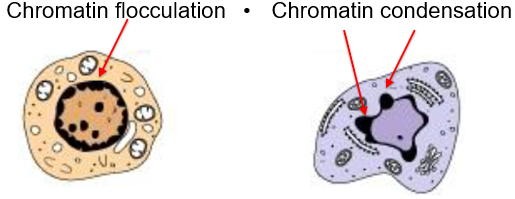
- Necrosis: Characterized by chromatin flocculation.
- Apoptosis: Characterized by chromatin condensation.
Energy Levels:
- Necrosis: Energy levels are rapidly depleted.
- Apoptosis: Energy levels are maintained or only depleted slowly, as the process requires energy to execute the death programme.Inflammatory Response:
- Necrosis: Elicits a significant inflammatory response due to the leakage of cellular contents into the surrounding environment.
- Apoptosis: No inflammatory response occurs. Apoptotic cells are rapidly engulfed by phagocytes before they can lyse and spill their contents.
The Apoptotic Pathway and its Biological Purpose
The Path of Apoptosis
Membrane Blebbing: Initial structural changes in the cell surface.
Chromatin Condensation: Packing of DNA into dense masses, membrane blebbing occurs.
Cell Fragmentation: The cell breaks into smaller fragments known as apoptotic bodies.
Phagocytosis: Phagocytic cells engulf these bodies, preventing the release of intracellular molecules.
Importance in the Nervous System
Preventing the release of intracellular molecules is critical in neural tissue.
The release of excitotoxic mediators, such as glutamate, from dying neurons can cause secondary injury to adjacent, healthy neurons.
Reasons for Committing Apoptosis
Metamorphosis: Regulated cell death during insect metamorphosis was described by Lokshin and Williams in 1964. Vogt (1842) noted physiological cell death during the resorption of the notochord (replaced by vertebrae) and the loss of tadpole gills.
Tissue Sculpting during Development: Elimination of cells that have served their purpose, such as the interdigital tissue (webbing) in a mouse paw, which disappears as the adult form is reached.
Viral Infection: Removal of cells infected by viruses to stop the spread of infection.
Cancer Prevention: Elimination of cells that could become malignant.
DNA Damage: Disposal of cells bearing excessive DNA damage.
Promote Self-Tolerance: Autoreactive lymphocytes (immune cells that might attack the body's own tissues) undergo apoptosis before they fully develop.
Biochemical Characteristics of Apoptosis
1. DNA Fragmentation
cells treated with an antibody that activates a death receptor
Endonuclease Activity: DNA is cleaved by endonucleases.
DNA Laddering: When resolved by electrophoresis, the DNA shows a "ladder" pattern. The fragments are distinct in size because cleavage occurs specifically in the linker regions between nucleosomes.
TUNEL Assay: Stands for "Terminal deoxynucleotidyl transferase mediated dUTP Nick End Labelling." This assay detects many new free ends of DNA generated by fragmentation. The transferase recognizes these ends and adds dUTPs labelled with a marker.
2. Plasma Membrane Changes
Phosphatidylserine (PS) Externalization: Under normal conditions, PS is located exclusively on the inner leaflet of the plasma membrane lipid bilayer. In apoptotic cells, PS "flips" to the outer leaflet.
Detection: Externalized PS can be detected using labelled Annexin V.
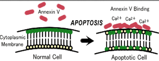
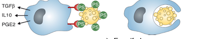
Eating Signals: Externalized PS acts as an "eat me" signal. Receptors on phagocytes bind to PS, stimulating the engulfment of the dying cell and the release of anti-inflammatory cytokines.
Finding Signals: Apoptotic cells also release "find me" signals to attract motile phagocytes.
leads to release of antiflammatory cytokines and engulfment of the dying cell
3. Mitochondrial Changes
Apoptotic cells lose the electrochemical potential exists across the inner mitochondrial membrane.
This change in membrane potential can be measured using positively charged fluorescent dyes.
Evolutionary Conservation and Caspases
Genetic Control in C. elegans
The hermaphrodite nematode Caenorhabditis elegans has somatic cells.
During development, exactly cells undergo apoptosis.
Four identified genes control this process, providing the first evidence that apoptosis is governed by a genetic programme.
Caspases: The Drivers of Apoptosis
the same genes encoding caspases (a class of protease) are found in humans where they play a similar role
proteases with cysteine at their acitve site which leave their substrates at specific aspartate sites
are the enzymes that drive apoptosis in multicellular eukaryotes
The human genome contains more than caspase genes.
Caspase Targets:
- ICAD (Inhibitor of Caspase-dependent Deoxyribonuclease): Caspases digest ICAD rendering the DNAase active. Nuclear localization once ICAD is digested, ICAD binds to catalytic site of CAD, scaffold proteins of the nuclear envelope (leads to nuclear shrinkage and fragmentation)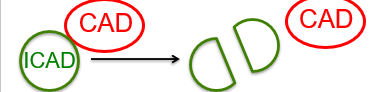
- Lamins: Scaffold proteins of the nuclear envelope. Cleavage leads to nuclear shrinkage and fragmentation.
- Gelsolin: A regulator of actin filament assembly/disassembly. Cleavage leads to membrane blebbing.Regulation of Caspases:
capases cause rapid cell death
premature activation would be lethal
robust mechanisms are in place to control activation, including:
synthesis of caspases as inactive zymogens
highly evolved upstream regulatory pathways including the presence of endogenous inhibitors
Apoptotic Signaling Pathways
I. The Extrinsic Pathway (Death Receptor Pathway)
Responds to extracellular signals indicating a cell is no longer needed for the well-being of the organism
Involves transmembrane death receptors belonging to the Tumor Necrosis Factor (TNF) receptor superfamily.
Binding of extrinsic ligands transduces intracellular signals to activate caspases.
II. The Intrinsic Pathway (Mitochondrial Pathway)
Responsive to internal stimuli such as DNA damage or cytotoxic drugs.
Mechanism: Stimuli cause mitochondrial membranes to become leaky, releasing Cytochrome c into the cytoplasm.
Cytochrome c activates specialized caspases.
UV Damage Example:
- UV-C (): Highly energetic and lethal, absorbed by the ozone layer, used for sterilization.
- UV-B (): Major mutagenic fraction of sunlight; induces thymine dimers (chemical bonds between adjacent thymines). This distorts DNA, causing replication errors and point mutations. Excessive damage leads to "sunburn," which is a mass apoptotic event to prevent cells from becoming cancerous (e.g., melanoma).
Pathway Integration
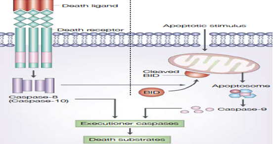
Both pathways converge on executioner caspases.
Extrinsic: Death ligand → Death receptor → Caspase-8/10 → Executioner caspases.
Intrinsic: Apoptotic stimulus → Mitochondria/Apoptosome → Caspase-9 → Executioner caspases.
The protein BID can bridge the two pathways when cleaved.
Apoptosis in Health and Disease
Excessive Apoptosis:
- Heart Attacks and Strokes: Characterized by significant cell loss through apoptosis. Blocking these pathways could save tissue.
- Type I Diabetes Mellitus: underlying cause is apoptosis of pancreatic B-cells (ability to prod insulin is now lost).Insufficient Apoptosis:
- Autoimmune Diseases: Characterized by large number of lymphocytes in spleen and lymph → stimulating loss of these cells by apoptosis could limit extent of the reaction against the individuals own tissues
- Cancer: Tumor cells often have defective apoptotic pathways, allowing the cancer to develop and progress.
Excitotoxicity
Definition: Coined by Olney in 1969, it describes cell death caused by excessive glutamate acting on excitatory receptors.
Glutamate Facts: It is the most abundant neurotransmitter in the brain but plays a pivotal role in pathogenesis of neuronal death due to increases in intracellular .
The Glutamate Cycle
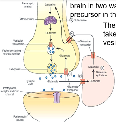
Glutamate is synthesized from a Krebs cycle precursor or recycled from glutamine.
It is taken up into exocytic vesicles in the neuronal terminal.
Upon release, nerve terminals and glial cells reuptake glutamate via membrane transporters.
Glutamine transported from glia into neuronal terminal via transporters in glial and neuronal terminal membranes
in neuronal terminal, glutamine is converted to glutamate
glutamate is taken up into vesicles and stored, then released by exocytosis
Pathogenesis of Excitotoxicity
Disturbance: During hypoxia or hypoglycemia, an excess of glutamate is released. leading to prolonged activation of receptors causing cell death
Concentration Gradient: Normally, intracellular glutamate is greater than extracellular glutamate (sequestered in vesicles).
Mechanism of Death:
1. Glutamate binds to NMDA (-methyl--aspartate) and AMPA (alpha-amino--hydroxy--methyl--isoxazole-propionic acid) receptors.
2. AMPA receptors allow entry, depolarizing the membrane.
3. Depolarization dislodges from the NMDA receptor, permitting entry of .Consequences of High Intracellular :
- Activation of -dependent enzymes that break down proteins, phospholipids, and nucleic acids.
- Activation of enzymes producing Reactive Oxygen Species (ROS), which further damage biomolecules.Associated Disorders: Excitotoxicity is the final destructive pathway for:
- Stroke and Trauma.
- Epilepsy.
- Neurodegenerative disorders: Huntington’s disease, Parkinson’s disease, and Alzheimer’s disease.