Neuroimaging
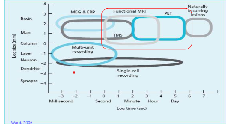
This diagram characterizes each neuroimaging technique into spatial and temporal resolution
Human neuroimaging:
Modern neuroimaging techniques have been intrinsic to the development of cognitive neuroscience
Allows us to view brain morphology and activity in vivo in awake behaving humans
Based upon the physical properties of tissue in the brain in the space (structural) and over time (dynamic)
Structural imaging produces static images of the brain’s physical structure
Functional imaging indexes dynamic aspects of brain function
How does activation in the brain change over time
Provides different types of information applicable to different types of questions
Structural neuroimaging:
Most common methods are:
Computerized tomography (CT)
Magnetic resonance imaging (MRI)
For each technique, the signals come from a different mechanism
CT constructs a 3D image of the brain based upon x-ray images
Different types of tissue eg grey and white matter have different absorption rates for x-ray material
Different densities generate different colours
CT scans are fast and cheap to acquire
Low spatial resolution, 0.5-1cm
Similiar absorption rates for grey and white matter makes it difficult to distinguish these on resulting images
Averaging in image means it is not possible to discriminate structures that are closer than 5mm
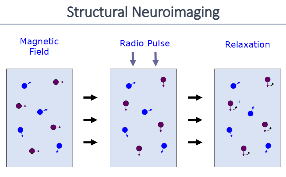
MRI scans exploit magnetic properties of living tissue
Signals come from protons in hydrogen atoms, they spin around the atoms axis producing a magnetic field
In normal circumstances the orientation of these protons is random
MRI scanner produces a powerful magnetic field (measured in tesla) to which these protons align
A radio pulse passed through the brain will momentarily return the protons to their original orientation
Once the radio pulse stops the protons realign with the magnetic field (T1 relaxation time)
Variations in the rate of this process differentiate different types of tissue in the brain
Radio pulses can be used to excite slices of the brain that are 1-3mm apart
MRI scans have high spatial resolution than CT scans
Differentiate white from grey matter as well as CSF and bone
Produce 3D images
Locations of these images are coded in mm on 3 axes with their origin centred upon the anterior commisure (Talairach coordinates)
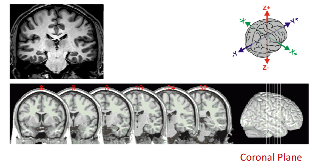
Coronal plane- front and back sections
Different spin rates across different types of tissue provide us with information
High spatial resolution images
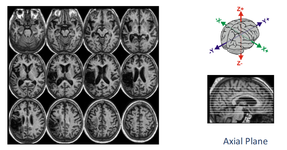
Axial plane- upper and lower sections
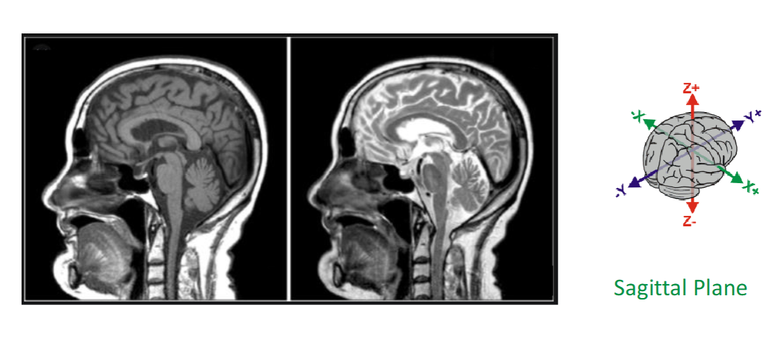
Sagittal plane- left and right sections
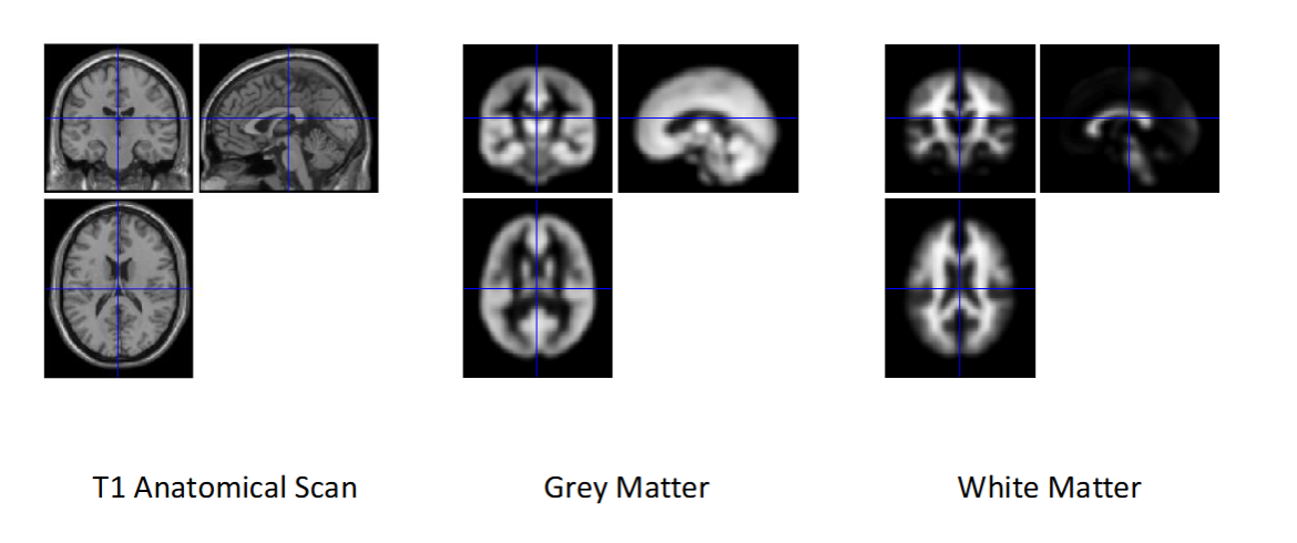
MRI distinguish really well between the different types of tissue within the brain
Functional neuroimaging:
Most common methods are:
Positron emission tomography (PET)
Functional magnetic resonance imaging (fMRI)
Functional imaging indexes the metabolic activity in the brain
Neural activity requires oxygen and glucose and these are supplied via the vasculature system of the brain
Allows us to measure dynamic changes in the brain metabolism under different situations
We can relate these to experimentally manipulated IVS
Neural indices of perception, cognition and emotion
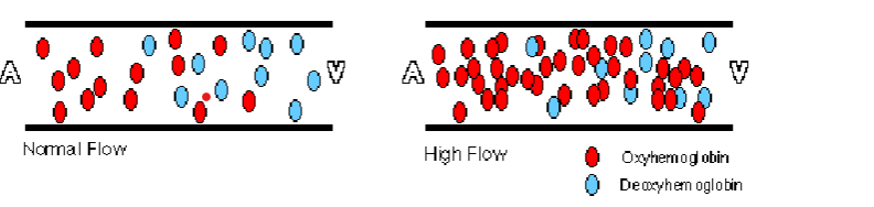
fMRI measures the ratio of oxygenated to deoxygenated blood
Deoxygenated haemoglobin distorts local magnetic fields and effects the spin rates of protons
Differences in the magnetic properties of oxygenated and deoxygenated blood forms the basis of fMRI contrasts (T2 spin rate)
The function of the BOLD signal against time is known as the haemodynamic response function
BOLD- blood oxygenated level dependent
Indirect measure of neuronal activity, which assumes a direct relationship between neuronal activity and haemodynamics
Simple designs eg subtraction provide information about the anatomical substrates of task perfomance
More complex techniques eg PPI provide information about task related activity across cortical networks
Allows us to measure dynamic changes in brain metabolism under different situations
We can relate these to experimentally manipulated IVS
Subtraction method:
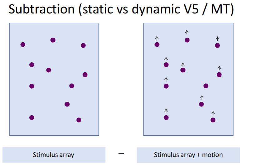
Everything is identical except the motion
Stimulus array- visual input
Stimulus array + motion involves movement
So the subtraction is (stimulus array +motion) - (stimulus array) = motion processing
Tells you which part of the brain is sensitive to motion
Subliminal priming (masked cues) :
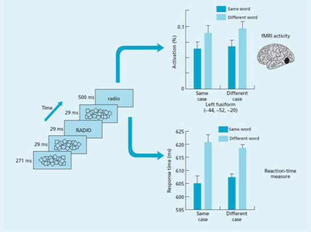
Tests whether the brain processes words without conscious awareness
Forward mask (≈ 500 ms)
A meaningless pattern
Prevents pre-activation of the word system
Prime word (≈ 29 ms)
Example: “RADIO”
Shown too briefly to be consciously perceived
Backward mask (≈ 29 ms)
Immediately wipes out conscious perception of the prime
Target word (≈ 271 ms)
Example: “radio”
This is consciously visible
Participant responds to this word (e.g., lexical decision)
Because of masking + short duration, the prime is subliminal.
Faster reaction times when the prime and target are the same word
Neuroimaging techniques provide unique access to the structure and function of the brain
Measures of morphology provide basis for comparisons across individuals or groups
Functional imaging provides a basis for structures to function deduction and induction
One of many convergent techniques that can be used to investigate links between the brain and behaviour