Bone Tissue - BIOL 2401 Lecture 7
Bone Tissue: Comprehensive Study Notes
Introduction to the Skeletal System
Skeletal system: Composed of bones, cartilages, and ligaments, forming the body's framework.
Osteology: The scientific study of bone.
Associated Tissues:
Cartilage: Acts as a forerunner for most bones during development and covers many joint surfaces in adults.
Ligaments: Connect bones together at joints.
Tendons: Attach muscles to bones.
Functions of the Skeletal System
Protection and Support:
Bones provide a rigid framework that supports the body and cradles soft organs.
Examples: The "thoracic cage" protects the heart and lungs; the skull protects the brain.
Movement:
Bones serve as levers, acted upon by muscles.
This lever system multiplies forces generated by muscle contraction, transforming them into movements.
Maintenance of Homeostasis:
Mineral Storage: Bone stores critical minerals like calcium () and phosphate (). These minerals are essential for electrolyte and acid-base balance within the body.
Blood Cell Formation (Hemopoiesis): Red bone marrow, a soft tissue found within bones, is the primary site for the production of all types of blood cells.
Fat Storage: Yellow bone marrow, predominantly found in adults, stores triglycerides (fat).
Bone (Osseous Tissue) and Bone Organs
Bone (osseous tissue): A specialized connective tissue where the extracellular matrix is hardened by the deposition of calcium phosphate and other minerals.
Mineralization or Calcification: The hardening process of bone tissue, involving the precipitation of minerals into the matrix.
Individual Bones (Organs):
An individual bone, such as the femur or a vertebra, is considered an organ.
Each bone organ consists of multiple tissue types: bone tissue, bone marrow, cartilage, adipose tissue, nervous tissue, and fibrous connective tissue.
Therefore, the term "bone" can refer either to the entire organ or specifically to the osseous tissue.
Bone Shapes and Examples
Flat Bones:
Description: Thin, curved plates.
Function: Primarily protect soft organs.
Examples: Occipital, parietal, frontal, nasal, vomer bones of the skull, sternum, scapula, ribs, ilium, ischium, pubis (hip bones).
Long Bones:
Description: Longer than they are wide. Act as rigid levers.
Function: Crucial for movement, acted upon by muscles.
Examples: Humerus, radius, ulna, femur, tibia, fibula, metacarpals, metatarsals, phalanges.
Short Bones:
Description: Approximately equal in length and width.
Examples: Bones of the wrist (carpals) and ankle (tarsals).
Irregular Bones:
Description: Bones with elaborate and complex shapes that do not fit into other categories.
Examples: Vertebrae and some bones of the skull.
Anatomy of a Long Bone
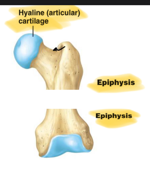
Articular Cartilage: A layer of hyaline cartilage that covers the joint surfaces of epiphyses, allowing for smooth, free movement within a joint. It is distinct from the periosteum in that it is avascular.
Periosteum: An external, tough sheath covering most of the bone surface, except for articular surfaces.
Outer Fibrous Layer: Composed of collagen, it connects the bone to surrounding tissues (muscles, tendons, ligaments).
Inner Osteogenic Layer: Contains bone-forming cells vital for bone growth in diameter and repair.
Compact (Dense or Cortical) Bone: The dense outer shell of a bone, providing strength and enclosing the marrow (medullary) cavity.
Spongy (Cancellous) Bone: Loosely organized bone tissue, less dense than compact bone.
Location: Found in the center of the ends (epiphyses) and the central shaft (diaphysis) of long bones, and in the middle of nearly all other bones.
Always covered by the more durable compact bone.
The skeleton is approximately compact bone and spongy bone by weight.
Medullary (Marrow) Cavity: The central cavity within the diaphysis of long bones, which contains bone marrow.
Nutrient Foramen: A tunnel that penetrates the diaphysis, providing access for the nutrient artery and/or vein, ensuring the bone organ is highly vascular.
Bone Marrow: Soft tissue occupying the medullary cavities of long bones and the small spaces within spongy bone.
Red Marrow (Myeloid Tissue):
Contains hemopoietic tissue, responsible for producing blood cells.
In children, it is found in nearly every bone.
In adults, it is restricted to the skull, vertebrae, ribs, sternum, part of the pelvic girdle, and the proximal heads of the humerus and femur.
Yellow Marrow: Primarily fatty marrow that replaces red marrow in most bones in adults.
Endosteum: A thin reticular connective tissue layer that covers all internal surfaces of the bone organ.
Lines the marrow cavity, covers all surfaces of spongy bone, and lines canal systems within compact bone.
Contains bone-dissolving cells (osteoclasts) and bone-depositing cells (osteoblasts).
Diaphysis: The shaft of a long bone, which provides leverage.
Epiphysis: The enlarged ends of a long bone.
Function: Strengthen joints and provide attachment points for ligaments and tendons.
Epiphyseal Line: A remnant of the childhood growth zone (epiphyseal plate), indicating where longitudinal growth has ceased.
Anatomy of Flat Bones
Structure: Flat bones have a "sandwich-like" structure.
Consist of an inner and outer "table" of compact bone.
Enclosing a middle layer of spongy bone called the diploe.
Diploe:
Function: Absorbs shock.
The marrow spaces within the diploe are lined with endosteum.
Examples: Occipital, parietal, frontal, nasal, vomer, sternum, ribs, ilium, ischium, pubis.
Microscopic Structure and Composition of Bone Tissue (Histology)
Bone is a connective tissue composed of cells, fibers, and ground substance.
Bone Cells
Osteogenic (Osteoprogenitor) Cells:
Description: Stem cells found in the endosteum and the inner layer of the periosteum.
Function: Multiply continuously and differentiate into osteoblasts.
Osteoblasts: Builders
Description: Bone-forming cells located in the endosteum and inner layer of the periosteum.
Function: Perform osteogenesis, which involves synthesizing the soft organic matter of the bone matrix and promoting its subsequent mineralization.
Stimulation: Mechanical stress stimulates osteogenic cells to multiply rapidly, increasing the number of osteoblasts to reinforce bone.
Osteocytes:
Description: Former osteoblasts that have become trapped within the matrix they deposited.
They reside in tiny cavities called lacunae.
Canaliculi: Little channels that connect lacunae, allowing cytoplasmic processes of osteocytes to reach out and contact neighboring cells.
Gap Junctions: Permit the passage of nutrients, wastes, and chemical signals between osteocytes.
Function: Some osteocytes can reabsorb bone matrix, while others deposit it. They also act as strain sensors; when bone is stressed, they produce biochemical signals that regulate bone remodeling (changes in shape and density).
Osteoclasts: cleave/break
Description: Bone-dissolving cells found on the bone surface.
Function: Perform osteolysis, the breakdown of bone, an essential part of bone remodeling.
Structure: Possess in-foldings that increase their surface area, forming a "ruffled border" facing the bone surface.
Location: Often reside in pits on the bone surface called resorption bays, which they etch out through their bone-dissolving activity.
Bone Matrix
The matrix of osseous tissue, by dry weight, is approximately organic matter and inorganic matter.
Organic Matter:
Synthesized by osteoblasts.
Components: Primarily collagen fibers, along with carbohydrate–protein complexes (e.g., glycosaminoglycans, proteoglycans, and glycoproteins).
Function: Collagen provides flexibility and tensile strength. It contains "sacrificial bonds" that can break and reform, absorbing shock.
Inorganic Matter:
Components:
hydroxyapatite (), which is a crystallized calcium phosphate salt.
calcium carbonate ().
Minor amounts of other minerals (e.g., fluoride, sodium, potassium, magnesium).
Function: Hydroxyapatite provides the hardness and rigidity of bone, allowing it to support body weight without sagging.
Bone as a Composite Material:
Bone is a composite of a ceramic material (hydroxyapatite and other minerals) and a polymer (collagen).
Ceramic portion: Provides compressive strength. A mineral deficiency can lead to soft, deformed bones, as seen in Rickets (in children) or Osteomalacia (in adults).
Polymer (protein) portion: Provides flexibility and tensile strength. A defect in collagen deposition results in Osteogenesis imperfecta (brittle bone disease).
Compact Bone Structure: The Osteon
Osteon (Haversian System): The functional unit of compact bone.
Description: A central canal surrounded by concentric layers (lamellae) of bone matrix.
Function: Serve as weight-bearing pillars, oriented longitudinally along the length of the bone.
Central (Haversian) Canal: A longitudinal canal at the center of each osteon, containing blood vessels and nerves.
Concentric Lamellae: Layers of bone matrix that form concentric rings around the central canal.
Perforating (Volkmann's) Canals: Transverse or diagonal passages that connect central canals to each other and to the periosteum and medullary cavity.
Circumferential Lamellae: Layers of bone that fill the outer region of dense bone, encircling the entire diaphysis just deep to the periosteum.
Interstitial Lamellae: Irregular fragments of lamellae found between intact osteons, often remnants of old osteons that have been partially reabsorbed during bone remodeling.
Spongy Bone Structure
Description: Consists of a lattice of bone covered with endosteum.
Spicules: Slivers of bone.
Trabeculae: Thin plates of bone.
Marrow Spaces: The spaces within the lattice of spicules and trabeculae are filled with red bone marrow.
Cellular Proximity: All osteocytes in spongy bone are close to the bone marrow, facilitating nutrient exchange.
Function: Provides strength with minimal weight. Supports and protects red bone marrow.
Adaptation to Stress: Trabeculae are built along lines of mechanical stress, demonstrating Wolff's Law at a microscopic level, providing optimal strength.
Bone Formation (Ossification)
Ossification (Osteogenesis): The process by which bone forms.
Bone formation occurs in four main situations:
Initial Formation of Bones in the Embryo and Fetus:
Intramembranous ossification: Forms flat bones of the skull and clavicle.
Endochondral ossification: Forms most of the body's bones (e.g., long bones, vertebrae).
Growth of Bones during Infancy, Childhood, and Adolescence.
Remodeling of Bone throughout Life.
Repair of Fractures.
Intramembranous Ossification (Mesenchyme to Bone)
Process: This method produces flat bones of the skull and clavicle in the fetus and thickens long bones throughout life.
Mesenchymal Condensation: Embryonic mesenchymal cells condense into soft sheets permeated with capillaries.
Osteoblast Differentiation: Mesenchymal cells differentiate into osteoblasts.
Osteoid Deposition: Osteoblasts begin to deposit osteoid tissue (unmineralized bone matrix).
Mineralization (Calcification): Shortly after osteoid deposition, mineralization occurs. Osteocytes become trapped in lacunae within the calcified matrix. Blood vessels become entrapped in the spaces between forming trabeculae.
Trabecular Formation: The developing bone forms a honeycomb appearance of trabeculae (spongy bone).
Periosteum Formation: Mesenchymal membranes surrounding the developing bone form the periosteum.
Compact Bone Formation: Outer and inner surfaces of the spongy bone are filled in with compact bone, creating a "spongy bone sandwich" structure.
Vascularization: Blood continues to perfuse the spaces between trabeculae, which become red bone marrow.
Endochondral Ossification (Hyaline Cartilage Model to Bone)
Process: Bone develops from a pre-existing hyaline cartilage model. This forms most bones of the body, including limb bones, vertebrae, ribs, sternum, scapula, and pelvic girdle.
Mesenchymal Chondroblast Formation: Mesenchyme differentiates into chondroblasts, which form a hyaline cartilage model of the future bone. This model is wrapped in perichondrium.
Cartilage Hypertrophy and Apoptosis: Chondrocytes within the center of the model hypertrophy (enlarge) and then undergo apoptosis (programmed cell death), leaving behind calcified cartilage.
Primary Ossification Center and Bony Collar: The perichondrium (now periosteum) forms a bony collar around the diaphysis. This area becomes the primary ossification center.
Vascular Invasion: Blood vessels invade the calcified cartilage, bringing osteoclasts (to break down cartilage) and osteoblasts (to deposit bone). This forms the primary marrow cavity.
Secondary Ossification Centers: The process repeats in each of the epiphyses, which become secondary ossification centers.
Metaphysis Formation: The metaphysis is a region of transition between cartilage and bone.
Epiphyseal Plate Persistence: Cartilage remains in the epiphyseal plate (growth plate) and on the ends of the bone as articular cartilage.
Epiphyseal Closure: In the late teens to early twenties, all epiphyseal cartilage is replaced by bone, and the epiphyseal plate closes. The epiphyseal line is the bony remnant, marking the cessation of longitudinal bone growth. The two marrow cavities fuse to form a single marrow cavity.
Bone Growth After Birth
Growth through Adolescence:
Long bones continue to elongate by endochondral ossification at the metaphysis.
Interstitial (Longitudinal) Growth: The increase in length of long bones.
Requires the presence of epiphyseal cartilage (epiphyseal plate).
Involves two major events:
Interstitial growth of cartilage on the epiphyseal side of the plate (chondrocytes multiply and enlarge).
Replacement of cartilage by bone on the diaphyseal side of the plate via endochondral ossification. This involves zones of the metaphysis:
Zone of Reserve Cartilage: Resting hyaline cartilage.
Zone of Cell Proliferation: Chondrocytes multiply and arrange in rows of small flattened lacunae.
Zone of Cell Hypertrophy: Chondrocytes cease mitosis, enlarge, and lacuna walls thin.
Zone of Calcification: Temporary calcification of the cartilage matrix between columns of lacunae.
Zone of Bone Deposition: Lacuna walls break down, chondrocytes die, and osteoblasts deposit bone, forming trabeculae of spongy bone.
Cessation of Longitudinal Growth: Near the end of adolescence, chondroblasts divide less often. The epiphyseal plate thins and is eventually replaced by bone (epiphyseal closure), forming the epiphyseal line. Lengthwise growth is then finished. This occurs at different ages for different bones.
Appositional Growth: The increase in bone thickness (diameter).
Occurs at the bone surface.
Osteoblasts of the inner periosteum deposit new osteoid tissue.
Layers are added to the circumferential lamellae, effectively adding compact bone to the superficial surface of the bone.
Simultaneously, osteoclasts dissolve bone tissue in the endosteum to enlarge the marrow cavity, preventing the bone from becoming too heavy.
Mineral Deposition and Resorption
Mineral Deposition (Mineralization):
Process where calcium, phosphate, and other ions are taken from the blood and deposited into bone tissue.
Osteoblasts produce collagen fibers that spiral the length of the osteon.
These fibers become encrusted with minerals as hydroxyapatite crystals () form at a specific solubility product.
Mineral Resorption:
Process of dissolving bone and releasing minerals (calcium and phosphate) back into the blood.
Primarily performed by osteoclasts at their ruffled border.
Bone Remodeling and Wolff's Law
Bone Remodeling: A continuous process of absorption (resorption) and deposition of bone that occurs throughout life.
Approximately of the adult skeleton is remodeled per year.
Functions: Repairs microfractures, releases minerals into the blood to maintain homeostasis, and reshapes bones in response to mechanical stresses.
Wolff's Law of Bone: States that the architecture of a bone is determined by the mechanical stresses placed upon it.
Bones adapt their shape, density, and trabecular patterns to withstand the stresses they regularly encounter.
Example: Orthodontic appliances reposition teeth by applying continuous pressure, causing osteoclasts to dissolve bone ahead of the tooth and osteoblasts to deposit new bone behind it.
Calcium Homeostasis
Calcium Levels:
The adult human body contains approximately of calcium, with stored in bones.
Most bone calcium is part of hydroxyapatite, but a small fraction is easily exchangeable with blood.
Normal calcium concentration in blood plasma ranges from .
Regulation: Calcium homeostasis depends on a balance between dietary intake, urinary and fecal losses, and exchanges between osseous tissue and blood plasma.
Three Hormones Regulating Calcium Homeostasis:
Calcitriol (Active Vitamin D):
Most active form of vitamin D, produced by sequential actions of the skin (UV exposure), liver, and kidneys.
Primary Effect: By far the most important effect is to increase blood calcium levels by enhancing calcium absorption from the intestines.
Other Effects: Necessary for bone deposition, helping to provide adequate calcium and phosphate for mineralization. Also stimulates calcium reabsorption in kidneys (weak effect).
Inadequacy: Inadequate calcitriol leads to abnormal softness of bones: rickets in children and osteomalacia in adults.
Parathyroid Hormone (PTH):
Secreted by the parathyroid glands, located on the posterior surface of the thyroid gland.
Release Trigger: Released when blood calcium levels fall below (low calcium).
Effects to Increase Blood Calcium:
Bone Response: Stimulates osteoclasts, accelerating their erosion of bone matrix and releasing stored calcium ions into body fluids.
Intestinal Response: Enhances the calcium-absorbing effects of calcitriol on the intestines, thereby increasing the rate of intestinal calcium absorption.
Kidney Response: Increases renal production of calcitriol and stimulates calcium reabsorption at the kidneys, conserving calcium and reducing its loss in urine.
Calcitonin:
Secreted by C cells (or clear cells) of the thyroid gland.
Release Trigger: Secreted when blood calcium levels rise too high (above ).
Effects to Lower Blood Calcium:
Bone Response: Inhibits osteoclasts, reducing bone resorption. It also stimulates osteoblasts to deposit calcium into bone.
Intestinal Response: Decreasing PTH or calcitriol levels (due to high calcium) result in a decrease in the rate of calcium ion absorption from the diet.
Kidney Response: Has an inhibitory effect on the kidneys, suppressing calcium ion reabsorption, leading to increased calcium loss in urine.
Significance: More important in children, where osteoclasts are more active due to faster bone remodeling. Its effect in healthy adults is relatively weak.
Summary of Influences on Bone Mass:
Increased Osteoblast Activity / Decreased Osteoclast Activity (Increased Bone Deposition):
Compressional load or exercise (tension on bone)
Testosterone
Adequate dietary intake of calcium and vitamins C, D, and K
Estrogen
Calcitonin (especially in children)
Increase in blood calcium ion concentration (triggers calcitonin)
Decreased Osteoblast Activity / Increased Osteoclast Activity (Increased Bone Resorption):
Inadequate exercise
Inadequate dietary intake of calcium or vitamins C, D, or K
Continuous pressure placed on bone (e.g., orthopedic braces)
Parathyroid hormone ()
Decrease in blood calcium ion concentration (triggers )
Bone Fractures and Repair
Fractures: Breaks in bone.
Stress Fracture: A break caused by abnormal trauma to a bone (e.g., a fall, athletic injury).
Pathological Fracture: A break in a bone weakened by disease (e.g., bone cancer, osteoporosis). Typically caused by stresses that would not break a healthy bone.
Types of Fractures (Categories):
Simple (Closed) Fracture: The bone break is completely internal and does not break through the skin. Only visible on X-rays.
Compound (Open) Fracture: The bone fragments project through the skin, exposing the bone to the external environment.
Types of Fractures (Alignment):
Non-displaced: The bone fragments retain their normal alignment.
Displaced: The bone fragments are shifted out of normal alignment.
Types of Fractures (Structural Characteristics):
Comminuted: The bone breaks into three or more pieces.
Greenstick: One side of the shaft is broken, and the other side is bent. Generally occurs in children whose bones are more flexible.
Compression: Occurs in vertebrae subjected to extreme stresses, causing the bone to be crushed.
Spiral: Produced by twisting stresses, where the fracture line spreads along the length of the bone.
Pott: A specific fracture at the ankle that affects both the tibia and fibula.
Colles: A specific fracture involving a break in the distal radius (wrist bone).
Bone Repair Process
Hematoma Formation (Immediate):
Torn blood vessels within the bone and surrounding tissues hemorrhage, leading to significant bleeding.
A clot (fracture hematoma) forms at the injury site.
The site becomes swollen, painful, and inflamed.
Soft Callus Formation (Days to Weeks):
Phagocytic cells (e.g., macrophages) begin to clear debris from the fracture site.
Fibroblasts secrete collagen fibers that span the break and connect the broken ends.
Fibroblasts, cartilage cells (chondroblasts), and osteogenic cells begin the reconstruction of the bone, creating a cartilage matrix of repair tissue (the soft callus).
Osteoblasts also start to form spongy bone within this matrix.
Bony Callus Formation (4-6 Weeks):
Osteogenic cells differentiate into osteoblasts.
The soft callus is converted into a bony (hard) callus of spongy bone via endochondral ossification.
During this phase, the bone must be immobilized (e.g., in a cast or traction) to allow proper healing.
Bone Remodeling (Several Months):
This remodeling process continues for several months.
Excess material on the exterior of the diaphysis and within the medullary cavity is removed.
Compact bone is progressively laid down to reconstruct the shaft walls.
The final structure of the repaired bone closely resembles the original bone because it responds to the same mechanical stresses, reinforcing Wolff's Law.
Osteoporosis
Definition: The most common bone disease, characterized by severe loss of bone density.
Effects: Bones lose mass and become brittle due to a reduction in both organic matrix and minerals.
Affected Areas: Spongy bone is most affected because it is more metabolically active and therefore more susceptible to rapid changes in density.
Clinical Significance: Patients are highly susceptible to pathological fractures, particularly of the hip, wrist, and vertebral column.
Role of Estrogen: Estrogen plays a crucial role in maintaining bone density in both sexes by inhibiting the activity of osteoclasts (bone resorption).
Risk Factors:
Postmenopausal Women: At the greatest risk due to a sharp decline in estrogen levels after menopause.
White women can begin to lose bone mass as early as age .
By age , the average loss can be of total bone mass.
Other Factors: Race (e.g., Caucasian and Asian women generally have higher risk), age, gender (women disproportionately affected), smoking, diabetes mellitus, and diets poor in calcium, protein, and vitamins C and D.
Young Female Athletes: Can also develop osteoporosis if their body fat levels are low enough to cause amenorrhea (cessation of ovulation) and decreased estrogen secretion.
‘