Lecture 15 – Forearm Flexor Aspect & Wrist
The Forearm as a Rotational Link
The forearm is the segment of the upper limb between the elbow and wrist joints. Its unique function is to rotate the hand through pronation and supination. This rotational capability, combined with movements at the shoulder, allows the hand to be positioned almost 360° around the long axis of the limb, which is essential for reaching and grasping objects in all direction.
The Bones of the Forearm: Radius & Ulna
unlike the arm, which has a single humerus, the forearm contains two long bones that work together:
radius
located on the lateral side
it’s the primary bone involved in rotation
during pronation, the radius crosses over the ulna
ulna
located on the medial side
it’s the stable bone
forms the main articulation at the elbow with the humerus
These two bones are connected along their length by the interosseous membrane, a strong fibrous sheet attached to the medial border of the radius and the lateral border of the ulna. This membrane:
acts as a hinge during rotation
provides an attachment surface for muscles
transmits forces form the hand to the arm
The Radio-Ulnar Joints: The Mechanism of Pronation and Supination
pronation and supination occur through the coordinated action of two pivot joints and the interosseous membrane:

summary: in supination, the radius and ulna are parallel, with the palm facing forward. In pronation, the radius crosses diagonally over the ulna, turning the palm to face backward
Compartment Organisation of the Forearm
the forearm is divided into two compartments by the interosseous membrane and medial/lateral intermuscular septa:
anterior (flexor) compartment
contains the flexor and pronator muscles
posterior (extensor) compartment
contains the extensor and supinator muscles
the bulk of the forearm muscles consists of muscle bellies whose long tendons cross the wrist to act on the hand and fingers
Naming the Forearm Muscles: A System for Understanding
There are ~20 muscles in the forearm. Their names often describe their function, location, or shape. Understanding these naming conventions makes memorisation easier

The Anterior Compartment: Muscles of Flexion and Pronation
the muscles of the anterior compartment are organised into three groups: superficial, intermediate, and deep
common flexor origin
five muscles share a common origin form the common flexor tendon
which attaches to the anterior aspect of the medial epicondyle of the humerus
this is the anchor point for most wrist and finger flexors
superficial group (4 muscles)
these muscles primarily act on the wrist and thumb

intermediate group (1 muscle)
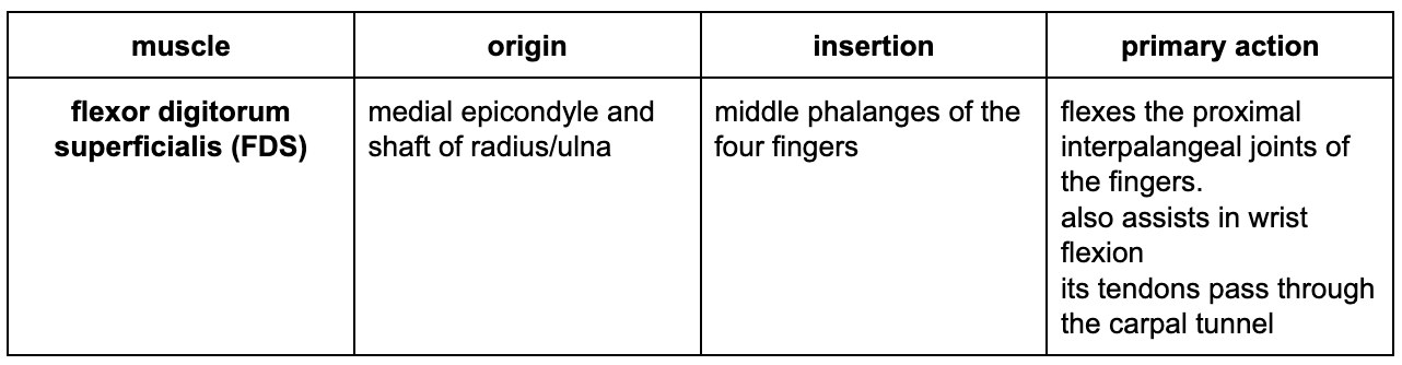
deep group (3 muscles)

The Wrist: Structure & Function
the wrist is a complex region composed of eight carpal bones arranged in two rows, forming three distinct joints
carpal bones

The 3 Joints of the Wrist Complex
radio-carpal joint (wrist joint proper)
articulation between the distal radius (and the articular disc/triangular ligament) and the proximal row of carpal bones (scaphoid, lunate, triquetral)
**The ulna DOESN’T articulate directly with the carpal bones; it’s separated by the articular disc
this joint allows flexion, extension, abduction, and adduction
midcarpal joint (transverse intercarpal joint)
articulation between the proximal and distal rows of carpal bones
allows some flexion and extension, contributing to overall wrist movement
carpometacarpal joints
articulations between the distal row of carpal bones and the bases of the metacarpals
fingers: very little movement; provides a stable base
thumb: a highly mobile saddle joint between the trapezium and the 1st metacarpal. This allows for opposition, flexion, extension, abduction, and adduction of the thumb
The Carpal Tunnel and Retinacula
flexor retinaculum
a thick band of deep fascia that spans the wrist
it attaches medially to the pisiform and hook of hamate, and laterally to the tubercles of the scaphoid and trapezium
carpal tunnel
the passageway formed by the carpal bones and the flexor retinaculum
it contains:
the long flexor tendons
the median nerve
synovial sheaths
lubricated coverings that allow tendons to glide smoothly
a common synovial sheath houses the FDS and FDP tendons, while FPL often has a separate sheath
palmar aponeurosis
a triangular, fan-shaped fibrous sheet that extends from the distal edge of the flexor retinaculum to the fingers
it’s continuous with the tendon of palmaris longus (if present) and helps protect underlying structures