Sliding filament theory
Structure of myosin and actin
myosin heads
hinge enabling movement
one site for binding to actin
another site for ATP binding
provides energy
actin filaments
sites for myosin head attachment
actin-myosin binding sites
tropomyosin and troponin proteins attached
regulatory role
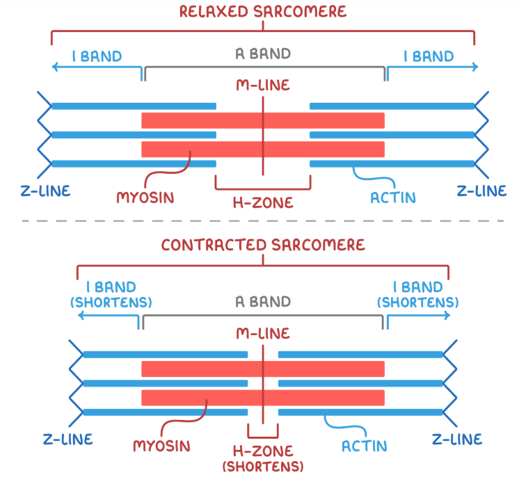
Muscle contraction
involves the actin filaments being pulled closer towards each other and towards the M-line in the centre of the sarcomere
the I band and H zone in sarcomere shorten due to increased overlap of actin and myosin filaments
A bands remain constant in length
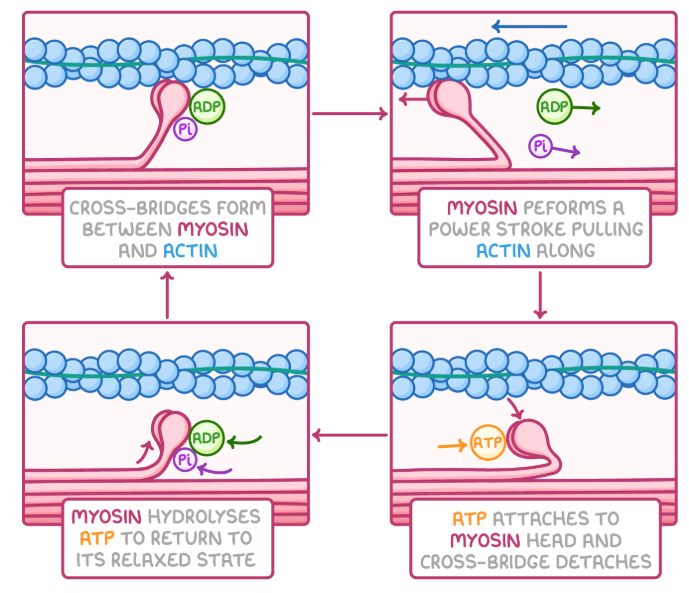
Sliding filament theory
Calcium ions bind to troponin - altering its shape
this change moves tropomyosin away from actin’s binding sites - making them available for myosin so myosin binding sites are exposed
myosin heads attach to these exposed actin filaments - forming actin-myosin cross-bridges
the myosin heads execute a power stroke - pulls the actin filament along and releasing ADP
an ATP molecule binds to the myosin head - leading to its detachment from actin
calcium ions activate myosin’s ATPase activity - breaking down ATP to ADP and phosphate thus releasing energy
this energy resets the myosin head to its original position
the myosin head reattaches to a new actin site further along the filament (10nm)
Energy sources for muscle contraction
requires ATP
generated via:
aerobic respiration
anaerobic respiration
ATP-creatine phosphate system
emergency usage
takes phosphate from creatine phosphate