Optics #2
Optics #6
Refraction
When light travels from one substance to another, its speed changes. This speed change can cause a ray of light to change its direction (refract or bend) if the ray is at an angle to the surface.
Index of Refraction
The ratio of the speed of light in a vacuum to the speed of light in a given medium.

c = 3 x 10⁸ m/s (speed of light in a vacuum)
How do we get an Index of Refraction?
Example: The speed of light in water is 2.26 x 10⁸ m/s
nwater = 3 x 10⁸ m/s
2.26 x 10⁸ m/s
nwater = 1.33
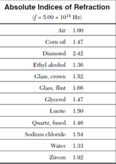
Refraction
When light travels from one substance to another its speed changes. This speed change can cause a ray of light to change its direction (refract or bend) if the ray is at an angle to the surface.
Optics #7
Refraction Phenomena
ainbows: When sunlight enters a droplet of water, it refracts and reflects back dispersing white light into its colours.
Shimmering: Hot air is less dense than cooler air. Hot air refracts light differently, resulting in the shimmering effect.
Mirages: Larger scale than shimmering. Sunlight refracts upward. Shimmering gives the illusion of water on the ground.
Partial Refraction
A phenomenon where light that is travelling from one medium into another is reflected and some is refracted.
Critical Angle
The angle of incidence that produces an angle of refraction of 90o When the critical angle is exceeded, the light is reflected back into the first medium following the laws of reflection.
Total internal reflection
A phenomenon where the incident light ray does not escape the medium and is fully reflected back from the boundary
Occurs when a ray is travelling from a “slow” medium to a “fast” medium in terms of the speed of light
Optics #8
Lenses
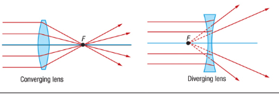
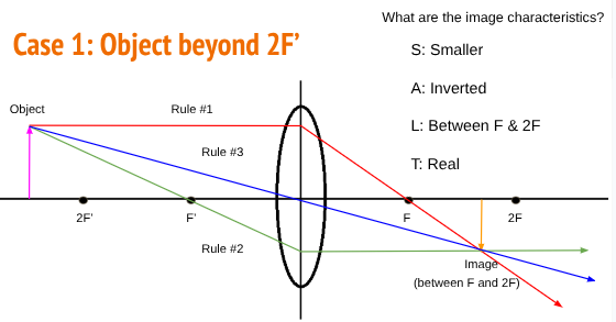
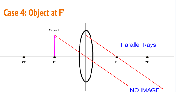
A lens is a curved piece of transparent material using the properties of refraction to form an image. There are two types of lenses:




optics #8
The Human eye
parts and functions
Location of the Eye:
The eyes sit in the orbital cavities
formed by the skull.
The orbital cavities are smaller than the eyeball which helps hold the eye in place
Openings on the back wall of the orbital cavity allows for the attachment of arteries, veins and the optic nerve
Muscles of the EyeThere are 3 pairs of opposing muscle groups that move the eye.
Lateral and Medial Rectus: side to side
Superior and Inferior Rectus: up and down
Superior and Inferior Oblique: rotation
Problems due to Muscle Imbalance

Protection for the Eye\
Eyelids: Keep out foreign particles, help spread tears to keep eye moist, block out light
Lashes: Heighten the protection against dust and foreign particles, and perspiration
Eyebrows: prevents sweat, water and debris from falling into the eye socket
Lacrimal Gland: produces tears to clean and lubricate the eye
Anatomy of the Eye
Vitreous Humour
Transparent jelly-like substance behind the lens that helps maintain the spherical shape
98% water
Refractive index of 1.33
Choroid
Blood vessels that provide nourishment for the retina
Sclera
Tough, white, outer layer
Provides protection
Continuous with the cornea
cornea
Thick, transparent membrane
Forms the front surface of the eye
Refractive value of 1.38
Provides 80% of the light refraction
Iris
Coloured part of the eye
Contains two muscles that can change the size of the pupil
Pupil
Opening that permits entry of light into the eye
Size can be adjusted depending on brightness and intensity
Lens
Transparent
Biconvex
Behind the pupil
Provides 20% of the refraction of light
n ranges from 1.40 in center to 1.38 at edges
Able to change shape to focus the light
Ciliary Muscles and Suspensory Ligaments
Control the shape of the eye to focus the light
Changes the focal distance for a sharp real image
Retina
Inner surface of the eye
Contains the light sensitive cells
Rods are more numerous, 120 million, not sensitive to colour
Cones are colour sensitive (red, green, blue), 6 to 7 million, concentrated in the central yellow spot (macula)
Real image is projected
Fovea
Most sensitive spot of the eye
Provides the sharpest vision
0.3 mm diameter rod-free area with very thin, densely packed cones, no rods
Optic Nerve
Transmits the electrical impulses to the optic centre of the brain
Information transmitted included brightness, perception and contrast (visual acuity)
How an Image is Formed
When the light passes into the eye, there are three points of refraction: at the cornea, the outer surface of the lens, inner surface of the lens. This directs the light to meet at the retina
Focusing an Image
The focal length changes depending on the distance of the object.
The cornea curvature is fixed, but the shape of the lens can change. This is called accommodation.
When an object is close, the lens is more rounded and thick. When the object is distant, the lens is longer and thin

Refractive Conditions
Myopia – Nearsighted (you can see nearby objects clearly e.g. reading a book)
– need glasses to drive and read signs
The eyeball is too long or the
refractive power is too strong
The image is focused in front of the retina
Myopia - near sightedness
This is corrected by using the biconcave (diverging) lens.
The light rays are spread out before they reach the eye.
The rays are essentially lengthened.
The refraction of the eye directs the light to the focus on the retina.
Hyperopia – Farsighted (you can see distant objects clearly, e.g. reading road signs)
– need glasses to read a book
The eyeball is too short or the refractive power is too weak.
The image is focused ‘behind’ the retina
This is corrected by using the biconvex (converging) lens.
The light rays are directed toward the lens, then the refraction of the eye focuses the light on the retina. The light is essentially shortened
Myopia: Objects close up are in focus, while distant objects are blurry
Hyperopia: Objects far away are in focus, while close objects are blurry
Describe the overall shape, colour and feel/consistency of the eye.
The eye was round and slightly oval in shape. The sclera was white with some red blood vessels visible. The eye felt firm but slightly soft and rubbery when pressed.
3. Provide two ways in which the eye was anchored to the skull/head.
The eye was attached by the optic nerve at the back.
The eye was held in place by several eye muscles connected to the surrounding tissues of the skull.
4. How was the movement of the eye controlled? Describe the appearance of these structures.
The movement of the eye was controlled by the extraocular muscles. These muscles appeared as thick, white, string-like bands attached to the outside of the eye. They pulled the eye in different directions to allow movement.
5. Note the fat surrounding the eye. What is the function of this fat?
The fat surrounding the eye acts as a cushion that protects the eye from injury. It also helps hold the eye in place within the eye socket.
6. Identify all evidence of blood vessels to and in the eye. What do they look like?
Small blood vessels could be seen on the surface of the sclera and within the surrounding tissues. They appeared as thin red lines or branching networks that supplied oxygen and nutrients to the eye.
7. Locate the optic nerve. To view the separate fibres that make up the nerve, pinch the nerve with your fingers. Describe the optic nerve. Where does the optic nerve lead?
The optic nerve was found at the back of the eye. It appeared as a thick, tough, white cord made up of many tiny fibres. The optic nerve carries visual information from the retina to the brain, where the images are processed and interpreted.