Morphological Unknown Guide
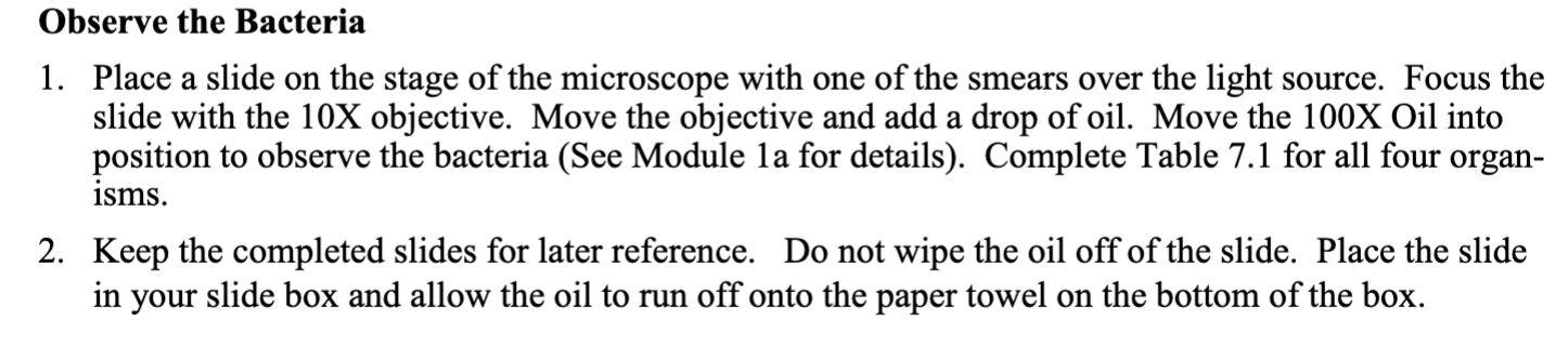
(All material is sourced from Dr.Gebler’s lab powerpoints/manuals)
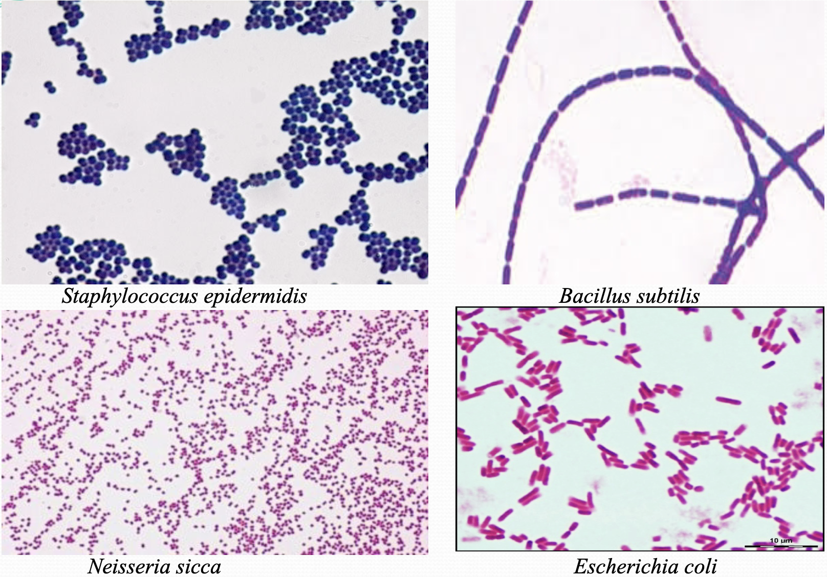
Possible Options: Staphylococcus epidermidis,Bacillus subtilis, Escherichia coli, Neisseria sicca
How to Complete a Bacteria Smear from a TSA (agar) Slant
Apply 1-3 drops of distilled water to slide
Aseptically transfer bacteria from the test tube after flaming both the loop and the tube.
How to Complete a Bacteria Smear from a TSB (broth) Slant
Do NOT add any water to the slide
Start flaming loop and test tube
Transfer 4-6 loop fulls of broth (make sure to flame both again before the next transfer)
Remember the microscope slides are not sterile
Bacteria Fixation
There are three options: 1. Air Dry, 2. Slide Warmer, 3. Bunsen Burner
Air Dry
Let the slide sit until the water evaporates at room temperature. However, it can take between 20-40 min & minimizes gram variability.
Slide Warmer Most recommended
Apply microscope slide on top at 60°C. There are no disadvantages this can take between 5-10 min. The advantage is keeping constant temp that does not damage cell.
Bunsen Burner
Using a clothing pin hold slide 12-18 inches above burner. The disadvantage is that it can lead to gram variability.
To avoid gram variability make sure to aseptically disinfect equipment by flaming loop and test tube.
Gram Stain Breakdown + Chemicals Needed
The sequence for a gram stain involves:
Crystal Violet (primary stain)
Grams Iodine (mordant)
Acetone alcohol (destain)
Safranin (counterstain)
Before starting allow the slides to cool, once cool take them to a sink or drain.
Then, coat the entire slide in crystal violet and allow it to sit for 1 minute.
After, use distilled water to gently rinse the slide, using too much force and water can cause the bacteria to wash off.
Next, coat the slide in iodine for 1 minute.
Proceed rinsing slide gently with distilled water once again. For the next step pay attention carefully.
You then want to spray the smear with acetone alcohol until no more purple is washed off the slide, this only takes a few (30) seconds.
Rinse the slide again with distilled water gently.
Then, place the slide back on the staining rack and apply safranin and allow to sit for 4 minutes.
Afterwards, rinse the excess with distilled water. Then use bibulous paper to blot the slide dry. DO NOT RUB THE SLIDE DRY.
To View Bacteria (Gathered from Lab Manual)
Acid Fast Procedure + Chemicals Needed
The following procedure involves:
Carbol Fuchsin (primary stain)
Acid alcohol (destain)
Loeffler’s Methylene blue (counter-stain)
Once you have your smear prepared and dry, apply carbol fuchsin to the slide and flood it.
Then, cover the flooded smear with white square shaped filter paper.
Either place slide over slide warmer or above the bunsen burner for 10 minutes, remember to constantly apply carbol fuchsin as needed to prevent it from drying out.
Afterwards, allow the slide to cool off for 3 min, and rinse with distilled water.
Then, you can flood the slide with acid alcohol and leave it for 30 seconds.
Next, add Loeffler’s methylene blue (counter-stain), and leave the stain on smear for 1 min.
After rinse the slide, and blot dry.
From then on you can observe it from a microscope.
Schaeffer-Fulton Procedure (Endospore) + Chemicals Needed
Conducting an endospore procedure calls for:
Malachite Green (primary stain)
Saffranin (counter-stain)
Remember out of all four bacterias Bacillus subtilis is the only one that produces spores.
To start, have a prepared smear of your sample, by from air drying or having it on the slide warmer.
Start with the malachite green stain and flood the smear.
Then, cover the smear with white squared shaped filter paper.
Apply it over a bunsen burner or on a slide warmer for 10 minutes, and make sure to reapply malachite green as needed.
Allow the slide to cool for 3 min, and rinse with distilled water.
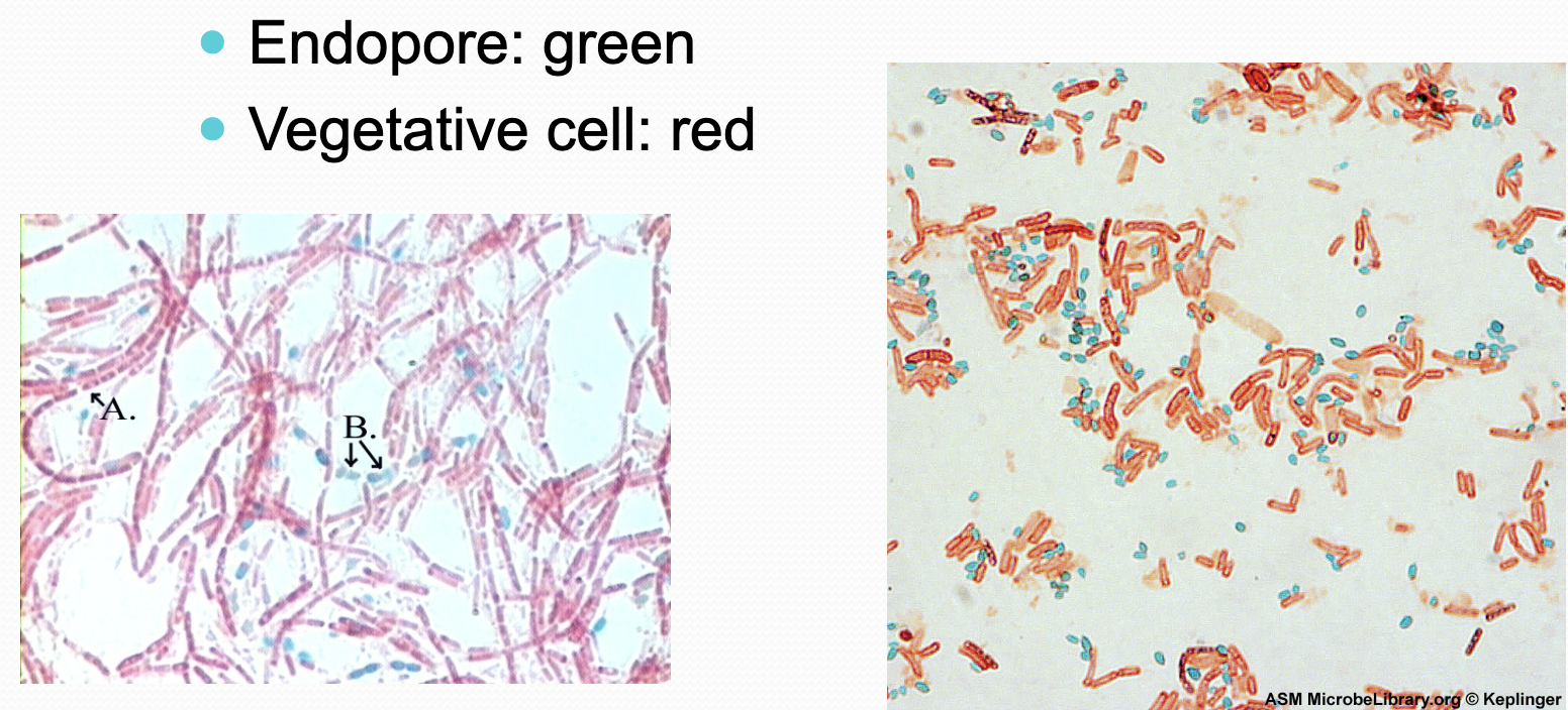
Then,add safranin stain (counter stain). This should add a green color to endospore cells, leave the stain on for 3 min.
Rinse the slide with distilled water, and blot dry.
You can then use oil immersion objective to view slide.
How to Label Sample
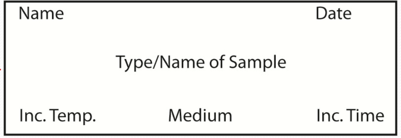
Image of Bacteria
Endospore Stain Result Image
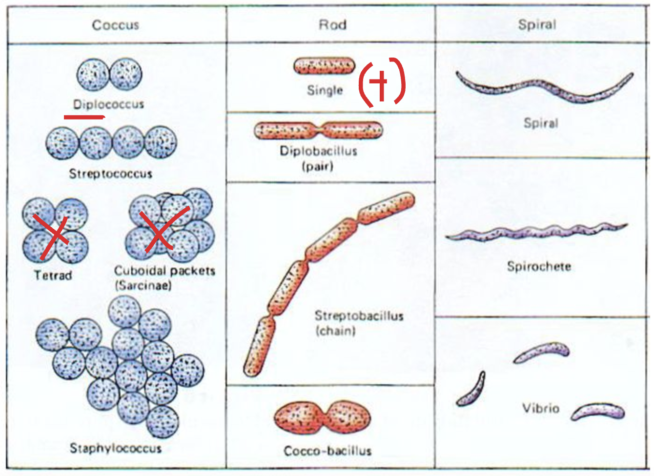
Bacteria Shape Morphology Configuration
There are three common types of shape for bacteria.
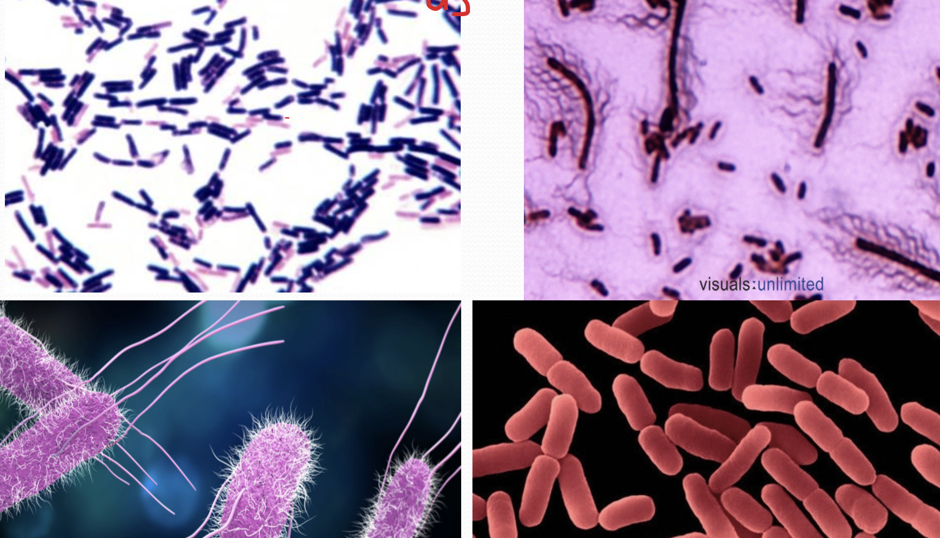
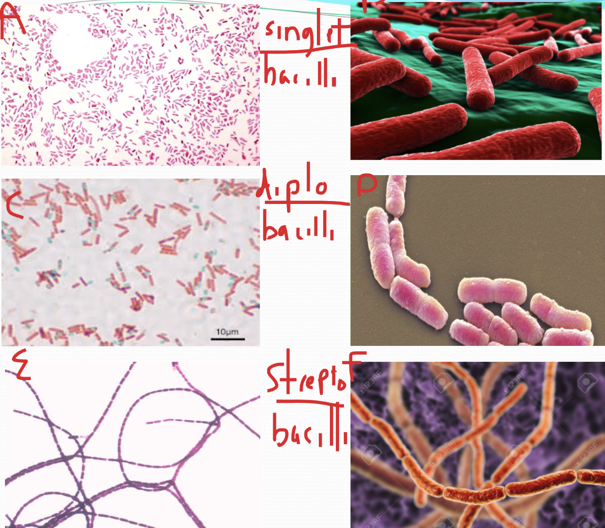
Bacilli (bacillid)
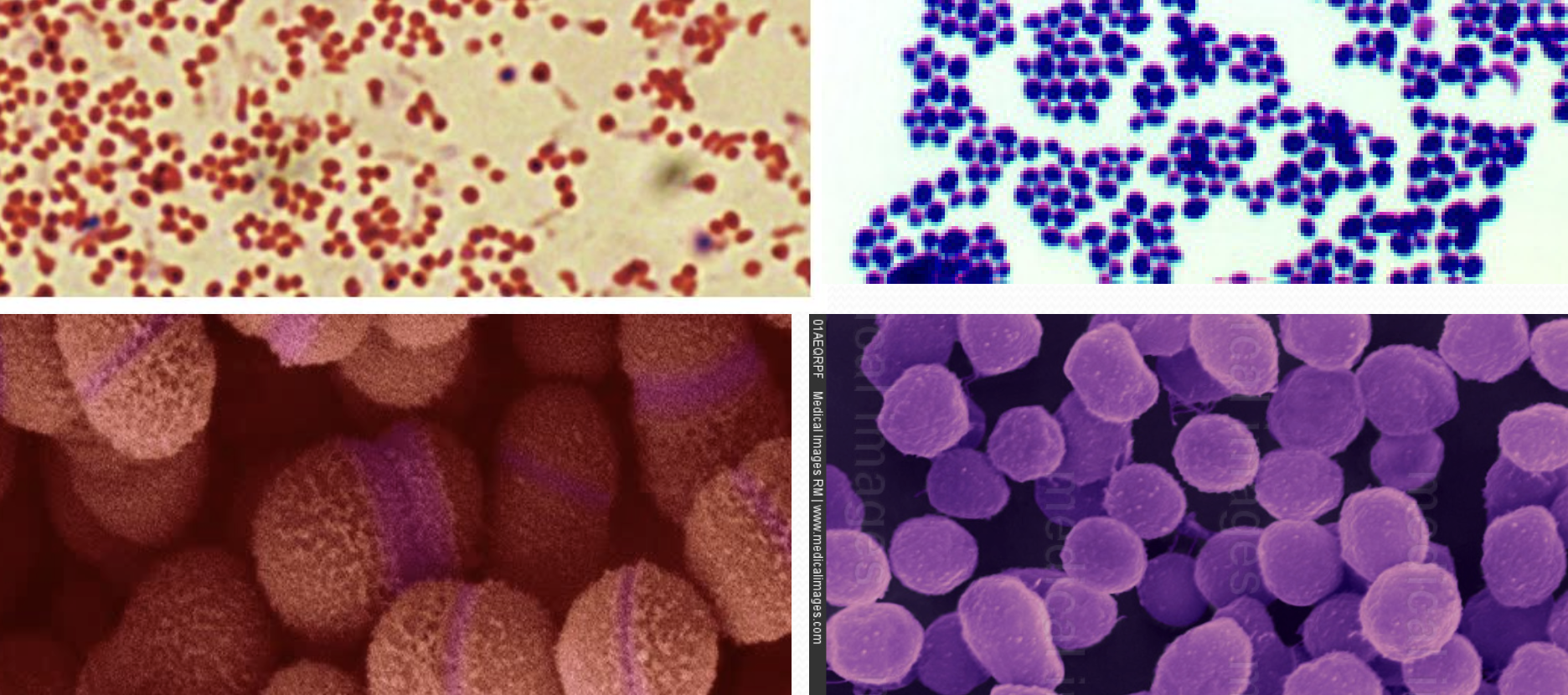
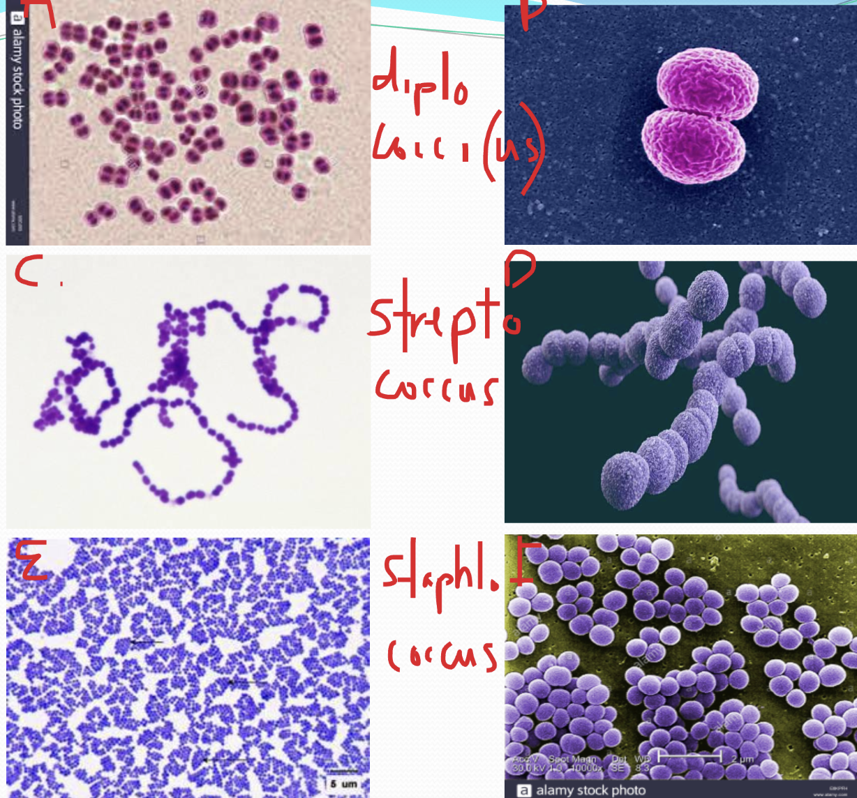
Coccoid (coccus)
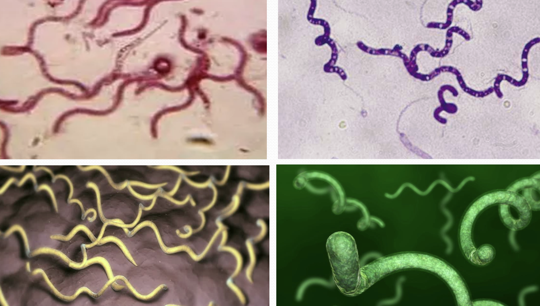
Spirilli (spirillid)
Bacillid- tube shaped
Coccoid- round shaped
Spirilli- spiral shaped
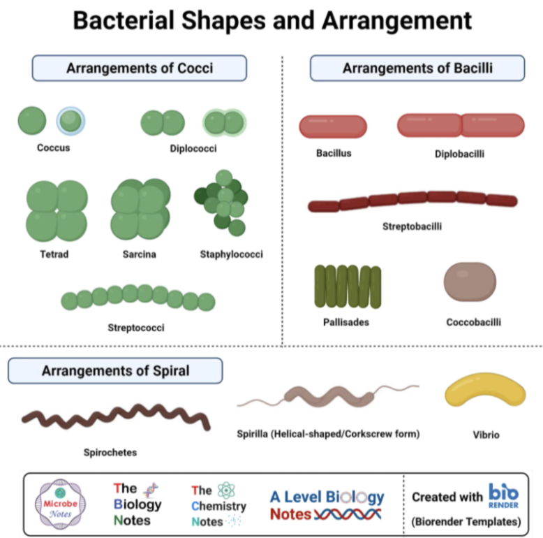
Actual Bacteria Shapes
How to Properly Label Title of Organism
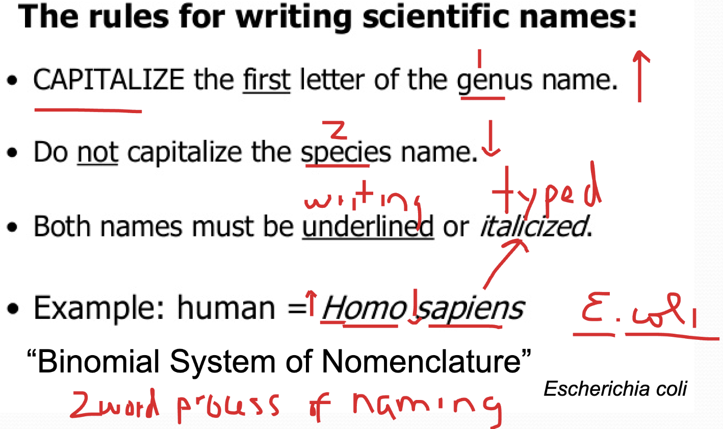
We are required to write out the full name for our unknown.