Protozoa
Protozoa
Protozoa: Amoebae
Subphylum Sarcodina
Single celled eukaryotes
Feed by engulfing particle
Move by Pseudopodia
Definitions
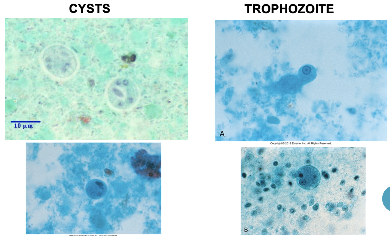
Trophozoite form
- Motile, reproductive, feeding stage
Cyst form
- nonmotile , resting stage that is resistant to environmental effects
Chromatoidal bars or bodies
- Dark-staining cytoplasmic inclusion of chromatin
Endosome or Karyosome
- Mass of organelles or mass of chromatin in the nucleus
Amoebae Diagnosis:
Motile trophozoites in fresh, warm, liquid stool
- Wet mounts and iodine preps
Trophozoites &/or cysts in fixed smears
- Trichrome stain
Trophozoites or cysts in other body tissues or fluids; serologic tests; ELISA
Pathogenic intestinal amoeba
- Entamoeba histolytica
Nonpathogenic intestinal amoebae
- Entamoeba coli
- Endolimax nana
- Iodamoeba butschlii
- Entamoeba hartmanni
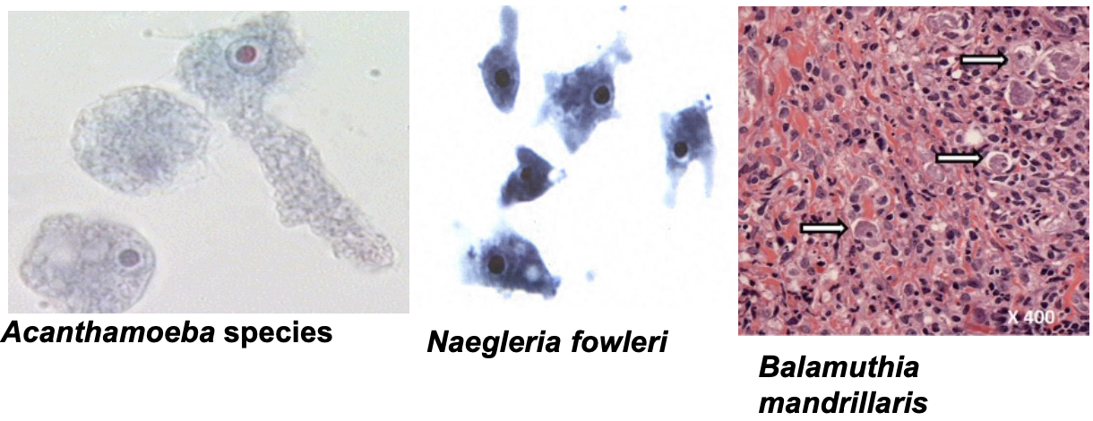
Free-living pathogenic amoebas
- Acanthamoaba castellani
- Naegleria fowleri
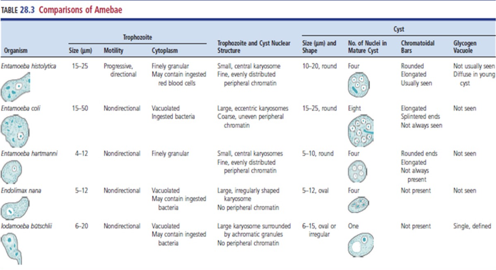
Differentiating the Amoebas
Shape of nucleus
Number of nuclei
Size
Shape
Inclusions
Entamoeba Histolytica
Amebiasis
Transmission by ingestion of cysts
- Fecal-oral route of contaminated food
Intestinal disease
- Asymptomatic or amoebic dysentery
- diarrhea, cramping, abdominal pain
Disseminated
- hematogenous spread to liver, spleen, lung, or brain
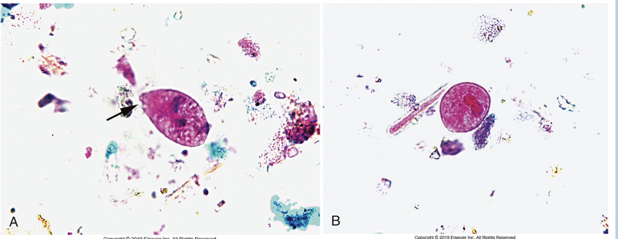
Microscopic Characteristics E. Histolytica
Trophozoite
- 10-20 um
- Irregular shape
- Single nucleus with central karyosome
- Ingested RBCs
Cyst
- 10-20 um
- Contain one, two or four nuclei with central karyosome
- Rounded chromatoidal bars
*Side Note
Entamoeba hartmanni
- Morphologically same as E. histolytica
- Much smaller size (less than 10 um)
Entamoeba dispar & E. moshkovskii
- Morphologically similar to E. histolytica
Non Pathogenic Amoebae
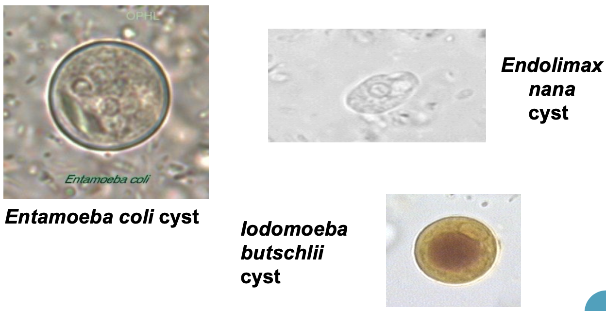
Entamoeba Coli
Very common commensal
Cysts
- >10 um
- Up to 8 nuclei with an eccentric karyosome
- Pointed, sharp chromatoid bars
Trophozoite
- Ingested bacteria
- NO ingested RBCs
*Blastocystis Hominis*
Some source consider Blastocystis hominis to belong to the genus Stramenopiles
Brown algae, water molds, and diatom also members of this group
The exact life cycle is not completely known and as such changes continue to occur
Status of being a “true pathogen” remains controversial.
Potential pathogen
- Recurrent diarrhea, abdominal cramping, anorexia, flatulence
Four forms
- Ameboid
- Granular
- Vacuolar
Most commonly Identified
- Cyst
Recovery of the vacuolar form
- Morphologic characteristics
- 5 to 15 um
- Empty central body (green on trichrome)
- Nuclear material in ring pattern between the central body and outer membrane
Comparison of Amebae
Free-Living Pathogenic Amoebas
Fresh and saltwater, soil, and decaying vegetation
Naegleria fowleri
- Gains entry through nasal mucosa
- Primary amebic meningoencephalitis (PAM)
- Ameboid form found in CSF and tissues
Acanthamoeba
- Inhalation of contaminated dust
- Granulomatous amebic encephalitis (GAE)
- Brain abscesses, keratitis, sinuses
Balamuthia mandrillaris
- Found in soil, inhaling cysts or direct inoculation and organ transplant
- GAE
Flagellates (Mastigaphora)
Flagella – provides locomotion
Axoneme – intracellular portion of a flagellum
Axostyle – rod that provides support in flagellates
Pathogenic Flagellates
Intestinal flagellates
- Giardia lamblia
- Dientamoeba fragilis
Vaginal flagellate
- Trichomonas vaginalis
Blood & tissue flagellates
- Trypanosoma sp.
- Leishmania sp.
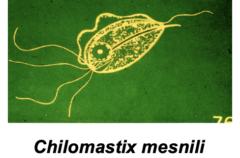
Non pathogenic Flagellates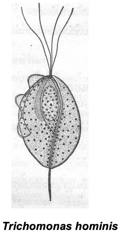
Ganstointenitnal flagellates
- Chilomastix mesnili
- Trichomonas hominis
Giardia Lamblia
Most common diarrheal disease transmitted by contaminated water in U.S.
Transmission: contaminated water, fecal-oral route, oral-anal sex, etc.
Only human protozoa that reside in the duodenum
Giardiasis: “Travelers diarrhea,” beaver fever,” backpacker’s diarrhea”
Symptoms: Mild to severe diarrhea
Diagnosis: trophozoites or cysts in stool and biopsies, ELISA
Microscopic Characteristics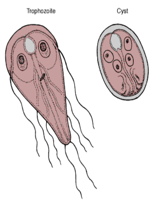
Trophozoites
- Bilateral symmetry
- Two nuclei
- Eight flagella
Cysts
- Oval
- Two to four nuclei
Dientamoeba Fragilis
Assoc. With cases of diarrhea
No cyst form, only trophozoite
Trophozoite
- Two nuclei (very dense central chromatin)
Trichomonas Vaginalis
Trophs live in vagina, urethra, epididymis, and prostate
No cysts stage
Transmission via sexual intercourse
Method of diagnosis
- Wet mount
- Urethral discharge, vaginal smear, or urine.
- Observe “jerky” pattern of motility
Non Pathogenic Intestinal Flagellates

Intestinal & Urogenital Flagellates
Blood & Tissue Flagellates
Trypanosoma (tsetse fly)
- T. brucei rhodesience
- East African sleeping sickness
- T. brucei gambiense
- West African sleeping sickness
*Trypomastigote the diagnostic from observed in the blood
Trypanosoma cruzi
Central, South America, Cuba, Mexico, and now Florida, TX, California
Vector is the Triatoma or reduviid bug
Amastigote form observed in the tissues (heart)
Trypomastigote (characteristics “C” shape) may be found in the blood
Chagas’ disease
The disease can be mild, causing swelling and fever, or it can be long lasting. LEft untreated, it can cause congestive heart failure
Treatment for Chagas disease focuses on taking medications that kill the parasite and on managing symptoms
Blood & Tissue Flagellates
Leishmania
Vector sandfly (Phlebotomus spp.)
L. tropica
- “Old world disease” boils
L. mexicana
- “New world disease” or espundia
L. braziliensis
*all three produce cutaneous lesions, amastigote forms found in tissues
Leishmania Donovani
Spread beyond the cutaneous lesions to the internal organs
- Spleen, liver, bone marrow
Amastigote forms (LD bodies) observed in tissues
Kala-azar
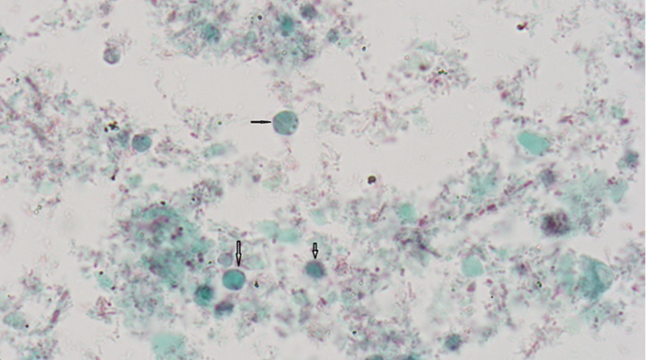
Ciliates: Ciliophora
Movement by cilia
Macronucleus & several micronuclei
Only pathogenic human ciliate
- Balatidium coli (commonly infects hogs)
- Largest human intestinal protozoa
Symptoms: asymptomatic or diarrhea with alternating constipation
Diagnosis
- trophozoites or cysts in feces
B. coli Trophozoite (A) & Cyst (B)