3.2 Histology blood vessels & capillaries
Blood and Cardiovascular System
Blood Vessel Functionality
Blood vessels form circuits that work with the heart.
Arteries: Transport oxygenated blood away from the heart (includes elastic arteries, muscular arteries, arterioles).
Veins: Return deoxygenated blood to the heart (includes venules, medium veins, large veins).
Comparison of Arteries and Veins
Arteries and veins have distinct structural differences:
Both have tunica intima, tunica media, and tunica externa.
Arteries contain more smooth muscle and elastic fibers.
Veins primarily consist of collagen fibers and have fewer smooth muscle layers.
Tunica intima in both types is thin and closest to the lumen.
Tunica media in arteries is thicker, regulating blood flow with elastic tissue.
Tunica externa is thickest in veins, consists of dense fibrous connective tissue, contains vasa vasorum.
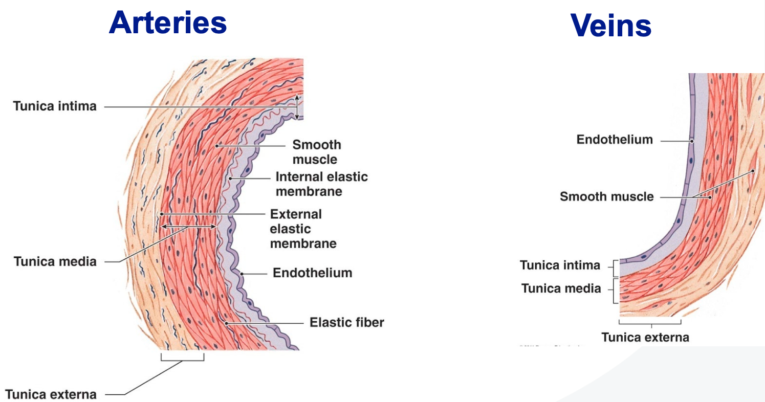
Wall layers in large vessels
Blood vessels are tubular structures with three primary layers:
Tunica interna/intima: Innermost layer composed of endothelium.
Tunica media: Middle layer primarily made up of smooth muscle.
Tunica externa/adventitia: Outermost layer providing structural support.
Vessel wall complexity varies with size and distance from the heart.
Vessel wall layers
Tunica interna/intima
inner most layer (thin)
Endothelium
Thin, simple squamous epithelium
smooth
antithrombogenic
Basement membrane
subendothelial connective tissue has fine elastic and collagen tissue, has fine elastic & collagen fibres
Internal elastic laminate-IEL
Tunica media
Middle layer, thickest in arteries
Variable quantities of elastic tissue and smooth muscles
Smooth Muscle
Smooth muscle is characterized by a concentric arrangement of fibers.
Contraction and relaxation of smooth muscle lead to variations in luminal diameter, which results in a decrease or increase in blood flow.
Elastic Laminae/Lamellae
Elastic laminae contain fenestrations that allow for flexibility and expansion of the blood vessel walls.
Tunica Externa/Adventitia
The outermost layer of the blood vessel wall.
Thickest in veins
Provides structural support to the blood vessel.
Composed of dense fibrous connective tissue.
Contains vasa vasorum, which are small blood vessels that supply the walls of larger vessels.
Specific Vessel Structures
Elastic arteries:
Large diameter blood vessels that have thick walls and a high content of elastin.
Function: Mitigate blood pressure fluctuations by stretching during ventricular systole and recoiling during diastole.
Media contains numerous concentric elastic laminae (sheets) making up more than 50% of this layer, interspersed with smooth muscle cells which help regulate vessel diameter and maintain blood pressure.
Muscular arteries:
Smaller than elastic arteries with pronounced internal elastic lamina.
Contain larger amounts of smooth muscle (25-40 cell layers).
Function: Allow for vasoconstriction and dilation, adjusting blood flow to various tissues and organs.
Tunica media is dominant, allowing for precise regulation of blood flow.
Vasa vasorum present in the dense connective tissue of the tunica adventitia.
Arterioles:
Small diameter, thick tunica media controls diameter.
<5mm diameter
Thick wall relative to luminal diameter
Tunica media (smooth muscle) are the most prominent
Constriction and dilation of smooth muscle vary the vessel diameter.
This variation reduces blood pressure for flow through capillaries.
Regulates blood distribution and flow through capillaries.
Arterioles regulate blood flow and pressure as they control the diameter of blood vessels.
They play a key role in reducing blood pressure before blood reaches the capillaries, allowing for efficient exchange of nutrients and waste.
Through constriction and dilation, arterioles adjust blood distribution to various tissues and organs based on metabolic needs.
Venules:
Collect blood from capillaries, thin walls, supported by pericytes.
Generally 8-100 micrometers in diameter
The tunica media is the middle layer of blood vessels, primarily composed of smooth muscle, which is the most prominent layer.
It allows for constriction and dilation, varying the diameter of the vessel, which helps regulate blood flow.
This variation in diameter serves to reduce blood pressure as blood flows through capillaries and plays a key role in the distribution of blood throughout the body.
Medium veins:
High capacitance, thin walls, irregular lumens, valves to prevent backflow.
Have thin walls compared to arteries, allowing for flexibility and expansion.
Characterized by irregular lumens that support blood flow.
Contain valves to prevent the backflow of blood, ensuring efficient circulation back to the heart.
Large veins:
Lack valves and internal elastic lamina, thinner tunica media, prominent adventitia with high collagen content.
No valves, no Internal elastic lamina (IEL), no External elastic lamina (EEL)
Summary of Vessel Structure Differences
Arteries:
High pressure
pressure reservoir
vessels at full blood capacity
No valves
circumferential muscle layer
Thinner adventitia
small lumens, thick walls overall, hold circular cross sectional shape
Veins:
low pressure
blood reservior
vessels contain 30-70% of blood capacity
presence of valves
reduced muscle in media, less compact, ‘patchy’ distribution
adventitia is usually thicker
Vessels have large lumens, thin walls, collapsed/irregular profiles in sections
Differences in large vessel structures
Large veins
Thinner walls
No valves large capacity reservoirs
Valves for unidirectional flow
Capacitance vessels
Large (elastic) arteries
Thicker walls
Conducting Vessels
Distributing vessels
Resistance vessels
Transition to Capillaries
Capillaries play crucial roles in substance exchange across the endothelium:
Types include continuous, fenestrated, and sinusoidal capillaries.
Continuous capillaries: Characterized by tight junctions and abundant vesicles.
Fenestrated capillaries: Allow extensive exchange through regulated fenestrations (holes), often with thin diaphragms.
Sinusoidal capillaries: Discontinuous structure, larger diameters, irregular pathways, optimize exchange.
Structural characteristics
Reduced complexity
Endothelium: Thin inner lining made of endothelial cells.
+/- Fenestrations: Small openings that allow for selective permeability and exchange of substances, present in some capillaries.
+/- Tight junctions: Cell junctions that regulate the passage of materials between endothelial cells, contributing to the barrier function of the capillary wall.
Pinocytotic vesicles: Small vesicles that facilitate the transport of materials across the endothelial layer, enabling larger molecules to move in and out of the bloodstream.
Basal lamina: A layer of extracellular matrix that supports the endothelium and regulates its function.
+/- Pericytes: Contractile cells associated with capillaries that help regulate blood flow and maintain capillary stability.
Smallest diameter vessels: Capillaries are the smallest blood vessels, measuring approximately 5-10 micrometers in diameter, facilitating efficient exchange of nutrients and waste between blood and tissues.
Endothelial cells
Thin (attenuated): Maximizes exchange between blood and tissues.
Anti-thrombogenic function: Helps to prevent blood clot formation.
Breakdown of lipoproteins: Converts lipoproteins into triglycerides and cholesterol for metabolism.
Intercellular tight junctions: Restrict passage between endothelial cells, maintaining barrier integrity.
Pinocytotic vesicles: Facilitate the transport of materials across the endothelium, aiding in nutrient uptake and waste removal.
Continuous capillary
Most widespread capillary type
Tight junctions
Many pinocytotic vesicles for exchange
No gaps between endothelial cells
Less common continuous type
Very tight junctions - more selective barrier
Few pinocytotic vesicles
No gaps between endothelial cells
restricted exchange between blood and tissues
Fenestrated capillary
Characterised by interruptions in endothelium: The endothelial cells have pores, known as fenestrations.
Endothelial cell cytoplasm is pierced by pores: These fenestrations extend through the full thickness of the wall.
Allow for extensive exchange: They facilitate exchange between blood and tissue, though with some limitations on particle size.
+/- Thin diaphragm: Some fenestrations may have a thin diaphragm, which is thinner than the cell membrane.
Continuous capillary vs Fenestrated capillary
Continuous: slower exchange more selective
Fenestrated: More rapid exchange, less selective
Sinusoidal Capillaries/sinusoids
Characterized by larger diameters: Wider compared to other capillary types.
Irregular pathways: Exhibit tortuous and non-linear arrangements, enhancing surface area for exchange.
Discontinuous endothelial lining: The endothelial layer is incomplete, facilitating greater permeability.
Numerous large fenestrations: Large openings present in the walls that allow for the passage of larger molecules and cells.
Discontinuous basal lamina: The supportive layer is also disjointed, promoting additional transport capabilities.
Maximum exchange: Designed to maximize the exchange of materials between blood and surrounding tissues.
Gaps enable transport of whole cells: Allow for the movement of entire cells, crucial for processes like immune response and tissue repair.
Location: Found mainly in organs such as the liver, spleen, and bone marrow, where significant exchange occurs.