Histology - tissues I
Histological technique
LSMU’s laboratory uses formalin-fixed samples that are embedded into paraffin.
- Fixation
Chemical processes in which tissues are preserved from disintegration and prepared for further processing.
Fixed in 10% formalin solution.
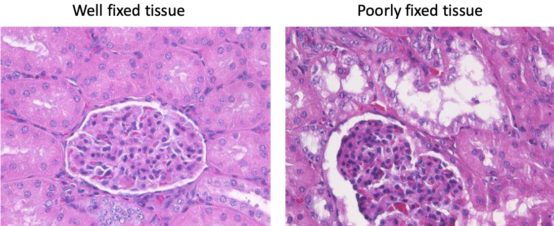
Factors determining fixation quality.
- Volume of fixation (10-20 times of the tissue volume)
- Sample size (fixative must be absorbed into all layers of the sample)
- Permeability of fixative (formaldehyde has fastest penetration 1mm/h)
- Start and duration of fixation time (immediately placed in fixative and stored 24h)
- Temperature (18-24 Celsius)
- Acidity (higher acidity 🡪 faster reaction, optimal pH 6-8).
- Removal of fixative material
- End of fixation, fixative is removed from tissue by washing in running water.
- Histological cartridges with date, name and number to avoid confusion.
- Dehydration
- Removal of free water because hydrophobic substances (paraffin/resin/waxes) will be used later.
- Ethyl alcohol, methanol, isopropyl alcohol, acetone
- Tissue dehydrated by increasing solution concentration 70% solution 🡪 80% 🡪 96%.
- Clearing
- Remove dehydration substance from tissue.
- Xylene, toluol or chloroform
- Refractive index of substances is close to protein index, which affects tissue making it translucent/transparent.
- To short clearing, may influence preparation: poorly impregnated, remain soft, fluffy and unsuitable for making thin incisions.
- To long clearing, the tissue solidifies, and protein desaturates.
- Impregnation
- With stiffening substances (paraffin), allowing the structures to be preserved ones cut thin.
- Preparation of paraffin sections
- Chilled paraffin block is cut by a microtome (a twisting handle) to 1-10 micrometers.
- Sections are placed in warm water and straightened, then placed on a glass and dried in a thermostat.
- Staining
- In laminar air flow chamber
- Different staining methods give rise to different highlighted structures.
- Golden standard: hematoxylin (pink-violet), eosin (blue)
- Xylene to remove paraffin.
- Rehydration with alcohol solution of decreasing concentration
- Stained 🡪 washed with running water 🡪 dehydrated 🡪 cleared with xylene.
- Histological glue on covering glass is applied.
Diversity of human cells
What to look for:
- Shape of cell
- Nucleus position
- Special features
- Location to other cells
Artifacts should be ignored.
- Folds
- Thick cuts
- Nucleus out of focus are not part of the focused cell.
- Different shape
- Reside between two cells.
Factors which determine morphology
- Environment
- Functions
- cytoskeleton
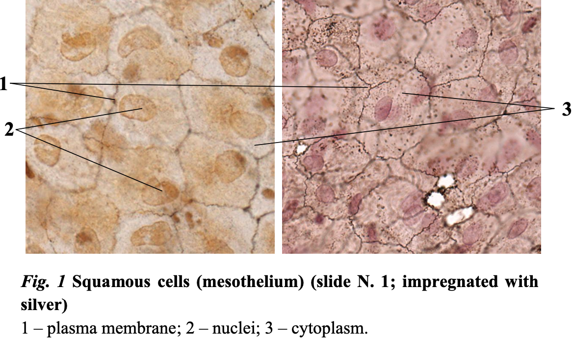
- Squamous cells/mesothelium/Epithelial tissue
- Flat cells
- Oval, central, middle nucleus
- Scale-like appearance
- No special features
- Reside side by side with other cells.
Avascular! Get their nutrition from connective tissue.
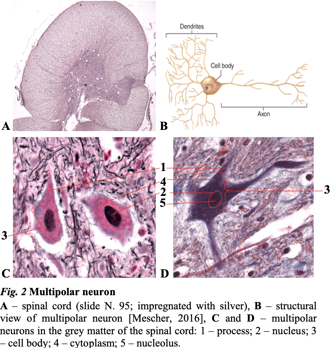
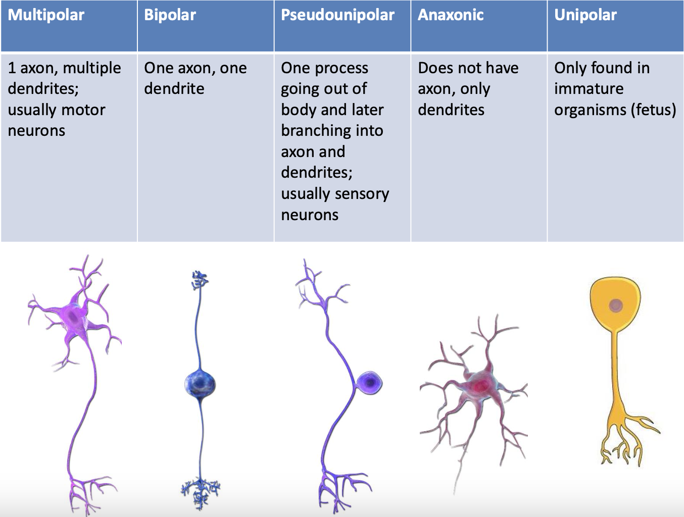
- Multipolar neurons
- Single axon extending from one end of the cell body.
- Several dendrites protruding from the other side of the cell body.
- Processes are either dendrites or axons.
- Posterior horn: Larger neurons
- Anterior horn: More in number
- Branched, oval cells.
- Large, oval, central nucleus with nucleolus
- Connections are not seen.
- Outer connective tissue is Neuropil with ganglia or other visible processes.
- Striated skeletal muscle fibers.
- Cylindrical shape
- Blunt ends
- Long
- Many nuclei peripheral
- Striations
- Vertically: y-band is lighter, A-band is darker
- Laterally: myofibrils
- Connective tissue
Cross-section
- Only one nucleus is seen.
- Myofibrils
- Cytoplasm is whiteish.
- Number of myofibrils determines blueness.
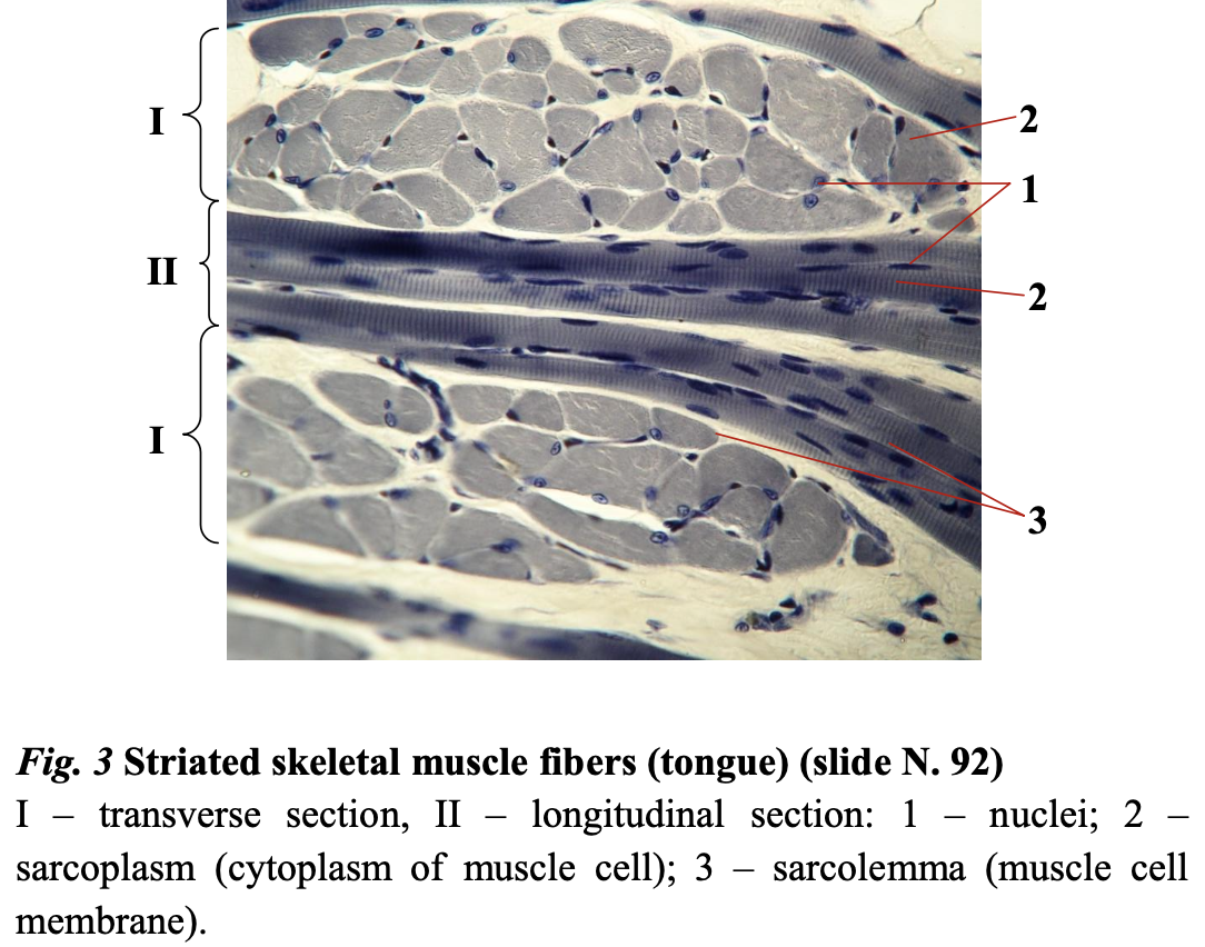
The tongue has striated skeletal muscle – epithelial tissue and connective tissue.
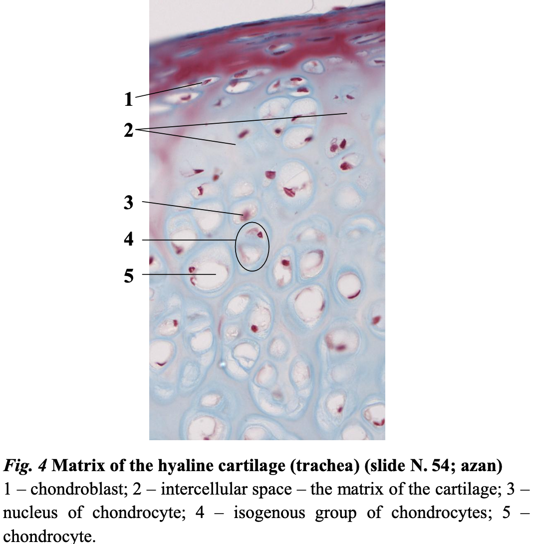
- Matrix of hyaline cartilage
- Fibers made up off type II collagen.
- Chondroblast/chondrocyte:
- Oval cells
- Oval peripheral nucleus
- Resides in clusters in even number: isogenous group.
- Territorial matrix in between cells in the clusters
- Interterritorial matrix is between cells.
- White cytoplasm because of lipids
- Connective tissue cells
- Maintain the formation of cartilage.
- Spherical cells
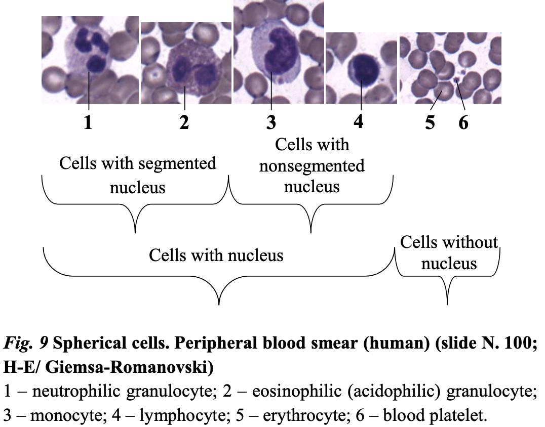
- Erythrocytes
- No nucleus
- Squamous center
- Biconcave
- 7,5 micrometers
- Most abundant
- Platelets
- Small
- Not cells
- Leukocytes
- Granulocytes:
- Basophils: bluer and granular
- Eosinophils: violet-pink-reddish, 2 segments
- Neutrophils: 2-5 segments, granulated, 15- 20 micrometers. Round
- Agranulocytes
- Monocytes: largest, round, horseshoe nucleus, 20-24 micrometers
- Lymphocytes: round, large nucleus, 8-11 micrometers
- Granulocytes:
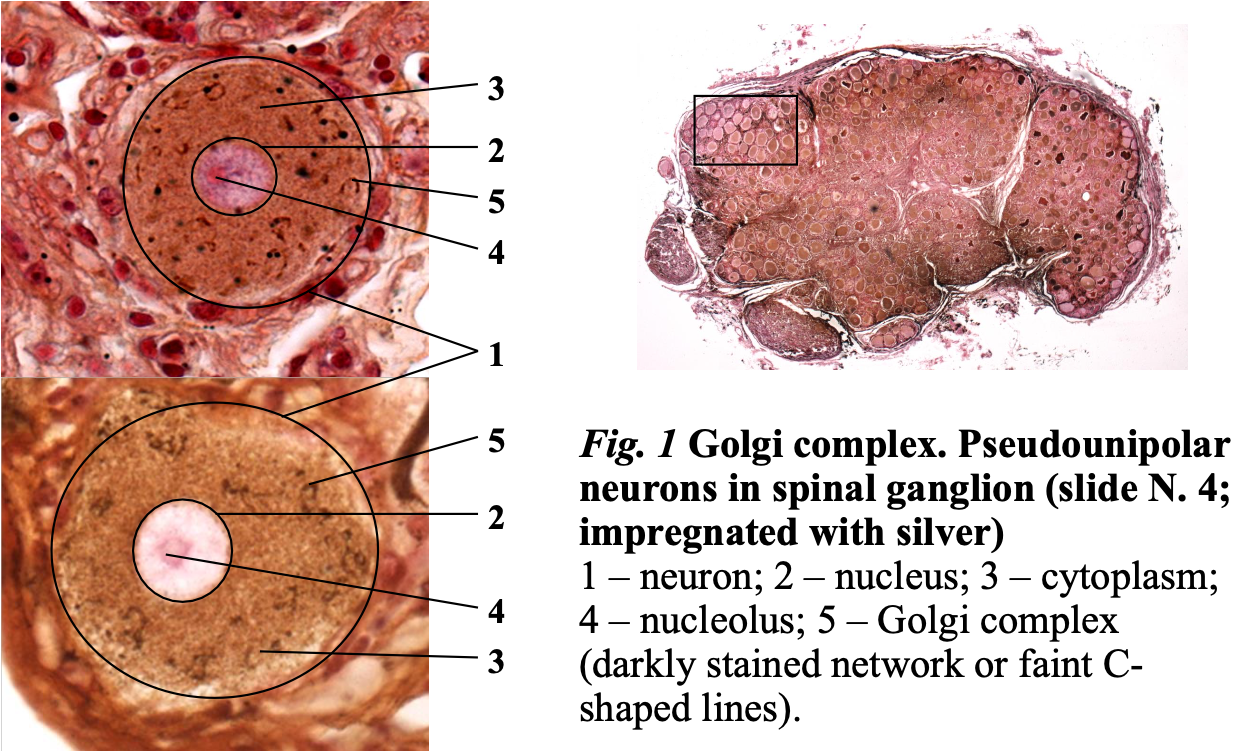
- Pseudounipolar neurons in spinal ganglion
Stained with silver salt 🡪 highlights membranous organelles, C-shaped lines are golgi
- On dorsal root ganglion
- Has one axon that projects a short period before splitting into two branches.
- Dark ganglion neurons: poor in neurofilaments
- Clear ganglion neurons: rich in neurofilaments
- Golgi complex is usually dark lines, C-shaped. This is seen because they produce neurotransmitters.
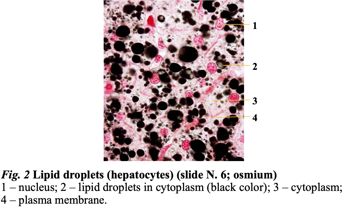
- Hepatocytes
Freezes in cryostat and stained with osmium.
- Lipid droplets, various in size
- polygonal cells (20-30µm), usually diploid cells arranged in lines.
- abundant granular eosinophilic (pink) cytoplasm,
- centrally placed round to ovoid nuclei,
- prominent nucleoli
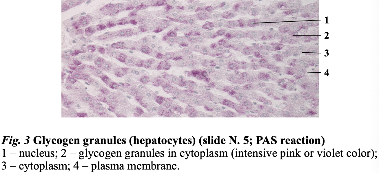
- Glycogen granules are the glucose storage. = nutritional inclusions
- Glycogen is shown with a PAS reaction.
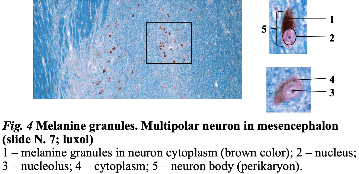
- Multipolar neuron in mesencephalon
- Melanin granules: dark brown pigment in dopaminergic neurons.
- Melanin granules, no staining needed.
- These neurons produce dopamine and then melanin is seen.
Organelles
Membranous:
- Nucleus
- ER
- Golgi
- Peroxisome
- Endosome
- Mitochondria
- Lysosome
Non-membranous:
- Ribosomes
- Centrioles
- Cytoskeleton
Cytoplasmic inclusion
- Nutritional inclusions
- Secretory inclusions
- Pigmental inclusions
- Endogenous (cells make melanin themselves)
- Exogenous (From the outside)
- Crystal inclusions, usually in cells that suffer a pathology, but Leydig cells have this.
Epithelial tissue
Tissue: an assemblage of similarly specialized cells united in performing a specific function
- Epithelial tissue
- Connective tissue
- Muscle tissue
- Nerve tissue
Lining of GI tract organs and other hollow organs and skin surface (epidermis)
Originates from all three germinal layers.
- Ectoderm: epidermis, sweat gland,
- Mesoderm: mesothelium
- Endoderm: Airways
Covering/lining group 🡪 simple or stratified
- Simple: mesothelium, endothelium
- Stratified: Epidermis
Glandular epithelium 🡪 exocrine glands or endocrine glands
- Exocrine: salivary glands
- Endocrine: thyroid or hypophysis
Pigmented,
spermatogenic,
sensory epithelium.
Properties
- Strictly polarized: Apical and basal poles and lateral surface
- Apical pole: covered in specialized structures, microvilli, stereocilia, cilia.
- Cells are always separated from other cells by basement membrane.
- Abundant junctions with other epithelial cells and with basement membrane
- Is avascular!
- Good regeneration, except sensory epithelium
Basement membrane
- Supports epithelial cells.
- Forms barrier, between epithelium and other tissues.
- Works as filter, not all molecules can pass.
- Composed of basal and reticular laminas.
- Basal: closer to the epithelium made of:
- Lucida: type IV, XVII collagen, integrins, laminins (Closest to epithelial cells)
- Densa: heparin, IV collagen, entactin
- Reticular: I, III, V collagen, proteoglycans, (closer to connective tissue: made of connective tissue cells) (closest to fibroblast)
- Basal: closer to the epithelium made of:
- Also composed of water!!
- No cells in basement membrane!!!
Intercellular junctions
- Hemidesmosomes
- cell to basement membrane with integrin and cytoskeleton (keratin filaments)
- Gap junctions
- not connected to cytoskeleton but have transmembrane proteins and allows transfer of small molecules between cells.
- Desmosomes:
- transmembrane proteins and have cytoskeleton (Keratin filaments), couple adjacent cells and strengthens tissue.
- Adherent junctions:
- transmembrane proteins and cytoskeleton filaments (actin filaments) and link cytoskeleton of cells and strengthen tight junctions. Not so strong as desmosomes
- Tight junctions: not all have this. Transmembrane proteins and actin filaments, seal adjacent cells and control molecule passage. Large molecules, bacteria can´t travel between cell membranes.
Specialized structures of apical pole
- Microvilli
- An extension of cytoplasm with cytoskeleton in them
- Increase surface area.
- Stereocilia
- huge microvilli,
- increase surface area.
- Cilia
- Motor proteins which can move this apical structure.
- Moves something.
General Functions
- Protect deeper tissue.
- Transports molecules and ions
- Secrete enzymes, mucus, hormones.
- Absorbs substances.
- Receives information from environment.
- Expels secretion of glands
Naming
Number of layers (simple or stratified) 🡪 cell shape (which is dominating in simple, and which is in the apical part in stratified?) 🡪 cell properties (has cilia or keratinized?) 🡪 Epithelium
Exception:
- Transitional epithelium
- Pseudostratified epithelium
- Simple squamous epithelium (the name does not clarify the function)
- One layer
- Squamous
- Irregular shape
- Alveoli
- Inner surface of cornea
- Hearth chambers
- Endothelium
- Mesothelium: peritoneum, pleura, pericardium
- Simple cuboidal epithelium
- One layer
- Basement membrane
- Bronchi
- Ovaries surface
- Excretory salivary gland ducts
- Kidney tubules
- Simple columnar epithelium
- One layer
- Basement membrane
- Can have microvilli, cilia or stereocilia.
- Mucosa of stomach and intestine
- Mucosa of gallbladder
- Mucosa of uterine tube and uterus
- Pseudostratified columnar ciliated epithelium
- Basement membrane and 4 types of cells
- All cells touch the basement membrane so one layer.
- Basal cells (basement membrane)
- Elongated cells
- Columnar cells (cilia which removes the debris into the mouth so we can cough or swallow)
- Goblet cells (secret mucus in the airways)
- Mucosa of airways
- Part of urethra
- Ductus deferens
Stratified epithelium
Only basal cells are attached to the basement membrane and the superior cells are attached by adherent junctions or desmosomes to other cells.
- Stratified squamous nonkeratinized epithelium (in all moist places)
- Basal layer (cuboidal or columnar)
- Intermediate layer (oval)
- Surface (squamous)
- Oral cavity
- Vagina
- Esophagus
- Cornea
- Anus
Lactobacteria in vagina converts glycogen (produced by epithelial cells) to lactic acid which lower pH to protect from other bacteria. Moistness and sugar attract bacteria.
- Stratified squamous keratinized epithelium (epidermis) Found on all the skin.
- Basement membrane and 5 layers (thick skin. hairless)
- Or basement membrane 4 layers (thin skin, hairy skin)
- Stratum basale
- keratin filaments
- Stratum spinosum
- polygonal cells
- Stratum granulosum
- squamous cells, few layers, start to lose organelles.
- Stratum lucidum
- not if there only are 4 layers.
- no organelles but keep the junctions.
- Stratum corneum
- thickest layer, with dead cells: keratinocytes.
- Start to lose their junctions and filled with proteins. The cells become a squam.
- They are impermeable and prevents water loss, protects from mechanical things and sun. No visible nucleus.
- Transitional epithelium
- Composed of basement membrane and basal and surface layers.
- Surface layer with specialized cells called umbrella cells.
- Prevent other cells from all harmful substances in the urine.
- When the organ extends 🡪 the cell layers will decrease because the epithelium also extends.
- Calyces of kidneys
- Ureters
- Bladder
Sensory epithelium
- Inner ear, tongue, olfactory epithelium
- Receive, convert and pass along information.
Spermatogenic epithelium (stratified)
- Testes – seminiferous tubules
Pigmented epithelium
- In retina to prevent light to affect deeper tissue, and bounce light to the receptor.
- Simple cuboidal epithelium which are pigmented
Glandular epithelium
- Endocrine (secrete hormones into the blood stream)
- Exocrine (have excretory ducts that go to the surface of something)
- Unicellular
- Goblet cell:
- In airway and intestine
- Merocrine/apocrine
- Secretes mucus.
- Microvilli
- Basal pole resides organelles and nucleus
- Secretory granules on the apical pole
- Goblet cell:
- Multicellular
- Simple glands (one duct + secretary portion)
- Compound glands (duct with branches + secretory portion)
- Secretory portion can be tubular, acinar, tubuloacinar
- Unicellular
- Merocrine: secretes but does not affect the cell
- Apocrine: secretes and loses part of the cytoplasm and plasma membrane
- Holocrine: disintegrates upon secretion
Connective tissue
- Fat and other soft padding tissue
- Bone
- Tendon
- Blood
- Cartilage
Classification
All connective tissue is derived from the embryo mesenchyme.
- Adult
- Fibrous (proper): in between the cells and structures without functions, “the one we imagen”.
- Collagen
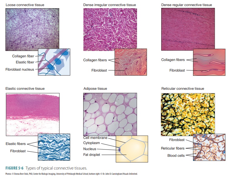
- Loose
- Dense
- Regular
- Irregular
- Elastic
- Collagen
- Specialized
- Blood & lymph
- Adipose
- White
- Brown
- Pigmented
- Reticular
- Skeletal
- Cartilage
- Hyaline
- Elastic
- Fibrous
- Bone
- Lamellar
- Woven
- Cartilage
- Fibrous (proper): in between the cells and structures without functions, “the one we imagen”.
- Embryo
- Mesenchyme
- Mucous
Connective tissue contains extracellular matrix and cells.
Extracellular matrix
- Ground substance (watery/gelatinous thing)
- Glycosaminoglycans (GAGs)
- Long chains of disaccharides longest is hyaluronan.
- Cells synthesize them directly and secrete them.
- Proteoglycans
- Chains of amino acids
- Synthesized in ER and get connected to GAGs in Golgi complex.
- Glycoproteins
- Strengthen and stabilize tissue and help cell migration thanks to areas which can join to other molecules.
- Glycosaminoglycans (GAGs)
- Fibers:
- collagen
- elastic
- reticular
Classification of fibers
- Fibrillar: type I, II, III collagen 90% of the human body
- I:
- bone tissue
- Fibrocartilage
- Pigmented CT
- Fibrous CT
- II:
- Cartilage
- III:
- Fibrous CT
- Glands
- Immune system
- Bone marrow
- I:
- Network/sheet forming: IV collagen.
- Linking/anchoring: Type VII collagen
Collagen (protein) is synthesized by cells (fibroblasts)
Fibers form only in ECM.
Reticular fibers
- III collagen
- Synthesized by reticular cells and fibroblast.
- Found in immune system and hematopoietic.
- Seen with silver salt staining.
Elastic fibers
- Composed of elastin and fibrillin (fibroblasts)
- Gives elasticity.
- Found in organs that change volume (lungs).
- Can form elastic membranes.
Cells in connective tissue proper
- Resident (true cells)
- Fibroblast, young cell 🡪 fibrocytes
- Actively synthesizes ECM components (build)
- Responsible for growth factors
- Oval euchromatic nucleus
- Developed ER and Golgi
- Fibrocytes
- Does not actively synthesize (does not build)
- Nucleus is tapered, heterochromatic.
- Can reverse to active form (fibroblast) when tissue is injured.
- Reticular cells
- In reticular connective tissue only synthesize type III collagen
- Partially cover fibers with their cytoplasmatic processes
- Pericytes
- Star shaped cells and can contract.
- Next to capillaries
- Mast cells
- Irregular shape
- Central, oval nucleus, not visible
- Basophilic granules
- Synthesize bioactive substances for immune response, inflammation and healing.
- Close to blood vessels and mucosa of organs
- Adipocytes
- In majority of organs
- Accumulate and storage lipids.
- Large groups are called adipose tissue.
- Pigmented cells (melanocytes)
- Fibroblast, young cell 🡪 fibrocytes
- Transient (traveling cells) All derived from blood.
- Granulocytes do not differentiate.
- Lymphocytes 🡪 plasma cells (look like monocytes)
- Derived from activated B lymphocytes by antigens.
- Produce immunoglobulins.
- 10 – 20 days life
- Cell is oval and heterochromatin clumps are visible in nucleus.
- Monocytes 🡪 macrophages
- Can phagocytose.
- Shape depends on activity.
- Mononuclear phagocytes system
Embryonic connective tissue
- Mesenchyme
- Mesoderm and partially from ectoderm
- Origin of all adult connective tissue
- Mesenchyme cells
- Mucous
- Found in umbilical cord.
- Does not found in adult because placenta is gone.
- Protects blood vessels of umbilical cord from strangulation due to movements.
- Mucocytes
- Hyaluronic acid in the ground substance attracts water.
Proper
Elastic connective tissue
- Elastic fibers prevail.
- Gaps can be filled with collagen, fibroblasts and fibrocytes.
- Ground substance is scarce.
- In ligaments and large arteries
- Fibrils are arranged in order.
Loose connective tissue
- Plenty of ground substance
- In submucosa of hollow organs and found almost everywhere, blood vessels.
- Fibroblasts prevails.
- Fibrils have no order.
Dense connective tissue
- Tons of densely packed tissue
- Ground substance and cells are scarce.
- Fibrocytes predominates.
- Irregular (no orderly arrangement)
- In dermis
- Regular (orderly arranged fibers)
- In tendons
- Irregular (no orderly arrangement)
Specialized connective tissue
- Reticular tissue
- Reticular cells
- Ground substance
- Reticular fibers (III collagen)
- Found in immune system and hematopoietic organs.
- Adipose tissue
- White
- Unilocular adipocytes and reticular fibers network
- Solitary cells are spherical.
- Nucleus and organelles are in the periphery.
- Secretes hormones (thermo-insulation) and pads, palms and soles.
- In adults
- Brown
- Small cells, polygonal, oval nucleus is central.
- Has lipid droplets in cytoplasm (multilocular)
- Have many mitochondria.
- Found in newborns vital organs.
- Well vascularized
- Production of heat which keeps the newborns temperature.
- White
- Pigmented tissue
- Variety of loose connective tissue (I, III collagen fibers, fibrocytes and fibroblasts + melanocytes)
- Irregular shaped cells with many branches and dark color, found in the choroid, iris and genital skin after puberty.
- Blood
- Composed of cells and extracellular matrix (plasma)
- Erythrocytes + leukocytes
- Granulocytes and agranulocytes
- Platelets (cytoplasmic processes of other cells, not cells)
- Red blood cells and platelets are never in the connective tissue but stay in the blood vessels.
- Neutrophils, secrete granules, segmented 2-5,
- Eosinophils have two segments, participate in allergic reactions and parasitic infections.
- Basophils have two segments but have high number of granules and that is why segments are not see. Allergy
- Lymphocytes
- T 🡪cytotoxins against cancers
- B 🡪 secrete immunoglobulins
- Monocytes travel to tissue and becomes macrophages which phagocytose.
Blood
Belongs to specialized connective tissue.
- 7-8% of body mass
- Erythrocytes 44%
- Leukocytes + Blood platelets <1%.
- Leukogram
- Neutrophils 50-70%
- Lymphocytes 20-40%
- Monocytes 2-8%
- Eosinophils 1-4%
- Basophils 0.5-1%
- Leukogram
- Plasma 55%
- Water 92%
- Proteins 7%
- Other solutes 1%
Hematocrit is percentage of red blood cells volume (40% women, 45% men)
Bright red is oxygenated.
Dark red is unoxygenated.
Denser than water
pH 7.35-7.45
Typical volume 4-6L
Functions:
- Distribution and transport of different materials
- O2 lungs 🡪 cells
- CO2 from cells 🡪 lungs
- Nutrients from GI
- Hormones
- Wastes from cells to excretory system.
- Regulation (maintenance of homeostasis)
- pH by albumin + bicarbonate
- Temperature
- Protection
- Platelets in coagulation seal vessel damage
- White blood cells
Plasma
- Water
- Albumins, globulins
- Clotting proteins (fibrinogen, prothrombin)
- Enzymes, hormones
- Nutrients
- Electrolytes
Serum
- Fluid that remains after blood clotting, lacks coagulation factors.
- Proteins not used in clotting, electrolytes, antibodies, antigens, hormones, exogenous substances.
Blood smear:
Erythrocytes
- Biconcave disc, thin middle, thick in periphery
- Spectrin allows shape shifting (elastic protein)
- No nuclei
- Few organelles: mainly hemoglobin carrier 33% of body.
- Anaerobic respiration
- 7,5 micrometer diameter
- Count: 4,1 – 6 x 10^12 per liter of blood (m), 3,9 – 5,5 x 10^12 per liter of blood (f)
- Live 120 days, phagocytosed in spleen, bone marrow and liver
Hemoglobin (Hb)
- Oxygen binding capacity
- Iron containing metalloprotein which transport oxygen.
- Bonds O2 to form oxyhemoglobin.
- Carries CO2 (30% carbaminohemoglobin), 10% is dissolved in blood plasma.
- 60 % CO2 is carried as bicarbonate buffer.
- Carbon monoxide reacts with hemoglobin 🡪 carbon monoxyhemoglobin which displaces O2 on hemoglobin, reducing the oxygen carrying capacity of blood.
Reticulocytes are young erythrocytes with remain of the chromatin.
Anemia
- Deficit of erythrocytes or hemoglobin in periphery blood
- Determine blood count, morphology, Hb concentration.
- Sickle cell anemia: damaged synthesis of hemoglobin and changes shape of red blood cells (autosomal recessive disease), attaches easier to blood vessel wall and can block blood passage.
Leukocytes
- 1% of blood volume
- Count 4-8 x 10^9 per liter
- Granulocytes: Cannot divide and differentiate 20 days life span
- Basophil (dark granules) 0,5 – 1%
- 2-3 lobes nucleus
- 12-15 micrometers
- 6 hours circulation
- Few days lifespan
- Histamine (increase permeability of blood vessels and causes edema), heparin (decreases blood clotting) granules.
- Acts on allergens and produce intense adverse systemic response (anaphylaxis)
- Eosinophil (red/orange granules) 2-4%
- 8-12 days lifespan
- 6-8 hours in blood
- 2 nucleus lobes
- Phagocytosis of antigen - antibody complex and enzymes destroy parasites.
- Eosinophilia: Increase during allergic reaction. Accumulate in tissues and may cause injuries of the cells, chronic inflammation, parasitic infections.
- Neutrophil (47-70%)
- Circulate 8-12 h in blood.
- Diapedesis: Exit to tissue and do not return
- 2-5 nucleus lobes
- Young stab cells increase during inflammation (2-3%)
- Phagocytose bacteria and remains of dead cells.
- When activated they restore tissue microenvironment by releasing chemokines which attract other leukocytes and cytokines.
- Release glycogen to use glucose for energy.
- Infected wounds: Pus is apoptotic neutrophilic granulocytes, degrading bacteria, tissue fluid.
- Neutropenia is an abnormal decrease in number (AIDS, drugs, genetic factors induce)
- 10-12 micrometers
- 1-4 days lifespan in connective tissue
- Female granulocytes show a Barr body (appendix with sex chromatin)
- Primary granules: major role in degrading and killing microorganisms.
- Secondary granules: smaller and less dense
- Basophil (dark granules) 0,5 – 1%
- Agranulocyte
- Lymphocyte 19-37%
- Normally in resting condition
- Rapid response to antigen 🡪 proliferates and differentiate to functional active state.
- Migrate to tissue.
- B-lymphocytes: humoral immune response
- Develop in bone marrow🡪
- Secondary lymph organs (spleen, lymph nodes, tissue)
- Differentiate into:
- B-memory cells (circulate many years and remember antigens)
- Plasma cells (effector cells participate in humoral response) increase in cytoplasm producing immunoglobulins. Peripheral nucleus and more cytoplasm.
- T-lymphocytes: cellular immune response
- Bone marrow
- Mature in thymus.
- Outspread in secondary lymphoid organs.
- Differentiate into:
- Helpers
- B-lymphocytes: humoral immune response
Activate other cells.
- Memory
Live long and remember antigens.
- Cytotoxic (killers)
Primary effector cells, destroy foreign cells.
- Suppressors
Suppress immune response when antigen is removed.
- Assist B-lymphocytes in performing humoral immune response.
- Natural killer cells
- Reside in spleen.
- Circulate in blood.
- Differentiate into:
- Cytotoxic
- Destroy other cells wall infected with virus, cancer.
- Release citokins and modulate immune response.
- Reside in resting state in lymphoid organs and circulation.
- When antigen is present, they will migrate to the particular area and start a immune response.
- Monocyte 3-8%
- Bean shape nucleus
- 12-20 micrometers
- Mitochondria, Golgi, RER granules with enzymes
- 5 days in blood
- In tissue 🡪 macrophages
- 60-90 days
- Accumulate in place of infection.
- Phagocytose
- Release cytokins that help to accumulate forces of the organism to fight infection.
- Interleukins attract lymphocytes, neutrophils.
- Antigen presenting cells to T-lymphocytes.
- Red bone marrow, spleen, lymph nodes, connective tissue: macrophages,
- Langerhans cells: epidermis,
- Lungs: macrophages,
- microglia in nerve tissue,
- Macrophages in synovial joints.
Functions:
- Protection: microbes, toxines, parasites, cancer cells.
- Diapedesis: can slip between capillary wall, reside short time in plasma after they are released from blood marrow.
- Ameboid motion
- Chemotaxis: moving in direction of a chemical
- Leukocytosis: increase number in response to infection
MHC major histocompatibility complex binds to peptide fragments from antigens for recognition by the appropriate T-cells and activate a response.
Differences
- B-cells recognize cognate antigen in native form, recognize free antigen in blood or lymph using membrane bound immunoglobulins.
- T-cells recognize cognate antigen in a processed form, peptide fragment presented by an antigen- presenting cells MHC molecule to the T-cell receptor.
Antigens: genetically foreign material that generate immune response (protein, virus, foreign cells, tumor cells)
Antibodies: proteins, plasma globulins which recognize antigens
- IgA, IgD, IgE, IgG, IgM
Blood platelets
- Originate from giant polyploid cells in bone marrow (megakaryocytes)
- 150 – 400 10^9 per liter
- 10 days life span
- 2-4 micrometers
- Central dark part (granulomere) initiates thromb process
- Hyalomere is brighter and helps degranulation.
Function
- Coagulation by attaching to blood vessel wall collagen in basal laminas.
- Release glycoprotein which induces further platelets to attach.
- Fibrinogen from plasma
- Von Willebrand factor from endothelium
- Platelet factor 4 from platelet granules
- Forms fibrin polymer network
Thrombocytopenia: reduced number in platelets
- Damaged megakaryopoiesis
- Side effect of chemotreatment
- Autoimmune disease
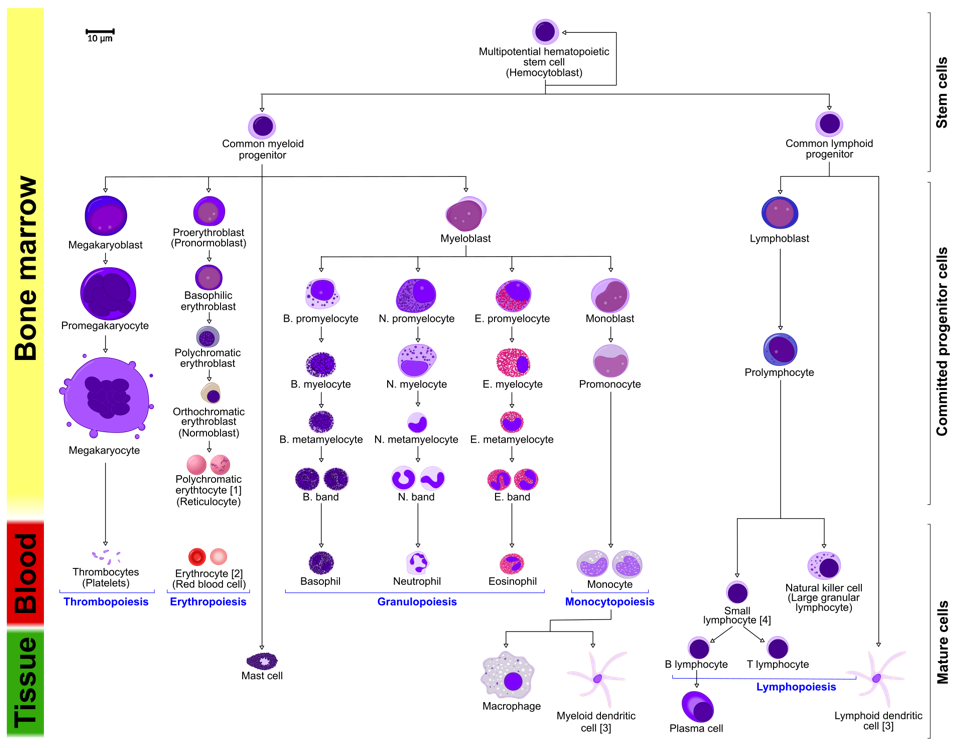
Hemopoiesis
- Mature blood cells have short life span and must be replaced.
- Yolk sac 🡪 spleen + liver 🡪 Red Marrow of specific bones in epiphysis: vertebra, sternum, rib, femur, tibia
Stem cells
- Unspecialized cell which has the ability to differentiate
- Asymmetric division
Properties:
- Proliferation – cell renewal
- Differentiation – develop to specialized cells
- Plasticity: ability of stem cells from one type of tissue to transform into stem cells of other tissue
- Lineage of stem cells: that retain ability to divide in vitro for long period without differentiation into specialized cell types.
In bone marrow are pluripotential hematopoietic stem cells
- Cell renewal and differentiation
Stem cells differentiate into:
- Lymphocytic stem cells
- Multipotent myeloid stem cells
- Give rise to major groups of blood cells (except lymphocytes)
- Burst-forming unit: erythrocytes
- Colony-forming unit: megakaryocytes
- Give rise to major groups of blood cells (except lymphocytes)
Bone marrow:
- Stroma: reticular connective tissue
- Modified fibroblasts 🡪 reticular cells
- Osteoblast
- Reticular fibers
- Parenchyma: hematopoietic cells arranged in irregular cellular cords or islands separated by thin-walled nervous sinusoids
- Red: active hematopoietic tissue
- Yellow (white):
Autologous transplantation from same patient
Allogenic transplantation from another patient
Erytrocytopoiesis is the development of erythrocyte from
- proerythroblast 🡪 erythroblast 🡪 reticuloblast 🡪 erythrocyte.
- Nucleus decrease in size and finally disappear.
- Stimulated by erythropoietin from kidney.
- Anemia and hypoxia stimulate the release of erythropoietin.
- Granulocyte
Myeloblast 🡪 promyelocytes 🡪 myelocytes 🡪 metamyelocytes (C-shaped nucleus)
- Megakaryoblast 25-50 micrometers (stimulated by thrombopoietin) 🡪 megakaryocyte 150 micrometers 🡪 Platelets.
- Monoblast 🡪 promonocyte 🡪 monocyte
Progenitors develop into B-cells in bone marrow, other progenitors travel to lymphatic organ systems and differentiate into B-cells, B-cells develop into plasma cells and release Ig.
Progenitor cells travel to thymus and maturate into T-lymphocytes.
Aplastic anemia
- Failure in bone marrow
- Bleeding (low platelets), fatigue (low erythrocytes), infection (low leucocytes)
- Pancytopenia is low count of erythroid and myeloid series.
- Caused by reduced hematopoietic stem cells.
Lymph
- Lymph plasm resembles blood plasma.
- Lymphocytes join lymph when it passes lymph nodes.
- Lymph capillaries are covered with endothelium.
Cartilage
- Hyaline cartilage
- Costal
- Articular cartilage of synovial joints
- Respiratory tract (trachea & bronchi)
- Elastic cartilage
- Auricle of Ear
- Walls of external auditory canal
- Epiglottis
- Fibrocartilage
- Meniscus
- Pubic symphysis
- Intervertebral discs
Matrix
Collagen or collagen & elastic fibres and ground substance
Cells (chondrogenesis)
Mesenchymal cells (mitotic proliferation and early differentiation)
🡪 Chondrogenic cells (form the cartilage in the embryo)
🡪 chondroblasts (produce ECM: elastic fibres + type II collagen)
🡪 chondrocytes (multiplication of cartilage cells give rise to isogenous groups)
Perichondrium
- Outer layer is fibrous.
- Dense connective tissue, less cells (fibroblasts + blood vessels)
- Inner layer is chondrogenic.
- Chondroblasts, which synthesize and secrete components of extracellular matrix.
Cartilage lacks vascular supply and receive nutrients from surrounding tissues capillaries by diffusion and they lack nerves.
Hyaline cartilage
- Cartilage matrix: collagen II + ground substance
- Articular cartilage (synovial joints) receives nutrition from the blood vessels of synovial membrane and from synovial fluid, lacks perichondrium.
- Isogenous groups of 4, 6, 8 chondrocytes
Molecular composition of hyaline cartilage
- Chondronectin 5%
- (structural multiadhesive glycoprotein) binds to GAGs, collagen and integrins mediating adherence of chondrocytes to the ECM.
- Aggrecan 9%
- Most abundant proteoglycan and binds to water.
- Cells 3 – 5%
- Collagen 15%
- II 80%
- Intercellular water 60 – 80%
Structure
Proteoglycans binds to collagen II which binds to Hyaluronan on the other end.
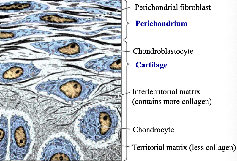
Territorial matrix has more proteoglycans.
Elastic cartilage
- Perichondrium
- Yellowish color (because of elastin)
- Cartilage matrix
- Elastic fibers
- Collagen II fibrils
- Ground substance
- Does not calcify.
Fibrocartilage
- Matrix of cartilage:
- Collagen I fibers
- Ground substance (less proteoglycans)
- Perichondrium is absent.
- General orientation of the collagen fibers depends on the stress on fibrocartilage.
- Chondrocytes are located inside the lacuna.
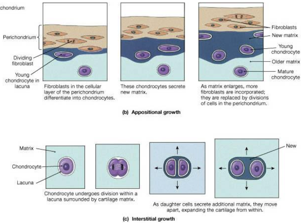
Cartilage growth
- Interstitial growth:
- Process forms within existing cartilage and is very active during endochondral ossification.
- Cells divide mitotically.
- Cells produce ECM and the mass increases.
- Appositional growth:
- Process forms new cartilage at the surface of an existing cartilage.
- Cells of chondrogenic layer differentiate into chondroblast.
- Chondroblast produce ECM and the new mass increase mass of cartilage.
- New layers of cells and ECM are added to the surface of cartilage.
- New cartilage is added beneath the perichondrium.
Bone tissue
Macroscopic:
- Spongy (cancellous)
- Compact bone
Microscopic:
- Primary (woven) bone
- Immature bone
- Collagen fibers with different directions, ground substance, osteocytes
- Lamellar bone (secondary, mature)
Bone matrix
Organic components 30%
- Collagen I (90%)
- Proteoglycans, enriched in chondroinsulfate, keratansulfate, hyaluronic acid.
- Bone sialoproteins
Inorganic components 70%
- Ca phosphate (hydroxyapatite crystals)
- Ions of Na, Mg, Fe
Mesenchymal cells
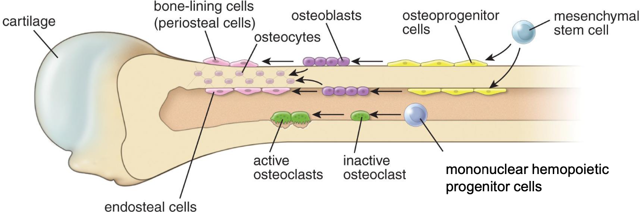
🡪 osteoprogenitor (osteogenic) cells, spindle shaped, pale and elongated nucleus.
🡪 Osteoblast, single nucleus in the periphery, tall (active) or flat (unactive) cells, produce material for general structure of bone.
- I collagen
- Osteocalcin
- Osteopontin
- Osteonectin
🡪 Osteocyte, cell body in the lacuna, elongated and branched processes forming a wide network in between cells. Communicate via gap junctions.
Osteoclast
- Multinucleated cells (20-40 nuclei), nuclei on the apical pole.
- Howships lacunae is the attachment point to the matrix.
- 100 micrometers
- Acidophilic cytoplasm
- cells are on the surface of the bone.
- Resorb bone matrix.
- Basal pole has ruffled border where enzymes and ions are released and participate in resorption of bone.
- Formed by diffusion of mononuclear hemopoietic progenitor cells.
Osteoblast
- Build the bone tissue.
- Cuboidal or columnar shape when active, less active are flatter.
- Produce young bone tissue (osteoid) distributed in the surface of woven bone.
- Polar: nuclei are oriented away from the bone
- Synthesize organic compounds
- Produce enzyme alkaline phosphatase which is needed locally for mineralization of osteoid
Osteocyte
- Embedded in lacunae in the matrix.
- Form mature bone
Bone
Diaphysis has the bone marrow and the compact bone on the periphery.
Epiphysis has spongy bone.
Compact bone:
Osteon with living osteocytes in the lamellae
- Canals with nerves and blood vessels
- Haversian canals go parallel and vertically.
- Volkmann canals are the bridges between haversian canals.
- Canals have concentric bone lamella around with parallel collagen fibers with different direction comparing with the earlier lamella. Stabilizes structure this way.
- Periosteum 🡪 Outer circumferential bone lamellae 🡪 osteon 🡪 inner circumferential bone lamellae 🡪 spongy bone
- Interstitial bone lamellae between each lamella
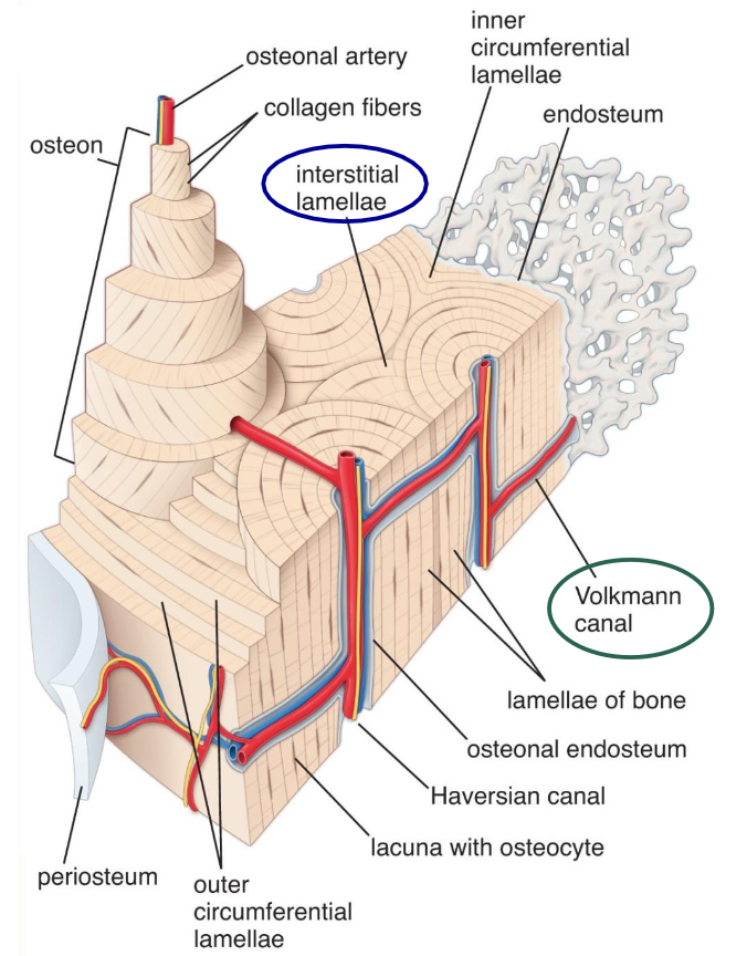
Bone canaliculus are tiny channels where osteocytes processes reside between the bone matrix of the lamellae. The osteocyte body reside in the lacuna between the lamellar layers.
Mature compact bone is actively replacing immature bone and remodeling the osteon.
Osteoblast regenerate new osteons once osteoclasts has dissolved old tissue.
Periosteum
- Fibrous layer: dense connective tissue of collagen fibers, fibroblasts, thick)
- Osteogenic layer: osteoprogenitor cells 🡪 osteoblasts: bone growth and repair
Endosteum
- Very thin layer of connective tissue on the cancellous bone
- Single layer
- Flattened osteoprogenitor cells, osteoblasts.
Osteogenesis
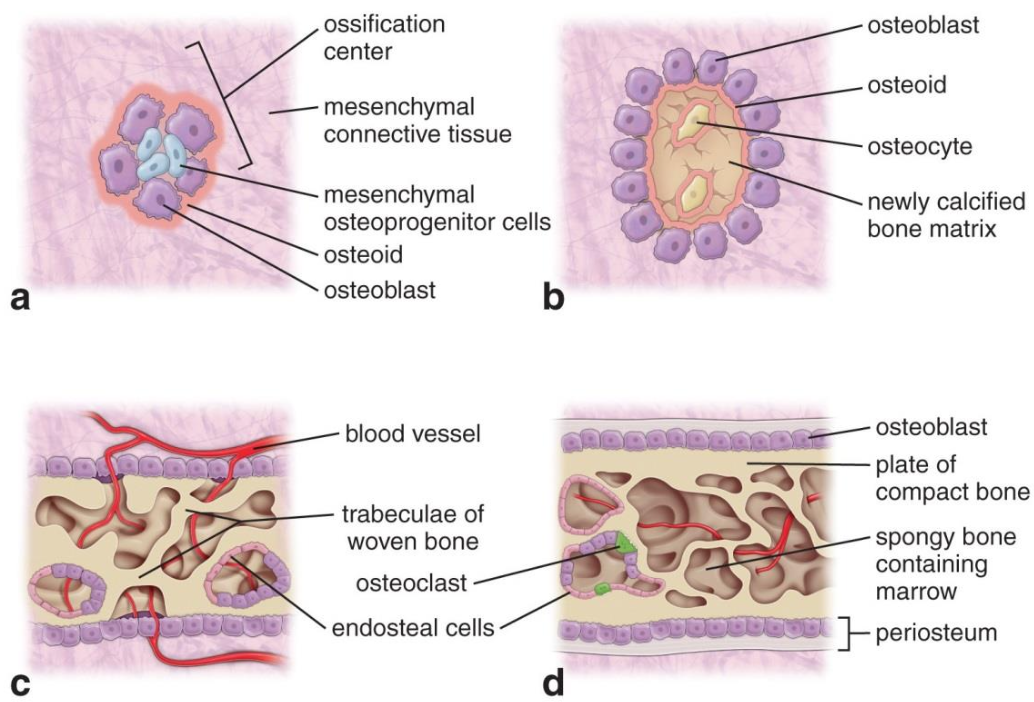
- Intramembranous ossification
- Most flat bones are produced, growth of short and thickening of long bones from mesenchymal cells.
- Development of ossification center
- Mesenchymal cells 🡪 bone blastema 🡪 osteoblast
- Formation of the bone matrix (osteoid), woven bone
- Unmineralized bone matrix (collagen I, ground substance) when osteoblasts secrete osteoid.
- Osteoblast line the surface of the matrix
- 🡪 osteocyte
- Mineralization/calcification of matrix
- Ca phosphate is deposited into matrix.
- Inorganic materials are transported by blood vessels.
- Remodeling of the bone
- Osteoclast initiate modeling of bone tissue.
- Conversion of woven bone (immature bone) 🡪 lamellar bone
- Lamellae arranged in rings around central blood vessels.
- Osteoblast produce bone lamellae on trabecular surface by recruitment of more osteoblasts to increase bone density.
- In some places spongy bone is replaced into compact bone
- Periosteum and endosteum are formed
- Development of ossification center
- Most flat bones are produced, growth of short and thickening of long bones from mesenchymal cells.
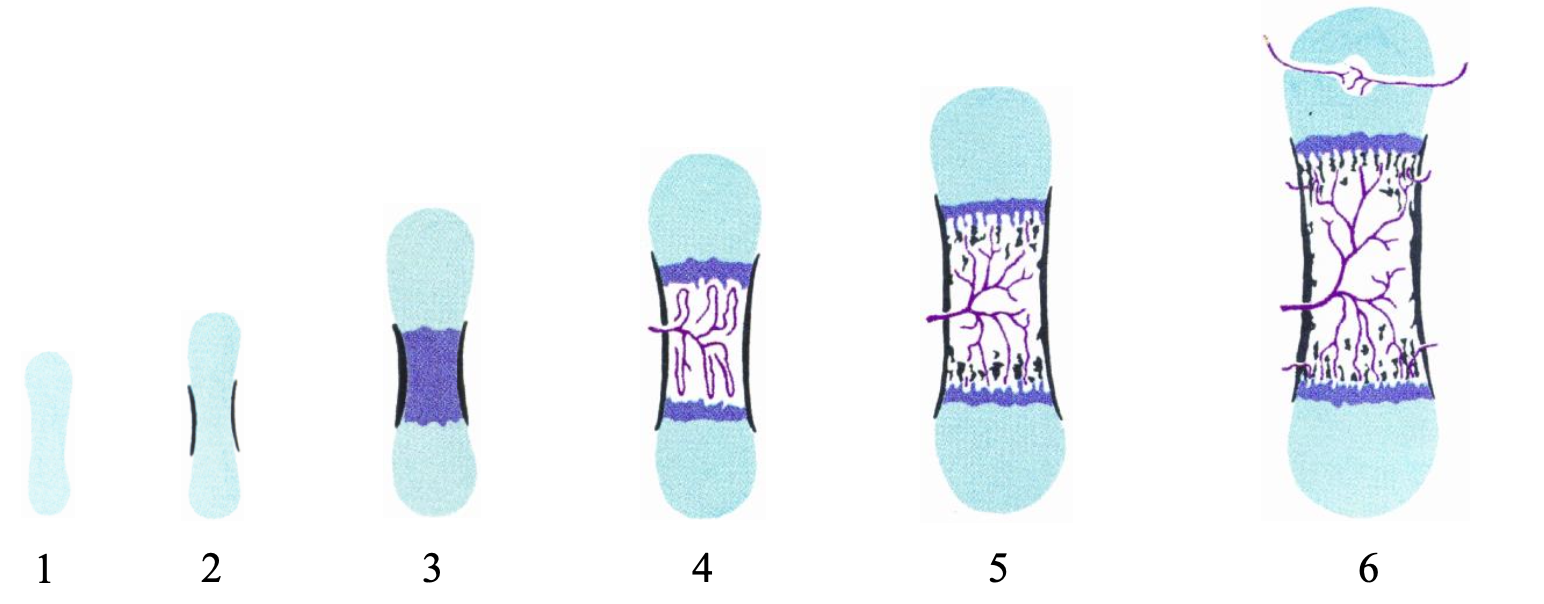
- Endochondral ossification (before birth)
- Formation of short and long bones
- Hyaline cartilage forms the model for the bone from chondroblasts 🡪 grows.
- Part of matrix calcifies, chondroblasts start to attract blood vessels when they “starve” when they are cut from the supply and attract progenitor cells which forms to osteoblast.
- Chondroblasts die and calcify the matrix
- Chondroclasts and other cells will eat the calcified cartilage and osteoblasts lay the bone on the remnants of calcified cartilage.
- Developing bone collar
- Primary (diaphyseal) ossification center where blood vessels are attracted.
- Bony trabeculae are formed.
- Secondary (epiphyseal) ossification center
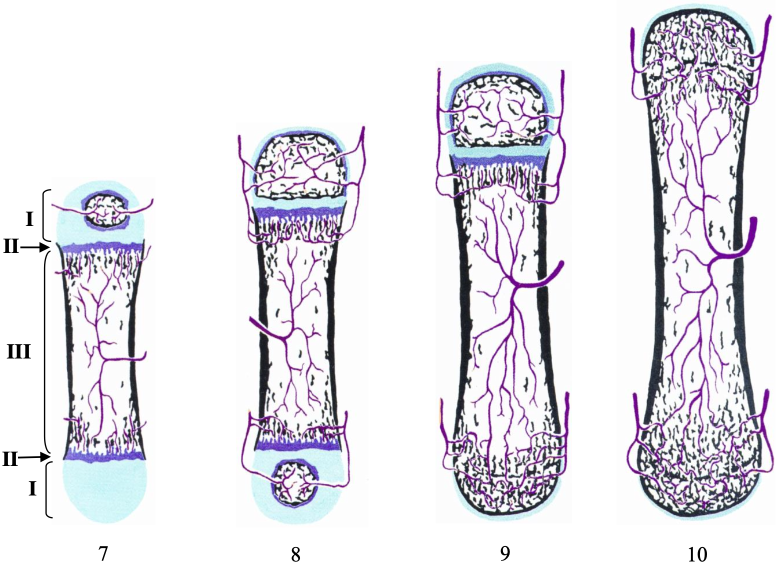
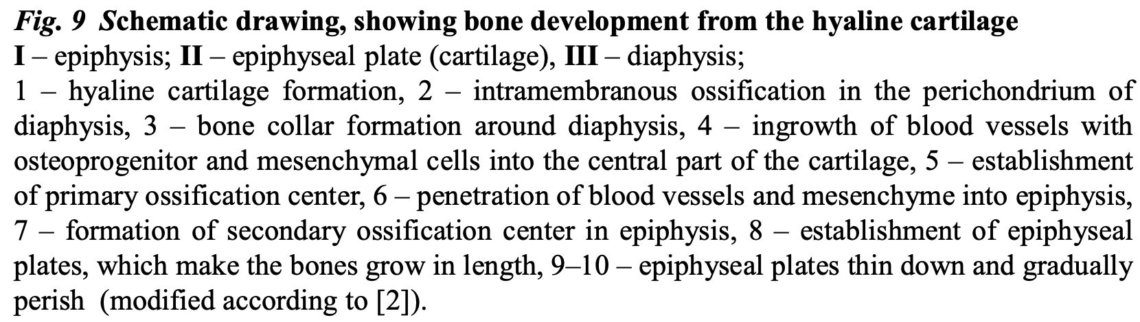
Epiphyseal cartilage plate is there as long as our bone grows and ossifies once we are done growing at about 20 years of age.
- Resting zone (reserve)
- Proliferation zone
- Hypertrophic zone
- Calcification zone
- Ossification zone
Hormones
- Parathyroid stimulates osteoclast activity (bone resorption). Raises low calcium levels.
- Calcitonin is antagonist and inhibits osteoclast activity.
Bones thicken in diameter as bone tissue is added beneath the periosteum through bone deposited by osteoblasts and they are reabsorbed by osteoclasts. As cartilage becomes bone at one side new cartilage is formed on the other site, lengthening bone.
Factors affecting bone growth.
- Lack of Ca affect bone growth.
- Vitamin D is necessary for absorption of Ca from intestine and can lead to softening of bone.
- Vitamin C necessary for collagen synthesis
Permanent bone remodeling
Needed because organs and tissues grow.
Cells age
Bones make mechanical work every day.
Bones contain reserves of Ca ions.
Osteoporosis
Deficiency in estrogen
Increase number of osteoclasts which increases reabsorption of old bone and makes cortical bone narrower and trabecular is thinner.
Muscle tissue
Forms from the mesoderm in the embryonic disc.
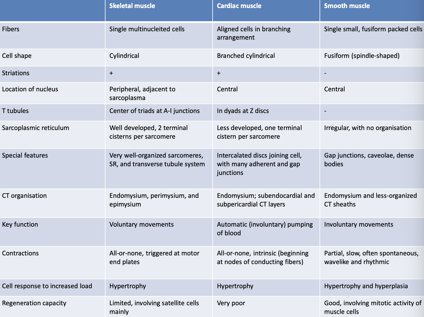
- Cardiac muscle
- Diameter: 10-20 micrometers
- Length: 50-100 micrometers
- Cylindrical shape with branches connected to each other (intercalated discs)
- Make a network.
- Uninuclear
- Myofibrils
- Smooth muscle
- Diameter: 3-15 micrometers
- Length: 20-200 micrometers
- Not striated but spindle shaped in longitudinal section.
- One elongated nucleus
- Small cells
- Skeletal muscle
- Diameter: 10-100 micrometers
- Length: 1mm-30cm
- Peripheral nucleus
- Large cylindrical shaped cells
- Striations
- Multinucleated
- Myofibrils
Skeletal muscle
- Composed of muscle fibers and connective tissue layers that separate and arrange fibers in certain directions.
- Epimysium:
- continuous with tendon
- dense irregular connective tissue
- Brings blood vessels and lymphatic vessels.
- Perimysium:
- Connective tissue layer covers the bundle/fascicles of fibers.
- Brings blood vessels and lymphatic vessels.
- Endomysium:
- Inner layer, loose connective tissue
- forms a network of capillaries around each individual muscle fiber, provides connection to nerve endings.
Muscle fibers
- Has organelles:
- Cytoplasm (sarcoplasma)
- Plasma membrane (sarcolemma)
- Nucleus
- Sarcoplasmic reticulum
- Specialized organelles:
- T-tubules
- Myofibrils
- All striated myocytes have myofibrils (structures made up of myofilaments).
- The location of myofilaments within myofibrils determines the striations of the cell.
- A-band is dark.
- I- band is light.
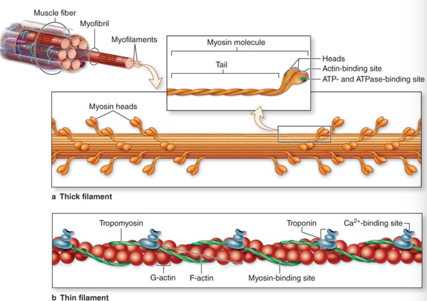
Myofilaments
- Threads made up of proteins.
- When these merge in a certain order 🡪 sarcomere is formed
- Do not change their length during contraction, they only overlap.
- Thick
- Myosin: composed of heavy and light merosine which twist into a chain.
- Light: shape of a stick
- Heavy: golf club shaped
- Myosin: composed of heavy and light merosine which twist into a chain.
- Thin
- Actin (G-actin forms a F-actin chain), 2 F-actin threads twist into a chain.
- Troponin (complex of 3 molecules on the tropomyosin)
- Tropomyosin (in between two F-actin chains)
- Thick
Sarcomere
- 2 I-bands with only thin myofibrils which only are seen as 1/2 of the I-band because of the Z-disc.
- A-band is the zone where thick and thin myofilaments overlap.
Muscle contraction
- Signal from nerve which releases acetylcholine neurotransmitter. Causes an impulse in the sarcolemma.
- Receptors from muscle cells receive the signal and travels to T-tubules and causes release of Ca ions from the sarcoplasmic reticulum to the sarcoplasm.
- Troponin binds to Ca ions and activate the thin filaments by moving tropomyosin exposing the areas of connection with actin.
- Myosin heads attach to these areas.
- Myosin pulls the thin filaments into the center of the sarcomere. Myosin heads uses ATP as energy and enables myosin heads to detach and further attach to next actin area.
- When pulse ends 🡪 Ca ions are transported to the sarcoplasmic reticulum. Tropomyosin covers the active area of actin, and the filaments return to their original positions
Triads are formed from t-tubules and sarcoplasmic reticulum.
Muscle fibers have connection with nerve cells.
- Dilated terminal axon, stores acetylcholine.
- Synaptic cleft (gap)
- Junctional folds of sarcolemma (postsynaptic surface) allow increase of surface area.
Different types of skeletal muscle fibers
According to speed of contraction and synthesis of ATP
- Slow, oxidative fibers (type I, red)
- aerobic activity and easily fatigued.
- Fast, glycolytic fibers (type IIb, white)
- aerobic activity
- Fast oxidative-glycolytic fibers (type IIa, intermediate),
- anaerobic glycolysis
Formation
- From paraxial mesoderm (somites) and somitomeres (cephalic area)
- Mesenchymal cells 🡪 myoblast 🡪 fuse to form myotubes 🡪 differentiate into Myofilaments due to the release of specific proteins.
Cardiac muscle
- Cardiomyocytes and connective tissue layers
- No epimysium and perimysium because there are layers of the heart (pericardial and periendocaridal CT).
- 3 types of Cardiomyocytes:
- Working cardiomyocytes
- Make a network by intercalated discs with gap junction to exchange ions and desmosomes so they keep connected.
- Only aerobic glycolysis (require ATP)
- Less developed sarcoplasmic reticulum with less cisternae
- Rhythmic contraction happens in cycles.
- Very vascularized.
- Contraction is controlled by conductive system of heart, sympathetic and parasympathetic nervous system.
- Formed from the visceral plate of lateral mesoderm.
- Myoblasts connect by special connections which turns into intercalated discs.
- Myofibrils are formed in the same way as in the skeletal muscle.
Smooth muscle
- Spindle shaped cells and connective tissue
- Cells connect by gap junctions.
- Adapted to slow, constant contraction which is regulated by the autonomic nervous system and hormones.
- Is a composition of several organs or tissues and have nu perimysium or epimysium
- Myocytes
- do not have T-tubules. Have caveolae instead which serve the same function.
- Superficial vesicles perform that function instead.
- Troponin and tropomyosin are replaced by calmodulin and myosin light-chain kinase.
- Able to produce components of extracellular matrix.
- Dense bodies (works as Z-lines) connect myofilaments and provides stability to the cytoskeleton and which explains why cells change the shape during contraction.
- Slow contractions and may stay contracted a long time.
Formation
Lateral mesoderm except:
- Iris muscle from ectomesenchyme
- Myoepithelial cells of some glands from the ectoderm
Nervous tissue
Developed from the ectoderm.
- Brain
- Spinal cord
- Nerves
In histology
These systems are composed of nerve tissue, which consists of neurons and glial cells.
Neuron structure
- Polarized
- Large body (perikaryon/soma) with a large euchromatic nucleus.
- A lot of chromatophilic substance (rET + free ribosomes) and may contain inclusions.
- Well developed cytoskeleton, all neurofilaments (actin + myosin) and microtubules (transport tracts) are called neurofibrils.
- Dendrites: small, short outgrowths branching off from the body of the neuron.
- Has many synapses with other neurons and can change (neuroplasticity)
- Entire surface of dendrite is covered with small spines that connect with axons.
- Axon: long outgrowth that branches off the body at the axon hillock.
- Axolemma and axoplasm
- Branch out at the end and form synapses with other cells
- Groups:
- tracts, fascicles (CNS)
- nerves, branches or roots (PNS)
- Constant exchange of organelles and substances between the body of the neuron and the axon, which is helped by motor proteins and neurofibrils. The neurofilaments attaches to the microtubules and can transport substances.
- Anterograde transport: from the body of a neuron to the axon
- Retrograde transport: from axon to the neuron body.
Synapses
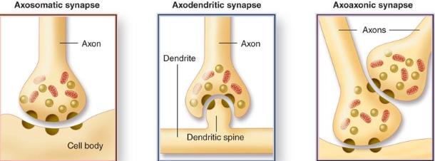
- Axosomatic synapses: axon connects to other cell body. Acts directly on a cell.
- Axodendritic synapse: axon connects to other cells dendrite.
- Axoaxonic synapse: one axon is connected to another axon that is connected to another cell body. Changes the output, can suppress or enhance signals.
Origin of neurons
CNS neurons originate from neuroepithelial cells from the neural tube. Everything is from the neuroblasts from the ectoderm.
PNS neurons originate from the cells of the neural crest.
Glial cells
- Help neurons survive and perform functions.
- About x10 more in number than neurons
- Neuropil is the network of neuronal and glial cells processes + little ground substance.
- CNS
- Ependymal cells
- Columnar
- Location; lining the ventricles of the brain and central canal of the spinal cord. Has cilia, microvilli. Attaches to neuropil via cytoplasmatic processes. No basementmembrane!
- Produce CSF (choroid plexus)
- Moves CSF (cilia)
- Absorbs CSF (microvilli)
- Choroid
- Ciliated
- Tanycytes
- Specialized cells of ependyme (simple columnar ciliated epithelium of the ventricles).
- Found in 3rd ventricle, next to nuclei important for regulation of metabolism.
- Connects with neurons, other glial cells and blood vessels.
- Columnar
- Astrocytes
- Covers/connects to synapses. Regulates plasticity!
- Regulates the concentrations of ions of the intercellular matrix around neurons.
- Helps neurons to move and orient themselves in the right direction during embryogenesis.
- Form blood-brain barrier (BBB)
- Capillaries are lined with a continuous endothelial, where cells connect with tight junctions.
- Pass only water, gas and fat-soluble substances.
- Astrocytes process covers the tight junction and stopping some molecules from passing.
- Endotheliocytes + basement membrane – astrocyte processes
- Forms the glial limiting membrane, separates brain tissue from pia matter (CT)
- Fills defects with astrocytic scar tissue after CNS lesions.
- Connects with other astrocytes.
- 2 types:
- Fibrous: white matter, long, thin outgrowths.
- Protoplasmic: gray matter
- Originates from gliablasts of the neural tube (ectoderm)
- Oligodendrocytes
- Mostly in white matter
- Function:
- Wrap the nearest axons forming the myelin sheath.
- Originates from gliablasts of neural tube (ectoderm)
- Microglia
- In gray and white matter.
- Small cells with many long branched outgrows
- Functions:
- Phagocytosis (dead cells/tissue)
- Cytokine synthesis (modulation of immune response)
- Antigen-delivering cells
- Origin from monocytes
- Ependymal cells
- PNS
- Schwann cells
- Forms the myelin sheath around the axons. Isolates and leads to faster propagation of the impulse.
- Originate from neural crest cells.
- Satellite cells
- Function:
- Nutrition
- Insulation
- Regulation of the environment around neurons
- Play a similar role as astrocytes.
- Originate from neural crest cells.
- Function:
- Schwann cells
Unmyelinated axons
- Schwann cell raps around multiple axons.
- Unmyelinated axons are enveloped by the Schwann cell, but there are no myelin sheath wraps around each axon.
Myelinated axon
- Schwann cell aligns along axon and extends a wide cytoplasmic process to encircle the axon.
- Forms a mesaxon
- Multiple spiral wrappings become compacted layers of cell membrane (myelin) as cytoplasm leaves the growing process.
- Mature Schwann cell myelin sheath has 100 lamellae, with most cytoplasm in the outermost layer with the cell body.
Myelin sheath
- Gaps between myelin segments: nodes of Ranvier.
- Loaded with large number of Na+ ion channels.
- Myelin cleft: oblique lighter streak where myelin plates are fluffier (important for axon metabolism
- Demyelinating: diseases only affecting the myelin sheath.
- Water 40%, dry mass: 70% lipids, proteins 30%
CSF
- Produced by choroid plexus (ependymal cells)
- Protects the CNS from injury, get rid of metabolites and volume correlates with blood pressure.
Nerve fibers = axon + Schwann cells
Nerve: an accumulation of nerve fibers surrounded by layers of connective tissue
- Endoneurium: loose CT surrounds each nerve fiber.
- Perineurium: surrounds bundle of nerve fibers, forms blood-nerve barrier
- Epineurium: covers the entire nerve, dense irregular CT.
Nerve endings (found in PNS)
- Unencapsulated
- Root hair plexus:
- feel hair movement.
- Free nerve endings:
- In papillary layer of dermis or epidermis layers and sense temperature, itch, touch, pain
- Tactile (Merkel) cells:
- Epidermis in basal layer, texture and sensation
- Root hair plexus:
- Encapsulated
- Meissner corpuscle:
- papillary layer of dermis, feel light touch.
- Neuromuscular spindles:
- in skeletal muscle: feels contraction or relaxation.
- Neurotendinous spindle:
- recognizes the stretching between muscle and tendon, preventing injury by suppressing muscle to contract to much.
- Ruffini corpuscle:
- dermis, feel tension or torsion of skin.
- Kraus end bulbs:
- Skin of genitals, feel low frequency vibration.
- Pacinian (lamellated) corpuscle:
- reticular layer of dermis, internal organs, feels course touch, pressure, tension and vibration.
- Meissner corpuscle:
Regeneration of nervous tissue
- CNS neurons do not regenerate, astrocytic scar is formed.
- PNS can regenerate damaged axon.
- Oligodendrocytes do not proliferate.
- All other glial cells can divide.