BIOL 117 Exam 1 Study Guide
HEART
Structures of the heart
- Exterior
- Auricles: right and left atria
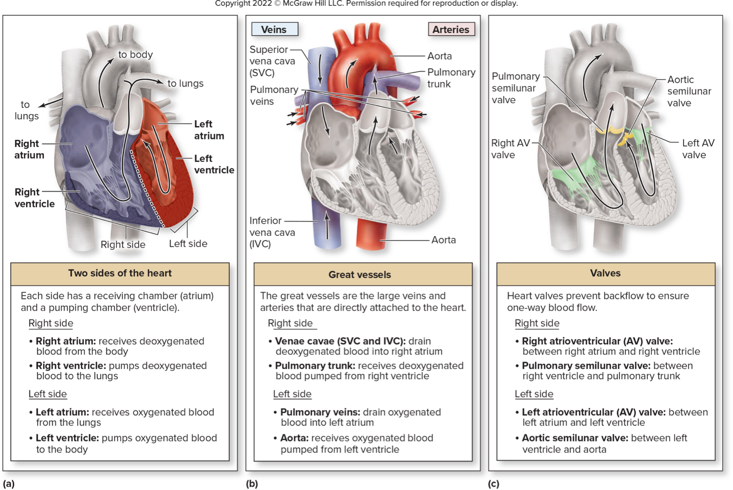
- Great vessels:
- Superior and Inferior Vena Cavae (drain deox blood into right atrium)
- Pulmonary Trunk (pumps deox blood from right ventricle to lungs)
- Pulmonary Veins (drain ox blood from lungs into left atrium)
- Aorta (pumps ox blood to body from left ventricle)
- Interior
- Chambers
- Right Atrium: receives deox blood from body
- Right Ventricle: receives deox blood from right atrium and pumps to lungs
- Left Atrium: receives ox blood from lungs
- Left Ventricle: receives ox blood from left atrium and pumps to body
- Valves
- Right Atrioventricular (AV) Valve: separates RA and RV; “bicuspid”
- Pulmonary Semilunar Valve: separates RV and pulmonary trunk
- Left AV Valve: separates LA and LV; “tricuspid” or “mitral”
- Aortic Semilunar Valve: separates LV and aorta
- Layers
- Endocardium: inner of heart and external of valves; simple squamous and areolar; lines vessels
- Myocardium: contracts for pump
- Epicardium: aka visceral pericardium; outer layer; simple squamous and areolar
- Fibrous skeleton: dense irregular CT; electrical insulator; so the atria and ventricles don't beat at the same time; attachment point for the valves to prevent backflow
- Cardiomyocyte: striated (has sarcomere), has intercalated discs (desmosomes and gap junctions) for cell communication; RMP = -90 mV
- Chambers
Route of cardiac conduction: Sinoatrial (SA) node → Atrioventricular (AV) node → AV bundle → right and left bundle branches → Purkinje fibers; nodal cell RMP=-60mV and threshold -40mV
ECG waves/intervals: P (atrial depolarization), QRS (atrial repolarization, ventricle depolarization), T (ventricular repolarization)
- Plateau phase: Ca2+ channels open and let ions in to maintain depolarization even with K+ channels open
- Ions going in and out: Na+ and Ca2+ going in and K+ going out
Cardiac cycle - phases
- Atrial Relaxation and Ventricular Filling
- 4 chambers at rest
- AV valves open
- passive ventricular filling
- Semilunar valve closed
- Atrial Contraction and Ventricular Filling
- SA node fires
- AV valves open
- ventricles filling untilEDV
- semilunar valves closed
- Isovolumetric Contraction
- AV valves closed
- semilunar valves closed
- ventricles contracting but not pumping blood away because ventricular pressure< arterial pressure
- Ventricular Ejection
- semilunar valves open
- ventricles pump blood into arteries bc ventricular pressure> arterial pressure
- AV valves closed
- Isovolumetric Relaxation
- AV valves closed
- semilunar valves closed
- 4 chambers at rest and blood is stagnant
Cardiac output: amount of blood pumped by 1 ventricle per min; L/min
- Formula: CO=HRxSV
- Stroke volume: amount of blood ejected in one heartbeat
- Factors affecting stroke volume:
- venous return (preload): determines preload; increased venous pressure and/or fill time (decreased HR) → increased SV
- inotropic agents: affect contractility
- afterload: resistance (pressure in arteries); increased afterload → decreased SV
- Chronotropic: pos (increases HR; EPI, NE, cocaine, nicotine, TH, caffeine) and neg (decreases HR; ACh)
- Inotropic: pos (increases SV; more Ca2+) and neg (decreases SV; less Ca2+)
BLOOD VESSELS
Differences between types of vessels
- Artery: away from heart; supplies; thick tunica media, narrow lumen, more elastic and collagen fibers; stays open w/o blood
- Elastic: bigger; closer to heart; lots of stretch and recoil (more elastic fiber)
- Muscular: middle size; vasoconstriction/dilation (thick tunica media)
- Arteriole: smallest; close to capillary
- Vein: towards heart; drains; thick tunica externa, wider lumen, less fibers; closed w/o blood
- Valves: prevent backflow
- Blood reservoir: hold a lot of blood for long time bc it doesn’t move very fast
- Capillary: gas exchange
- Continuous: most common; complete w tight junctions w intercellular clefts (borders) to allow small substances
- Fenestrated: complete w tight junctions and intercellular clefts + fenestrations (small pores) for small proteins
- Sinusoid: incomplete w large openings; more permeable
- Precapillary sphincter: open (flows through true capillaries), closed (bypass via thoroughfare channel)
- Capillary bed: web of capillaries working together
- Portal system: two capillary beds bc blood goes to second organ before going back to heart
- NFP (net filtration pressure): higher hydrostatic pressure on arterial end (go out of capillary) and higher osmotic pressure on venue end (go into capillary)
- Vasa Vasorum: vessels that supply blood to vessels
- Tunics
- tunica intima: endothelium (continuous w endocardium) and subendothelial layer (areolar CT)
- tunica media: smooth muscle and elastic fiber
- tunica externa: areolar CT; anchors vessels to other structures
- Differences in pressure in each: highest in arteries (about 120/80)—> capillaries(sys=dia)—> lowest in veins(0 by the times it reaches vena cava)
Angiogenesis: growing new vessels in tissues that need more perfusion; return to normal is called regression
Blood pressure regulation: blood volume, cardiac output, TPR
- Neural: in medulla oblongata; short term
- vasomotor center: regulates resistance through SNS only
- cardiac center: cardioacceleratory (increase HR&SV→ CO; SNS) and cardioinhibitory (decrease HR&SV→CO; PSNS) centers
- Hormonal: long term
- ANP (atrial natriuretic peptide): vasodilation → decrease resistance and increased urine output → decreased blood volume → decreased BP
- Renin/Angiotensin II: vasoconstriction → increased resistance→ increase BP; thirst center stimulation and decreased urine formation → increased blood volume → increased BP
- Aldosterone: from adrenal cortex; increased Na+ and water in kidney → blood volume maintenance→ increase BP
- ADH (antidiuretic hormone): from posterior pituitary; thirst, kidney water absorption → maintain bloo volume → increase BP
- Peripheral resistance: friction due to blood contact w vessel wall
- Blood viscosity: usually constant; thickness due to particles (WBC, RBC, platelets) and fluid (plasma); high viscosity→ high resistance
- Blood vessel length: constant besides angiogenesis; longer vessel → more resistance
- Vessel diameter: changes often; laminar flow says fluid moves faster in middle; vasodilation → less resistance and vasoconstriction → more resistance
- Afterload: increase AL→decrease SV→decrease CO→ decrease BP
- preload: increased PL→ high SV→higher CO→higher BP
- Baroreceptors: if BP decreases…
- decreased stretch detected by baroreceptors
- decreased signals to cardiovascular system
- cardioacceleratory center increases signals along SNS to SA node, AV node, and myocardium/ cardioinhibitory center decreases PSNS signals (increase CO)
- vasomotor SNS signals cause vasoconstriction (increase TPR)
- GOAL: increase BP
- chemoreceptors: if high CO2, low pH, or low O2
- chemoreceptors increase vasomotor signals
- vasoconstriction (increase TPR)
- venous reservoir blood moves to venous return (increase SV→CO)
- GOAL: increase BP
- better respiratory gas exchange :D
Measurements
- Arterial Blood Pressure: sys/dia
- Pulse pressure: sys-dia; measure arterial elasticity
- Mean Arterial Pressure: dia + ⅓(pulse pressure)
Routes of blood vessels
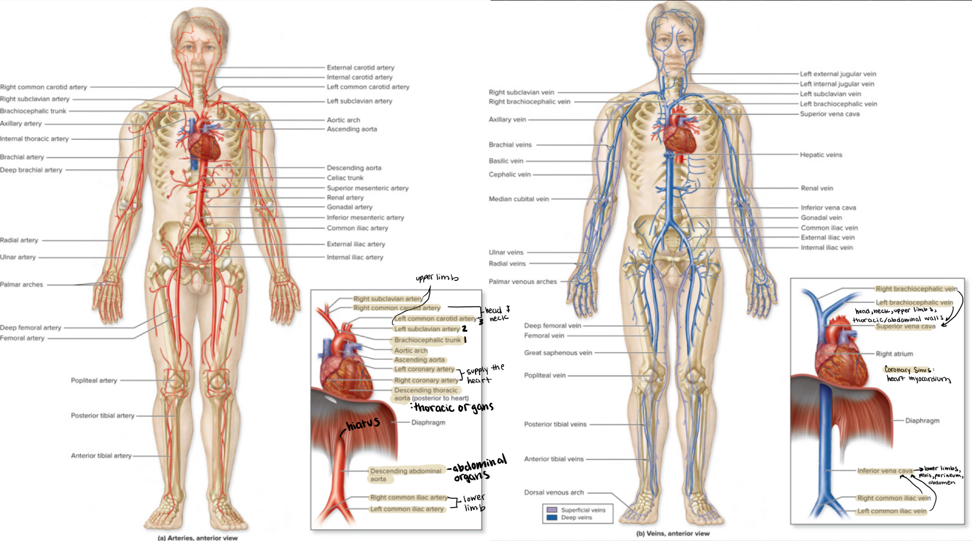
- Vessels coming off the heart
- Upper limb
- General route down to foot
- Pulmonary vs. systemic vs. umbilical
- Pulmonary: arteries carry deox blood and veins carry ox blood
- Systemic: arteries carry ox blood and veins carry deox blood
- Umbilical: arteries carry deox blood to placenta and veins carry ox blood from placenta; placenta works as the capillaries