W2: Animal Models of Myopia
Historical Discoveries & Paradigm Shift
1977–1978 “serendipitous” studies overturned the pure-genetic view of refractive error.
Wiesel et al. (1977): monocular form-deprivation in monkeys ⇒ axial elongation & myopia.
Sherman et al. (1977): lid-suture in tree shrews produced myopic changes.
Wallman et al. (1978): chicks wearing translucent plastic goggles developed large refractive shifts.
these discoveries challenged the dominance of the genetic theory, highlighting "active emmetropia" and "visual dependence on environmental factors."
Goals of Animal Research
To perform research that cannot be performed using human subjects (in an ethical manner)
To understand the process of emmetropization
To determine why this process can be disturbed
To determine the biological substances (transmitters/growth factors) which mediate eye growth
To determine if these substances could be used to correct anomalous eye growth (proof of concept studies)
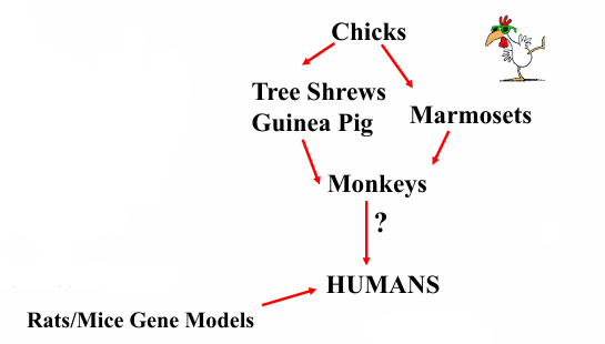
Animal Models & Research Strategy
Visual cues
Retinal control
Choroidal and scleral changes
Biochemical cascades
Effect of pharmacological
agents on refractive errors
Proof of concept, safety,
efficacy
Lead to large clinical trials
Measurement Techniques
Refraction
Infra-red photoretinoscopy, streak retinoscopy, modified autorefractors.
Axial length
-scan ultrasonography, OCT, post-mortem calipers.
Choroidal thickness
Spectral-domain OCT (EDI), swept-source OCT, histology.
Emmetropization: key points
Emmetropization is the biological process by which the eye grows and adjusts during early development to achieve low hyperopia/ emmetropia
Finds:
Nearly all neonates start farsighted; rapid axial elongation reduces refractive error in first weeks–months.
The eye uses visual cues—especially retinal defocus—to guide growth.
the most significant changes in axial length and refractive error occur in the first few weeks to months after birth.
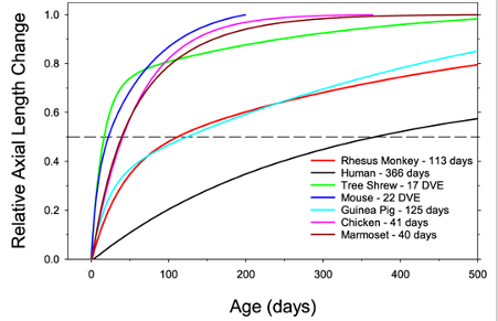
Axial Length growth in animals
Relative axial length changes for different species. The functions were normalized to the total change in axial length that occurred from birth or eye opening and adulthood.
Mice & tree shrews plotted by days of visual experience; chicks, guinea-pigs, primates by chronological age.
Form-Deprivation Myopia (FDM)
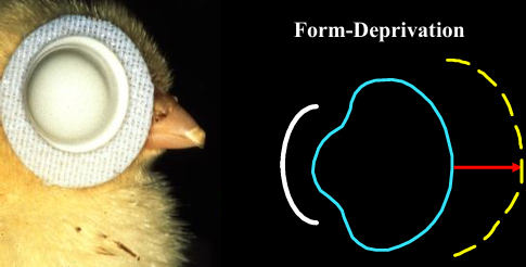
Method: translucent diffusers, lid suture ⇒ low luminance, attenuated high-spatial frequencies, poor retinal image contrast.
Mechanism: open-loop; visual feedback removed; eye cannot “know” it is growing too long.
Outcomes
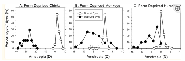
Large axial enlargement, high myopia, choroidal thinning.
Reduced retinal dopamine levels.
Temporal rules
Earlier & longer deprivation ⇒ greater myopia.
Removal early can reverse much of induced error; late removal minimally effective.
Short deprivation bursts in older animals produce negligible change.
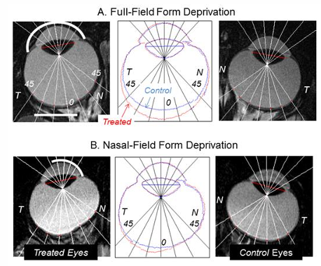
Local form deprivation causes local vitreous expansion—proved by MRI in monkeys.
Lens-Induced Myopia/Hyperopia (LIM & LIH)
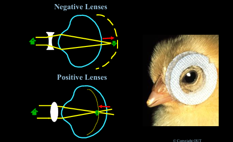
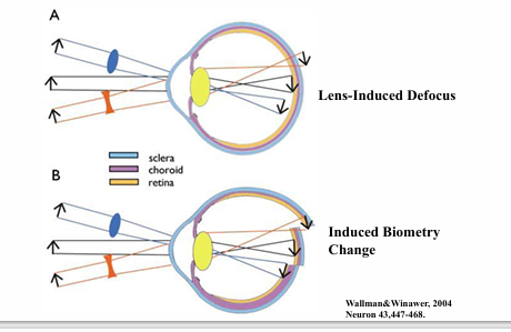
Negative lenses supply hyperopic blur ⇒ LIM.
Positive lenses supply myopic blur ⇒ LIH.
mechanism: emmetropization system responds to the blur
Visual system detects sign & magnitude (not just amount) of defocus.
Key findings
Induced refractive effects primarily via vitreous chamber depth changes.
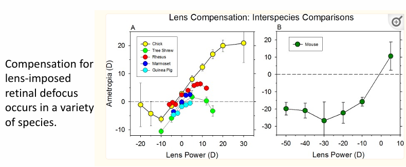
Chicks can fully compensate up to of defocus
Lower lens powers accurately compensated for in monkey and tree shrew.
The older the animal the less the change in eye growth and refractive error that can be induced.
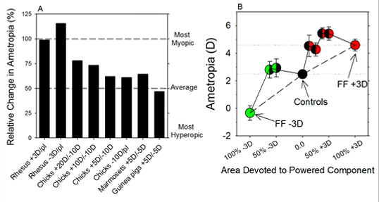
When lenses alternate or are dual-focus, positive blur has the greatest effect
Intermittent wear
Removing a −3D lens for four × 15 min per day reduced final myopia vs full-time wear in infant rhesus monkeys during the daily 12-hour lights on cycle.
Multifocal lenses:
Multifocal lenses (50:50 Fresnel): animals compensated for mean power; changing powered-area ratios shifts outcome.
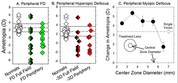
Peripheral defocus
Full field deprivation - heads towards myopia
Peripheral deprivation - less myopia than full field but shift still seen.
When using lenses for peripheral vs full field deprivation, the results are similar with a myopia shift seen in both
show the periphery is important for eye growth.
Effects of Light Modification
Mechanism: Altering ambient light parameters influences eye growth
Findings: Flicker (temporal modulation) disrupts emmetropization.
Constant light: corneal flattening + hyperopia
constant darkness: corneal flattening + eye enlargement + hyperopia.
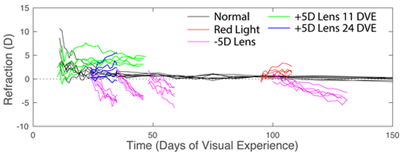
Spectral effects
Short-wave (blue) light retards axial growth, promotes hyperopia.
Long-wave (red) light promotes myopia in chicks/guinea-pigs but hyperopia in tree shrews/monkeys.
Tree-shrew study: narrow-band red light at 95 days visual experience vs monocular lenses and binocular lenses (starting 11 or 24 DVE) illustrate competing stimuli.
Myopia and Vision
-0.50 = 6/9
-1.00 = 6/12 (300 um)
-1.50 = 6/24
-2.00 = 6/36 (600)
-2.50 = 6/60
-3.00 = worse than 6/60 (1mm)
-4.00 = worse than 6/60 (1.4mm)
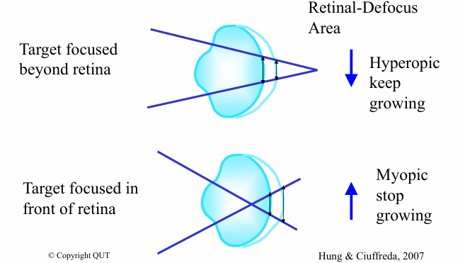
Change in retinal image defocus area theory
Retinal-defocus area (size of the retinal blur circle) provides magnitude but not directional information
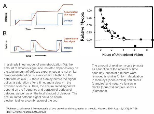
Add in time factor
Time-integrated changes in area of retinal defocus provide the critical information for directional modulation of eye growth
Hung & Ciufreda, 2007
Requirements for emmetropization:
Emmetropization is dependent on:
requires access to relatively clear images (lenses > + 40D cause myopia)
spatial frequency
contrast limited
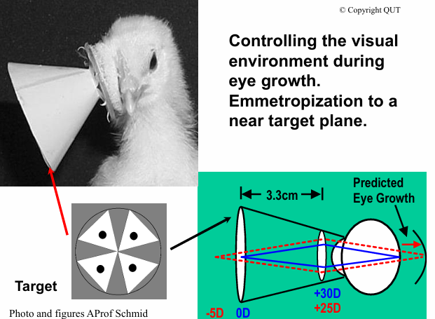
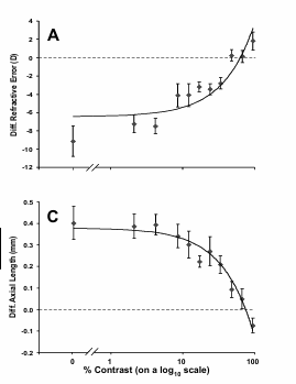
Image contrast
if contrast is too low → myopia
if contrast is at the right level → guides growth
contrast-limited syste
Spatial frequency
Form-deprivation myopia was attenuated optimally by a 1.0 cpd square-wave target, and minimally by the extreme high and low spatial frequency gratings.
mid spatial frequency that are most sensitive to changes in blur without the image being blank (too high or too low).
Evidence for Retinal Local Control
Partial (localised) deprivation results in localised vitreous elongation
FDM not affected by optic nerve section in chick
FDM not affected by blocking ganglion cell activity in tree shrew & chick
Normal retinal function fundamental to normal eye growth
Anterior and posterior segments of the eye differentially regulated
Central feedback required for fine-tuning of refractions
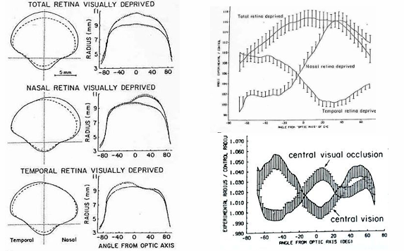
The retina controls the growth of the eye: localised or regional growth changes occur just where the retina is deprived or blurred.
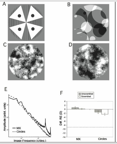
Noise target where the spatial frequencies are not in phase can control eye growth and maintain emmetropia. This is a target that the retina but not the visual cortex prefers. Scrambled vs unscrambled images very similar → retina drives emmetropization.
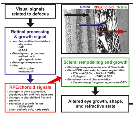
Signalling Cascade: Retina → RPE → Choroid → Sclera
Retina: secretes modulators
Dopamine: light-activated, growth inhibitor.
Retinoic acid: alters gene transcription.
Nitric oxide: vascular & signaling roles.
RPE: Acts as a mediator, transmitting signals from the retina to the choroid
Choroid: Responds by changing thickness (e.g., thinning in response to hyperopic defocus). May act as a short-term buffer to adjust retinal position
Sclera: Final target of the signaling cascade. Undergoes extracellular matrix remodelling, affecting axial elongation and refractive state.
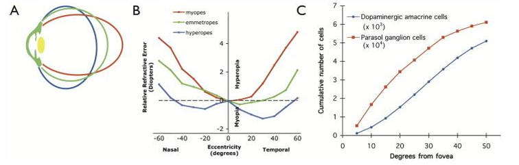
Retinal cell number across central retina:
Questions to consider:
Do you know immediately which way to move the microscope dial to clear the slide image?
How does an autofocus camera work?
How does the accommodation system know to relax or stimulate accommodation?
How does the eye growth system know what ocular parameters need altering for emmetropia to be achieved?
some sort of temporal comparison and it knows if it’s gotten worse and better in order to compensate. Integrated signal about vision and how its changing across time.
trial and error vs a signal (debated in myopia research)